VASCULAR DEVELOPMENT
During embryogenesis, the first blood vessels are formed through vasculogenesis, defined as the differentiation of undifferentiated angioblasts into endothelial cells that form a primitive vascular network.
Angioblasts originate from blood islands in the extraembryonic mesoderm of the yolk sac. Blood islands are clusters of epithelioid cells; the outer cells differentiate into angioblasts, whereas the inner cells differentiate into primitive hematopoietic cells, suggesting that both cell types originate from a common embryonic precursor cell, the hemangioblast (Fig. 102.6). Hemangioblasts are derived from earlier precursor cells under the influence of fibroblast growth factors (FGFs), and they already express the high-affinity receptor for vascular endothelial growth factor-A (VEGF-A) – vascular endothelial growth factor receptor-2 (VEGFR-2; KDR [human]; Flk-1 [murine]).
Angioblasts develop under the influence of stimulation by VEGF-A and express several vascular endothelial cell markers, including the Tie-1 and Tie-2 receptors, vascular endothelial (VE)-cadherin, and the high-affinity VEGF receptors 1 (Flt-1), 2 (KDR), and 3 (Flt-4). Within the embryo, angioblasts that appear along the anterior intestinal portal and the lateral edges of the somites coalesce to form the dorsal aorta and the large vessel primordia of the body. Angioblasts are highly migratory “prevascular” endothelial cells and are found throughout the embryo with the exception of tissues such as cartilage and epithelia.
When angioblasts form a lumen, they become endothelial cells with a polarized phenotype and production of a basal lamina. Interaction of integrin receptors on the endothelial surface with the basement membrane is critical for the formation of the primordial vascular plexus, while vascular endothelial (VE)-cadherin is required for the formation of interendothelial adherence junctions. The early vascular system consists of sinusoidal capillaries arranged in a polygonal honeycomb pattern (see Fig. 102.6). This primordial vascular plexus then becomes surrounded by mesenchymal cells that give rise to pericytes and vascular smooth muscle cells. In a next step, profound remodeling of the primordial vascular plexus takes place under the influence of VEGF-A, angiopoietins, and ephrins (see below). Remodeling involves both the formation and regression of blood vessels as well as apoptosis of vascular endothelial cells.
Angiogenesis, the sprouting of new blood vessels from existing primordial vessels, represents the major mechanism for new blood vessel formation. During this process, specialized endothelial cells, so-called tip cells, extend filopodia and sense the environment for guidance cues such as VEGF-A (Fig. 102.7). Tip cells are followed by stalk cells which extend the sprout by proliferation and form a lumen. Tip cells can connect with neighboring tip cells to form new vascular branches. The specification of tip and stalk cells is regulated by Notch signaling. Activation of VEGFR-2 on tip cells by VEGF-A results in increased expression of the Notch ligand Delta-like 4 (Dll4), which binds to Notch expressed on neighboring stalk cells. Notch signaling in stalk cells leads to reduced VEGFR-2 expression, rendering these cells less responsive to VEGF-A (see Fig. 102.7). This is a highly dynamic process in which endothelial cells compete for the tip cell position. In addition, glycolysis plays a vital role in the regulation of vascular sprouting, in parallel with genetic cues.
The Tie-2 receptor tyrosine kinase present on endothelial cells also plays a crucial role in vascular sprouting and remodeling during early embryonic angiogenesis. Pericyte-derived angiopoietin-1 (Ang-1) activates the Tie-2 receptor, whereas Ang-2 can act as an inhibitor of Ang-1 under angiogenic conditions (Fig. 102.8). In addition, Eph–ephrin receptor interactions, via bidirectional signaling, play an important role in vascular remodeling and in the determination of vascular identity. Ephrin B2 is specifically expressed by arterial endothelium, whereas its receptor, Eph-B4, is expressed by venous endothelium. Both molecules are involved in the definition of boundaries between arterial and venous endothelial cells.
Additional molecules are involved in the control of embryonic angiogenesis and vascular remodeling, including semaphorins, transforming growth factor-β (TGF-β), endoglin (the low-affinity receptor for TGF-β), and activin receptor-like kinase-1. Dysfunction of the latter two proteins is seen in hereditary hemorrhagic telangiectasia (see below). Platelet-derived growth factor subunit B (PDGF-B), secreted by endothelial cells, plays a critical role in the recruitment and differentiation of pericytes and periendothelial smooth muscle cells that contribute to the stability of mature blood vessels.
Regulation of Skin Angiogenesis
Angiogenesis is a common feature of developing skin during embryogenesis. In contrast, the blood vessels in healthy adult skin are predominantly quiescent, except for the expansion and involution of perifollicular blood vessels during the hair cycle. However, adult skin retains the capacity for brisk initiation of angiogenesis in the setting of wound healing, inflammation, and neoplastic growth. Angiogenesis occurs as: (1) the sprouting outgrowth of new capillaries from pre-existing postcapillary venules; or (2) non-sprouting remodeling of pre-existing blood vessels either through circumferential growth/vascular enlargement or through the formation of intravascular endothelial cell pillars (intussusception). Angiogenesis is a multistep process that often involves as a first step the induction of microvascular hyperpermeability, i.e. increased vascular leakage, leading to extravasation of plasma proteins such as fibrinogen and prothrombin (Table 102.2). Fibrinogen is then further processed to fibrin and serves as a provisional matrix for migrating endothelial cells, whereas the generation of thrombin acts as an initiator of the extrinsic coagulation cascade, leading to cleavage and activation of several matrix proteins. Together, these processes lead to the generation of a highly angiogenic stroma.
A major function of pericytes appears to be the maintenance of mature and quiescent blood vessels (a function which is interrupted during
Activation of VEGFR-2 by VEGF-A in vascular tip cells promotes filopodia formation and increased expression of the Notch ligand Delta-like 4 (Dll4). Dll4 activates Notch signaling in neighboring stalk cells, leading to reduced VEGFR-2 expression and diminished sensitivity to VEGF-A.
angiogenesis). In normal adult skin, pericytes secrete Ang-1, which leads to phosphorylation of the Tie-2 receptor (a receptor exclusively expressed on endothelial cells) and maintenance of the mature vascular architecture with low vascular permeability (see Fig. 102.8). In turn, endothelial cells secrete growth factors that mediate the recruitment of pericytes, including PDGFB that binds to the PDGFR-β on pericytes. During angiogenesis, endothelial cells express high amounts of Ang-2, a functional antagonist of Ang-1 in activated endothelium, thereby blocking the maturation-inducing effect of Ang-1 and leading to detachment of pericytes from vascular endothelial cells. In the presence of VEGF-A, endothelial cells then undergo the consecutive steps of angiogenesis, whereas in the absence of VEGF-A, vascular endothelial cells undergo apoptosis (see Fig. 102.8). Both VEGF-A and Ang-2 expression are upregulated in angiogenic skin diseases, including psoriasis, and keratinocyte-derived VEGF-A and placental growth factor (PlGF; see below) both potently induce the expression of Ang-2 in dermal microvascular endothelial cells. Bone marrow-derived progenitor cells also contribute to the angiogenic response, with most of these cells differentiating into perivascular macrophages that secrete angiogenic factors, including VEGF-A.
The vascular basement membrane serves as a natural barrier that prevents sprouting of endothelial cells from quiescent vessels, with pericytes assisting in the maintenance of the quiescent state. An early step in the angiogenic response involves the degradation of vascular basement membrane components such as collagen type IV by the concerted action of activated proteases, including matrix metalloproteinase (MMP)-2 and MMP-9. Integrin receptors on the surface of activated endothelial cells, in particular the αβ, αβ, and αvβ integrins, then interact with either native or degraded matrix molecules (e.g. collagen type I, fibronectin) to facilitate endothelial cell migration. The migration of endothelial cells is directed towards a gradient of angiogenesis factors that are usually produced by the epidermis, but may also be secreted by macrophages and other stromal cells. Endothelial cells then undergo a series of cell divisions, promoted by potent angiogenesis factors such as VEGF-A and basic fibroblast growth factor (bFGF). Finally, the endothelial cells form vascular lumina and mature new blood vessels with basement membranes and pericytes.
In normal skin, vascular quiescence is maintained by the dominant influence of potent endogenous angiogenesis inhibitors over angiogenic stimuli. Angiogenesis is induced by increased secretion of angiogenic
factors and/or by downregulation of angiogenesis inhibitors. The major skin angiogenesis factor, VEGF-A, was originally discovered as vascular permeability factor (VPF) due to the activity of tumor cell-conditioned media to induce accumulation of ascites fluid. VEGF-A is a homo dimeric, heparin-binding glycoprotein with at least four isoforms of 121, 165, 189, and 201 amino acids (due to alternative splicing) found in human skin. VEGF-A binds to two type III tyrosine kinase receptors that are expressed predominantly on vascular endothelial cells: Flt-1/ VEGF receptor-1 (VEGFR-1) and KDR/Flk-1/VEGFR-2 (Fig. 102.9). In addition, VEGF-A165 also binds to the neuropilin-1 receptor on endothelial and other cells.
In vitro, VEGF-A acts as a specific mitogen for human dermal microvascular endothelial cells and induces endothelial cell migration. In vivo, VEGF-A enhances microvascular permeability and angiogenesis. VEGF-A is expressed at low levels in normal epidermis, whereas in healing wounds and in skin diseases associated with angiogenesis and vascular hyperpermeability, particularly psoriasis, VEGF-A expression by epidermal keratinocytes is markedly increased. The expression and biologic activity of VEGF-A are controlled by several distinct molecular mechanisms. Hypoxia results in transcriptional activation (via hypoxia-inducible factors) and stabilization of VEGF-A mRNA. In the skin, hypoxia leads to upregulation of VEGF-A expression by epidermal keratinocytes, dermal fibroblasts, and dermal endothelial cells. This mechanism is likely important for the induction of epidermal VEGF-A expression during wound healing and in hypoxic areas of skin cancers.
Keratinocyte VEGF-A expression is also induced by growth factors that mediate epidermal hyperplasia. In psoriasis, healing wounds, and squamous cell carcinomas (SCCs), TGF-α and other ligands of the epidermal growth factor receptor (EGFR) are released by suprabasal keratinocytes. In an autocrine loop, these growth factors induce hyperplasia of the epidermis. Simultaneously, they induce VEGF-A gene expression and protein secretion by epidermal keratinocytes, which
leads to paracrine induction of angiogenesis via interaction with VEGF receptors on cutaneous microvessels (see Fig. 102.9). This mechanism links epidermal hyperplasia with increased vascularization, thereby providing enhanced vascular support to meet the enhanced nutritional needs of proliferating keratinocytes. Several other growth factors that stimulate keratinocyte proliferation, including keratinocyte growth factor (KGF), bFGF, and hepatocyte growth factor (HGF), can also promote VEGF-A production by epidermal keratinocytes.
The biologic importance of epidermis-derived VEGF-A for cutaneous angiogenesis in vivo has been confirmed in genetic mouse models designed to have overexpression of VEGF-A in epidermal keratinocytes. VEGF-A transgenic mice are characterized by elongated and tortuous dermal blood vessels that are hyperpermeable to circulating plasma proteins. These mice are unable to downregulate experimentally induced cutaneous inflammation and develop chronic inflammatory skin lesions that closely resemble those of human psoriasis. Many human cancers are characterized by increased expression of VEGF-A by tumor cells and by increased expression of VEGF receptors by tumor-associated blood vessels. VEGF-A also promotes skin carcinogenesis, in that selective transgenic overexpression of VEGF-A in epidermal keratinocytes resulted in enhanced tumor development and metastasis.
Placental growth factor (PlGF) is a member of the VEGF family and two isoforms, PlGF-1 (149 amino acids) and PlGF-2 (170 amino acids), are expressed in human skin. Similar to VEGF-A, PlGF expression is upregulated in cutaneous SCCs, epidermal keratinocytes at the advancing wound edge, and hyperplastic psoriatic epidermis. In contrast to VEGF, PlGF does not activate VEGFR-2 (KDR) but selectively binds to VEGFR-1 (see Fig. 102.9). In addition, the heparin-binding isoform PlGF-2 binds to the neuropilin-1 receptor. Evidence from genetic mouse models indicates that PlGF and VEGF-A act in synergy to induce both angiogenesis and vascular leakage and that PlGF exerts proinflammatory effects. A number of additional pro-angiogenic molecules are upregulated in angiogenic skin conditions, including interleukin-8, PDGF, bFGF, and several angiogenic chemokines.
Thrombospondin (TSP)-1 and TSP-2 are major inhibitors of cutaneous angiogenesis that are expressed in normal human skin. These matricellular proteins mediate interactions between extracellular matrix molecules and cellular integrin receptors. TSP-1 inhibits endothelial cell proliferation in vitro and angiogenesis in vivo. In normal human skin, TSP-1 is expressed by dermal cells and by epidermal keratinocytes. It is deposited within the dermal–epidermal basement membrane zone, contributing to the barrier that prevents ingrowth of blood vessels into the epidermis. TSP-1 expression is downregulated in cutaneous SCCs, and reintroduction of TSP-1 into SCCs inhibits tumor growth in mice, in association with inhibition of tumor angiogenesis and enhanced tumor cell death. Overexpression of TSP-1 in the skin of transgenic mice results in impaired granulation tissue formation and wound vascularization, delayed and reduced skin carcinogenesis, and diminished photodamage from chronic UVB irradiation. Interestingly, UVB irradiation of human skin results in an angiogenic switch with upregulation of VEGF-A and downregulation of TSP-1. The mechanisms by which TSP-1 inhibits skin angiogenesis include the induction of endothelial cell apoptosis through specific interactions with the endothelial CD36 receptor, the activation of latent TGF-β, and the inhibition of matrix metalloproteinase activity.
TSP-2 also acts as an endogenous inhibitor of angiogenesis. Mice deficient in TSP-2 show increased skin vascularization, enhanced and prolonged inflammatory reactions, and increased skin carcinogenesis, confirming the important role of endogenous TSP-2 in controlling skin angiogenesis. Accordingly, targeted overexpression of TSP-2 within murine epidermis protects from skin cancer development and inhibits the growth of established skin cancers. Antiangiogenic activity has also been reported for several cleavage products of molecules that are expressed in the vascular basement membrane, including fragments of collagens type IV (tumstatin), XV, and XVIII (endostatin).
Angiogenesis and Wound Healing
Capillary growth from pre-existing vasculature is an essential component of the proliferative phase of cutaneous wound healing (see Fig. 141.3). After exposure to thrombin, endothelial cells release matrix metalloproteinase-2 (gelatinase-A), which causes local dissolution of the basement membrane and extracellular matrix (ECM), allowing endothelial cells to migrate through the basement membrane. Capillary sprouts then invade the ECM and the fibrin clot in the wound bed, and within a few days, a new stroma forms. This so-called granulation tissue is characterized by the proliferation of blood vessels, fibroblast migration, and collagen synthesis. In this neostroma, the angiogenic sprouts organize into a dense microvascular network, thereby supporting cells with nutrition and oxygen.
The process of vascular sprouting within the premature wound matrix is controlled by several factors including hypoxia (due to microvascular damage) as well as various growth factors and cytokines released from platelets or produced by neutrophils, macrophages, fibroblasts, and keratinocytes during the inflammatory stage of wound healing (Fig. 102.10). The low oxygen gradient between injured and healthy tissues induces the transcriptional activator hypoxia-inducible factor-1 (HIF-1) which promotes angiogenesis via multiple mechanisms. The latter include: (1) upregulation of a large number of proangiogenic target genes; and (2) production of pro-angiogenic growth factors such as VEGF-A and stromal cell derived factor-1 (SDF-1 or CXCL12; see Fig. 102.10). In addition to hypoxia, cellular disruption also initiates angiogenesis via release of fibroblast growth factors (FGF-1/2). Endothelial progenitor cells are also recruited to the wound site, where they differentiate into mature endothelial cells.
During these initial phases of wound healing, the pro-angiogenic environment creates an immature microvascular network characterized by high vessel tortuosity, often resulting in dead-end flow pathways. The newly formed microvascular network has a density of blood vessels that exceeds that of normal skin. However, after this maximum density of blood vessels during the initial phases, microvascular regression is observed due to the release of several antiangiogenic factors including pigment epithelium-derived factor (PEDF), IFN-γ-inducible protein-10 (CXCL10), or Sprouty-2.
At the same time, vascular interstitial cells, including smooth muscle cells and pericytes, stabilize the newly formed microvascular capillaries. The major growth factor that supports the stabilization of this new vascular network is PDGF-B which stimulates pericyte differentiation and has been used therapeutically in the treatment of diabetic wounds. Furthermore, the angiopoietins Ang-1 and Ang-2, together with their receptor Tie-2, regulate the angiogenic response throughout cutaneous wound healing. Additional soluble factors that have been reported to
be involved in the orchestration of the angiogenic phase of cutaneous wound repair are depicted in Fig. 102.10. Defects in cutaneous angiogenesis impair the formation of granulation tissue and delay healing, leading to the development of a non-healing environment in several types of chronic cutaneous wounds.
Regulation of Leukocyte–Endothelial Cell Interactions
Accumulation of leukocytes is a prominent feature of acute and chronic inflammatory skin diseases and represents a rate-limiting step in the inflammatory response. Leukocyte recruitment is a multistep process controlled by the sequential activation of a number of adhesion molecules on both leukocytes and vascular endothelial cells, as well as by several inflammatory cytokines and chemokines. Leukocyte extra vasation occurs primarily in the postcapillary venules of the skin as they are particularly sensitive to the induction of adhesion molecules. The specific cell-type expression, ligands, and function of the major adhesion molecules involved in leukocyte–endothelial cell interactions are summarized in Table 102.3.
As the first step in leukocyte extravasation, relatively weak adhesive interactions between leukocytes and endothelial cells, involving distinct selectins and their ligands, lead to tethering and rolling of leukocytes on the vessel wall (Fig. 102.11). In a second step, leukocytes become firmly attached to the vessel wall, which is mainly mediated by the interaction of adhesion molecules of the immunoglobulin superfamily and their ligands. The switch from the rolling phase to firm luminal adhesion and crawling requires activation of integrins, in which they switch from a low-affinity to a high-affinity conformation. This activation is mediated by signaling via chemokines that are either expressed by endothelial cells themselves and presented on their surface or expressed by perivascular cells and transcytosed by the endothelium.
Finally, leukocytes migrate into the dermis through spaces between adjacent endothelial cells. Junctions between endothelial cells are usually very tight due to interactions of vascular endothelial (VE)-cadherin, junctional adhesion molecules (JAMs), and others, and they need to be opened transiently for leukocytes to be able to transmigrate. During this process, vascular endothelial-protein-tyrosine phosphatase (VE-PTP) plays a major role. VE-PTP is normally associated with VE-cadherin but becomes transiently separated from it during leukocyte transmigration.
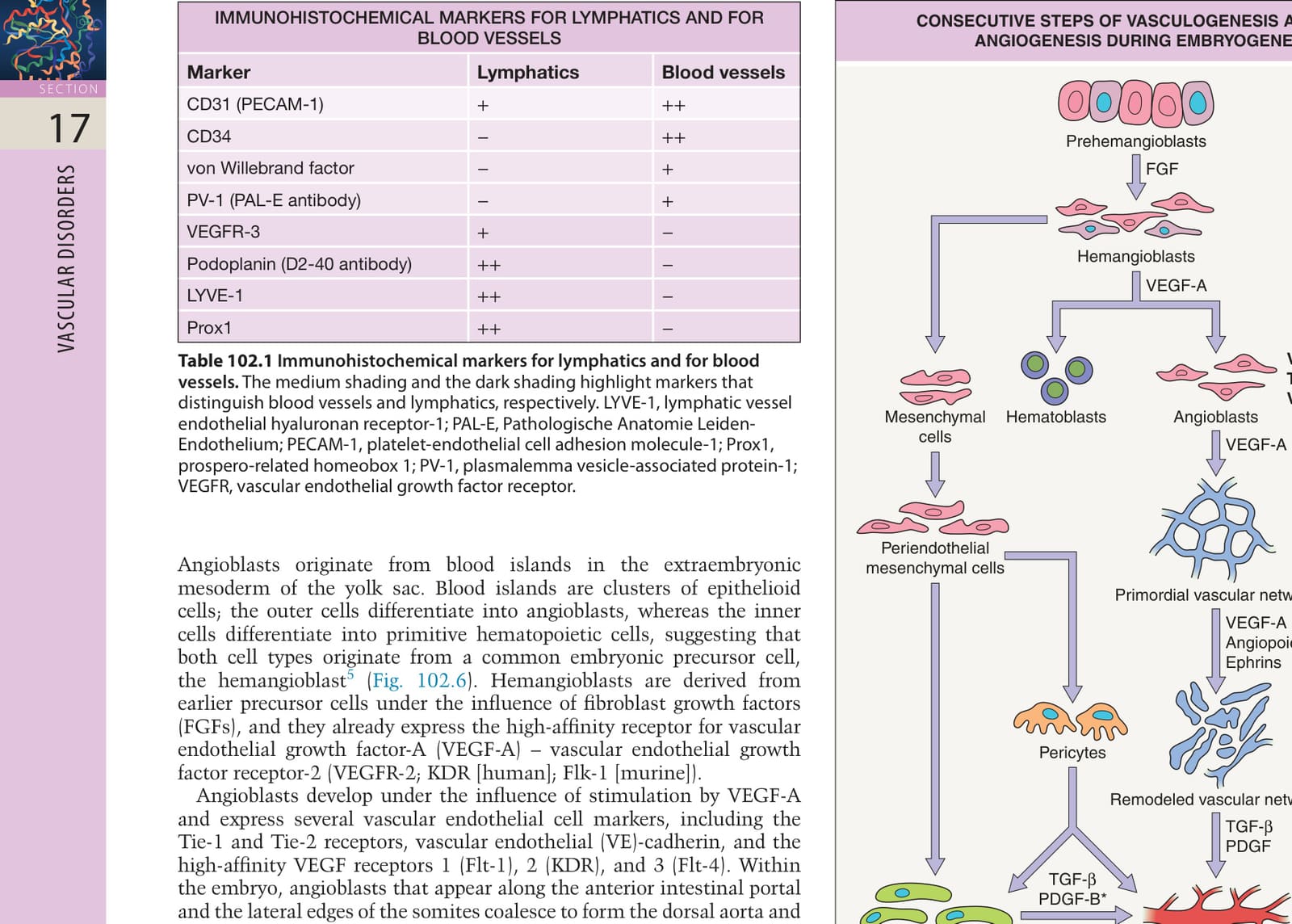
Fig. 102.6 Consecutive steps of vasculogenesis and early angiogenesis during embryogenesis: formation and remodeling of primordial vascular networks.
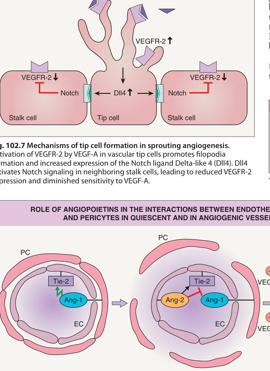
Fig. 102.7 Mechanisms of tip cell formation in sprouting angiogenesis.
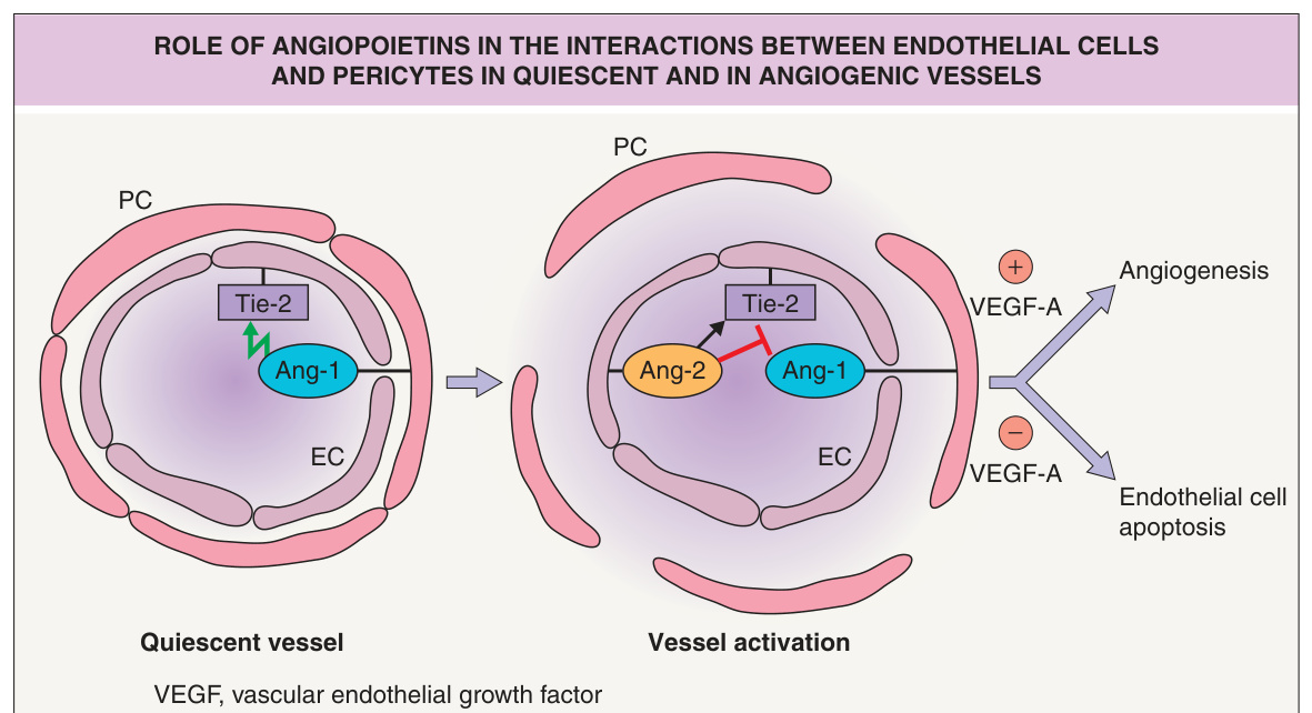
Fig. 102.8 Role of angiopoietins in the interactions between endothelial cells and pericytes in quiescent and in angiogenic vessels. Activation of the endothelial Tie-2 receptor by pericyte (PC)-derived angiopoietin-1 (Ang-1) maintains vessel maturation. Upregulation of angiopoietin-2 (Ang-2) in angiogenic endothelial cells (EC) blocks the vessel-stabilizing effect of pericyte-derived Ang-1 via the endothelial Tie-2 receptor. Both VEGF-A and Ang-2 expression are upregulated in angiogenic skin diseases, including psoriasis.
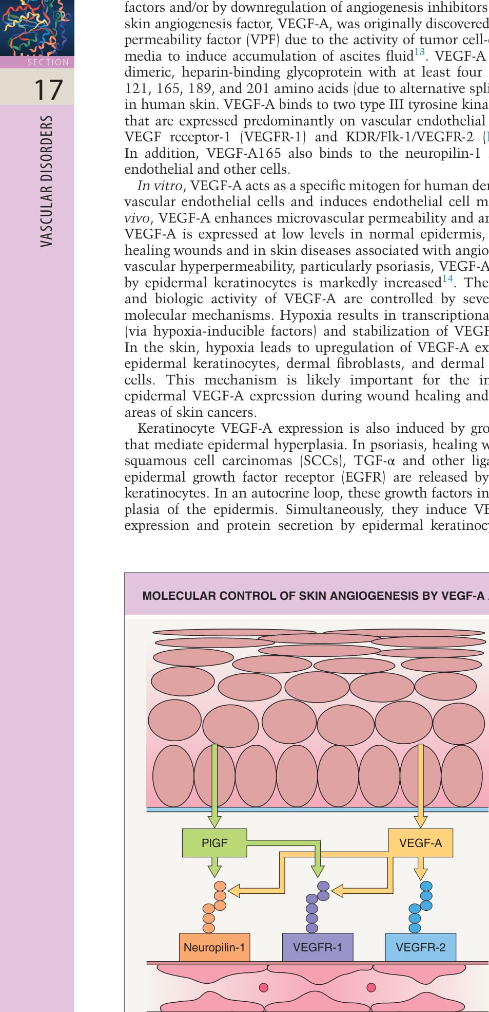
Fig. 102.9 Molecular control of skin angiogenesis by vascular endothelial growth factor-A (VEGF-A) and placental growth factor (PlGF).
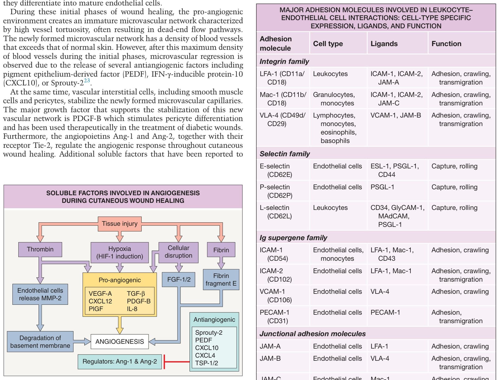
Fig. 102.10 Soluble factors involved in angiogenesis during cutaneous wound healing. Following tissue injury, multiple pro-angiogenic as well as antiangiogenic factors modulate cutaneous wound healing. Ang-1/2, angiopoietin-1/2; CXCL4/10/12, C-X-C motif chemokine ligand 4/10/12; FGF-1/2, fibroblast growth factor-1/2; HIF-1, hypoxia-inducible factor-1; IL-8, interleukin-8; MMP-2, matrix metalloproteinase-2; PEDF, pigment epithelium-derived factor; PlGF, placental growth factor; PDGF-B, platelet-derived growth factor-B; TSP-1/2, thrombospondin-1/2; TGF-β, transforming growth factor-β; VEGF-A, vascular endothelial growth factor A. Stromal cell derived factor-1 (SDF-1) is also known as CXCL12.
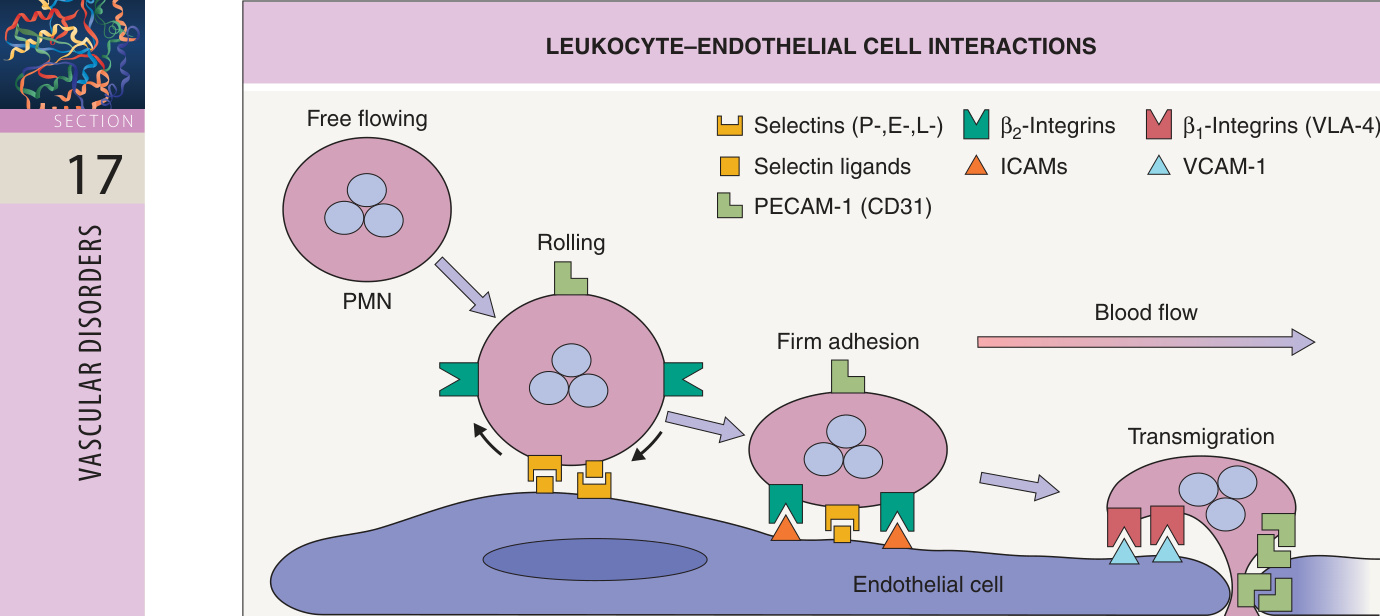
Fig. 102.11 The multistep process of leukocyte– endothelial cell interactions that leads to leukocyte recruitment into inflamed skin. Leukocyte extravasation involves tethering and rolling, largely mediated by selectins and their ligands. This is followed by firm adhesion and transmigration, largely mediated by adhesion molecules and their integrin ligands. Selectin ligands on leukocytes include E-selectin ligand-1 (ESL-1) and P-selectin glycoprotein ligand (PSGL-1).
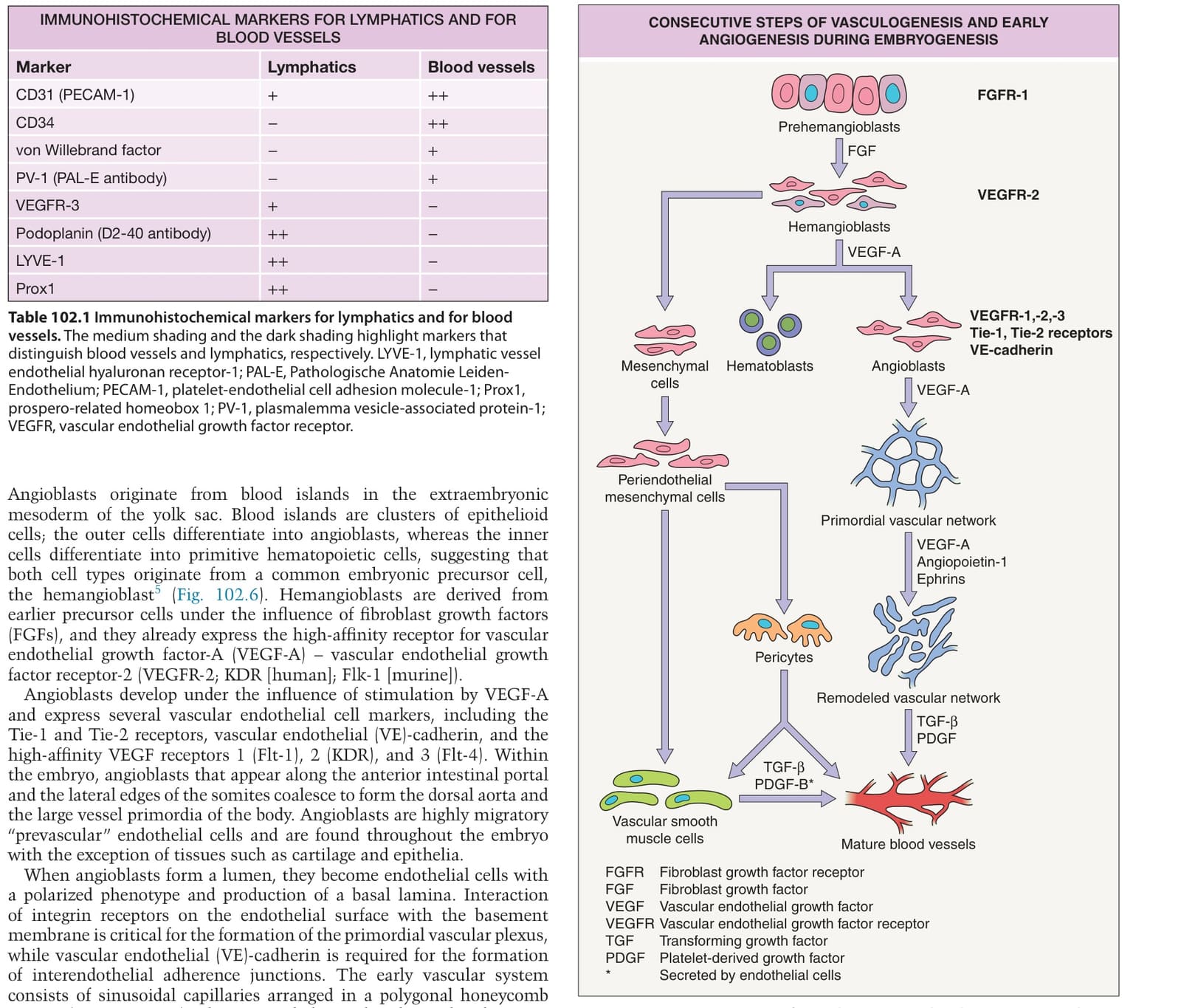
Table 102.1 Immunohistochemical markers for lymphatics and for blood vessels. The medium shading and the dark shading highlight markers that distinguish blood vessels and lymphatics, respectively. LYVE-1, lymphatic vessel endothelial hyaluronan receptor-1; PAL-E, Pathologische Anatomie Leiden- Endothelium; PECAM-1, platelet-endothelial cell adhesion molecule-1; Prox1, prospero-related homeobox 1; PV-1, plasmalemma vesicle-associated protein-1; VEGFR, vascular endothelial growth factor receptor.
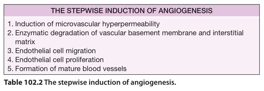
Table 102.2 The stepwise induction of angiogenesis.
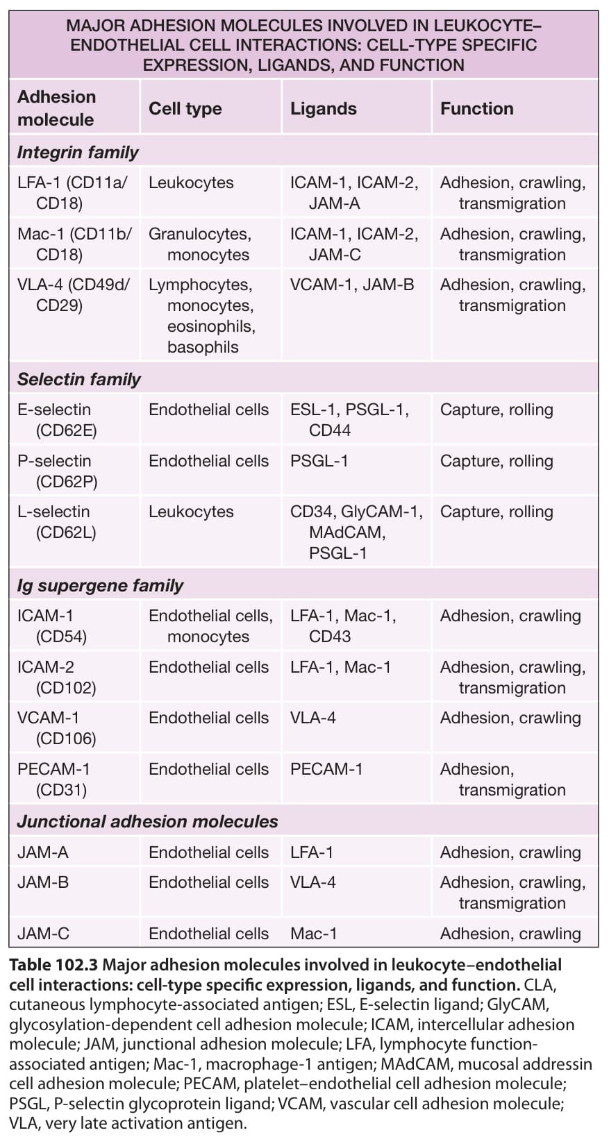
Table 102.3 Major adhesion molecules involved in leukocyte–endothelial cell interactions: cell-type specific expression, ligands, and function. CLA, cutaneous lymphocyte-associated antigen; ESL, E-selectin ligand; GlyCAM, glycosylation-dependent cell adhesion molecule; ICAM, intercellular adhesion molecule; JAM, junctional adhesion molecule; LFA, lymphocyte functionassociated antigen; Mac-1, macrophage-1 antigen; MAdCAM, mucosal addressin cell adhesion molecule; PECAM, platelet–endothelial cell adhesion molecule; PSGL, P-selectin glycoprotein ligand; VCAM, vascular cell adhesion molecule; VLA, very late activation antigen.