RADIOLOGIC FEATURES
Most superficial and mixed IHs are diagnosed based upon their clinical features. Ultrasonography or MRI may be helpful in confirming the diagnosis and extent of deep IHs that lack superficial changes. It is important to consider the phase of growth when assessing
Collections of endothelial cells forming vascular lumens, separated by fibrotic septae. The inset shows positive GLUT1 staining of endothelial cells. Courtesy Matthew Kuhar, MD.
hemangiomas radiographically, because proliferating hemangiomas demonstrate different characteristics than their involuting and involuted counterparts.
MRI with contrast is the most useful imaging technique for helping to define the extent and tissue characteristics of vascular anomalies, and the use of contrast can help to differentiate a hemangioma from other tumors. If MRI does not confirm the diagnosis of hemangioma or there are concerns regarding a possible malignancy, histologic examination of the tumor is recommended. MRI and/or MR angiography (MRA) can also be used to determine the presence of hemangiomas within internal organs and to detect other structural abnormalities, including CNS malformations and arterial anomalies.
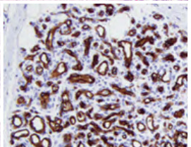
Fig. 103.21 Histologic features of a proliferating infantile hemangioma.