COMPONENTS OF PDT
A range of photosensitizers have been developed and evaluated along with a multiplicity of different light sources and delivery systems (Table 135.3). The two basic pro-photosensitizers that are utilized are ALA and mALA. They were originally used with either artificial blue or red light devices. Additional light sources have been adopted and include natural daylight and investigative garments that incorporate low irradiance light-emitting diodes and are placed over the skin.
Photosensitizers
Although topical and systemic photosensitizers based on the full porphyrin structure have been developed and evaluated, to date none have become practical, either on a clinical or commercial level, for dermatologic use. Thus, for practical purposes, topical PDT relies primarily on the “ALA prodrug” approach.
ALA and mALA represent topical prodrugs that are converted within the skin into PpIX. A familiarity with porphyrinogenesis in mammalians aids in understanding the pharmacology of ALA and its congeners. ALA (5-aminolevulinic acid, δ-aminolevulinic acid) is synthesized in all nucleated cells, and it is an intermediate compound in the heme biosynthetic pathway (see Fig. 49.1); ALA synthase catalyzes the formation of ALA from succinyl-CoA and glycine. The latter reaction represents the rate-limiting step in this pathway and is inhibited by heme. When pharmacologic quantities of ALA are supplied to cells as in PDT, this regulatory step is bypassed, leading to an intracellular accumulation of PpIX, which is photodynamically active. Of note, PpIX is synthesized within mitochondria (see Fig. 135.1) and then diffuses to cellular membrane systems such as the endoplasmic reticulum and the plasma membrane.
Unlike fully formed porphyrin molecules that are relatively large, ALA is small and readily crosses the cutaneous permeability barrier via active transport mechanisms. Within the skin, the absorbed ALA induces porphyrinogenesis, thereby inducing “porphyria” at the cellular level. In some PDT applications, the skin is first prepared by gentle curettage to remove excess surface scale and crust or to remove the stratum corneum; this preparatory step can promote topical drug absorption. Within the skin, PpIX accumulates in the epidermis and sebaceous glands due to its lipophilicity.
The carboxyl moiety of ALA can be esterified by a variety of side chains, the simplest of which results in mALA. Substituted ALA congeners are absorbed into the skin by passive and active mechanisms different from those of ALA. Upon their uptake into cells, these substituents are hydrolyzed to yield free ALA (see Fig. 135.1). Both ALA and mALA are considered prodrugs or pro-photosensitizers since the skin cells themselves generate the active photosensitizer PpIX following application of these exogenous compounds.
ALA has been compounded in a variety of different vehicles for topical application and, in North America, it is currently commercially available in 10% and 20% preparations. The FDA-approved indication for ALA is treatment of AKs, although multiple off-label uses have been explored and described (Table 135.4). An incubation time allows ALA to be absorbed and converted to PpIX. When utilizing artificial light with ALA, many clinicians typically use a shorter incubation period of 1–4 hours, instead of the recommended 14–18 hours employed in the original controlled clinical trials. Pretreatment with microneedling or ablative fractional laser therapy can allow for even shorter incubation times while retaining similar efficacy in reducing AKs.
Mechanistically, mALA is similar in principle to ALA; it is available in a 16.8% cream formulation that must be refrigerated. The recommendation is to apply mALA for 3–4 hours under occlusion prior to exposure to red light from an LED device. It has been used for multiple different skin diseases, but depending upon the country, the approved indications include treatment of AKs and/or superficial keratinocyte carcinomas (see Table 135.4). Currently, mALA is not commercially available in the US.
Newer ALA formulations have recently been developed, including a nanoemulsion gel and an ALA-impregnated adhesive topical patch, with the latter not requiring pretreatment with curettage (see Table 135.3).
Light Sources
Choosing a light source for PDT depends fundamentally on three considerations. First, the spectral output of the light should include a waveband that corresponds to one or more of the peaks in the absorption spectrum of the photosensitizer (see Fig. 135.3). Next, the desired depth of effect determines which photosensitizer absorption peak should be chosen. For PpIX, red light activation will provide a deeper clinical effect as compared to blue light. Finally, the surface area to be treated must be considered. For example, while lasers can provide precise irradiation, they may be less practical when treating broader, extensive surfaces.
Artificial light sources (conventional PDT)
The use of artificial lamps can be referred to as “conventional PDT”. Light-emitting diodes (LEDs) and blue fluorescent lamps have been the most commonly employed and approved artificial light sources for PDT (see Table 135.3). In general, these commercial devices are relatively simple to use and can be powered by standard electrical outlets.
Off-label light sources have also been used for PDT. Lasers (e.g. copper vapor, KTP, pulsed dye) may be as effective as non-coherent artificial lights for PDT. However, they are significantly more expensive, more complex to operate, and less studied. Intense pulsed light (IPL) has also been used as an artificial light source for PDT. Adhesive light-emitting patches and fabric garments with incorporated lights have been designed to be worn by patients, and because they operate at low irradiances, there may be less pain associated with these devices.
Natural outdoor light (daylight PDT; D-PDT)
Studies have shown that natural outdoor light may be used for “daylight PDT (D-PDT)”. Drug activation occurs because this light includes all the appropriate activating wavelengths. The main apprehension with daylight activation is the inability to precisely control the light dose delivered to the skin, with the possibility of phototoxicity from overexposure or lack of efficacy due to underexposure. However, randomized controlled trials have demonstrated both safety and efficacy of daylight-activated PDT (DA-PDT). Specifically, non-inferiority for effective treatment of AKs and superiority in degree of associated pain were demonstrated in a split-face trial of daylight- versus red light-activated PDT.
Although not yet FDA-approved, D-PDT has several practical and safety advantages. There is no requirement for a specific topical drug incubation time since patients are typically exposed to outdoor light immediately after drug application. Patient self-exposure to ambient outdoor light eliminates the requirement for staff to operate a specialized device and, perhaps most importantly, the level of PDT-related pain is much reduced with D-PDT. Published measurements of daylight doses in various cities can help clinicians identify months in which there is sufficient outdoor light to effectively administer D-PDT in their locale. As a general rule, if the latitude is <45° (as in most of the US, southern Europe, South America, and Australia), PDT can be done year-round depending on weather conditions. At higher latitudes in the northern hemisphere, it might be preferable to avoid daylight PDT from October to April. Of note, direct sunlight exposure to the skin is not necessary for daylight PDT. Lastly, non-mineral, untinted sunscreens applied to provide daylight UV protection do not interfere with the absorption of visible light by PpIX.
Light delivery
Regardless of the specific light source, the PDT light dose is measured and delivered in a manner analogous to conventional phototherapy. The irradiance of the light source is specified in units of W/cm, which represents the power density per unit area. Irradiance depends on the light source itself, and it can vary based on the lamp’s age, overall operating conditions, and the distance from the lamp to the skin target. For D-PDT, irradiance is affected by geographic location, season, time
of day, cloud coverage, humidity, and air pollutants. The light dose used to activate the photosensitizer is also known as the fluence and is measured in J/cm. The exposure time in seconds required to deliver a specified fluence can be determined from the formula:
exposuretime(seconds)= fluence(J/cm) irradiance(W/cm)
PDT Dosimetry
Although PDT is deceptively simple in concept, there are multiple considerations when determining the appropriate doses of photosensitizer and light. Current clinical PDT dosing guidelines have been determined by systematic as well as semi-empiric clinical investigations; nevertheless, they are based on several basic concepts unique to PDT. The net production of singlet oxygen depends on both the amount of photosensitizer and the fluence of light delivered to the target. A certain degree of reciprocity (i.e. inverse relationship) exists between these two components, in that a high photosensitizer level combined with a low fluence can in theory produce an equivalent amount of singlet oxygen as a low concentration of photosensitizer activated by a high fluence.
Another important limit to the principle of drug–light dose reciprocity relates to the irradiance of the activating light. At very high irradiances, there is significant heat production due to both non-radiative photosensitizer absorption/relaxation and nonspecific light absorption by other tissue chromophores. This leads to photothermal effects super-imposed on the PDT process itself. At lower irradiances, however, there is evidence that the pain associated with PDT might be reduced. In D-PDT, the reduced pain may be due to the lower irradiance and longer gradual exposure to natural outdoor light as compared to artificial lamps (i.e. hours versus minutes).
An interesting and useful variation in light delivery is to fractionate the total light dose into a series of two or more separate exposures, each punctuated by defined pauses. The potential net effect of fractionating the light dose is to achieve a greater overall PDT effect without applying more drug. Mechanistically, there are several reasons why this can happen. During PDT, molecular oxygen is consumed, and a pause in light exposure allows oxygen to be replenished via vascular reperfusion and diffusion. Likewise, this pause may also allow more PpIX photosensitizer to be synthesized de novo from the residual ALA that may still be present within viable skin cells; unlike PpIX, ALA itself does not undergo photobleaching.
Other factors that can influence the PDT effect include the duration of incubation between drug application and light exposure. A longer incubation interval for topical ALA or mALA leads to greater accumulation of active photosensitizer in the skin prior to light exposure. With D-PDT, the incubation period is intentionally minimal since patients are typically instructed to go outdoors immediately after the topical agent is applied. In this way, PpIX is gradually activated as soon as it is synthesized in the skin, which is another reason why there is less overall treatment pain with D-PDT.
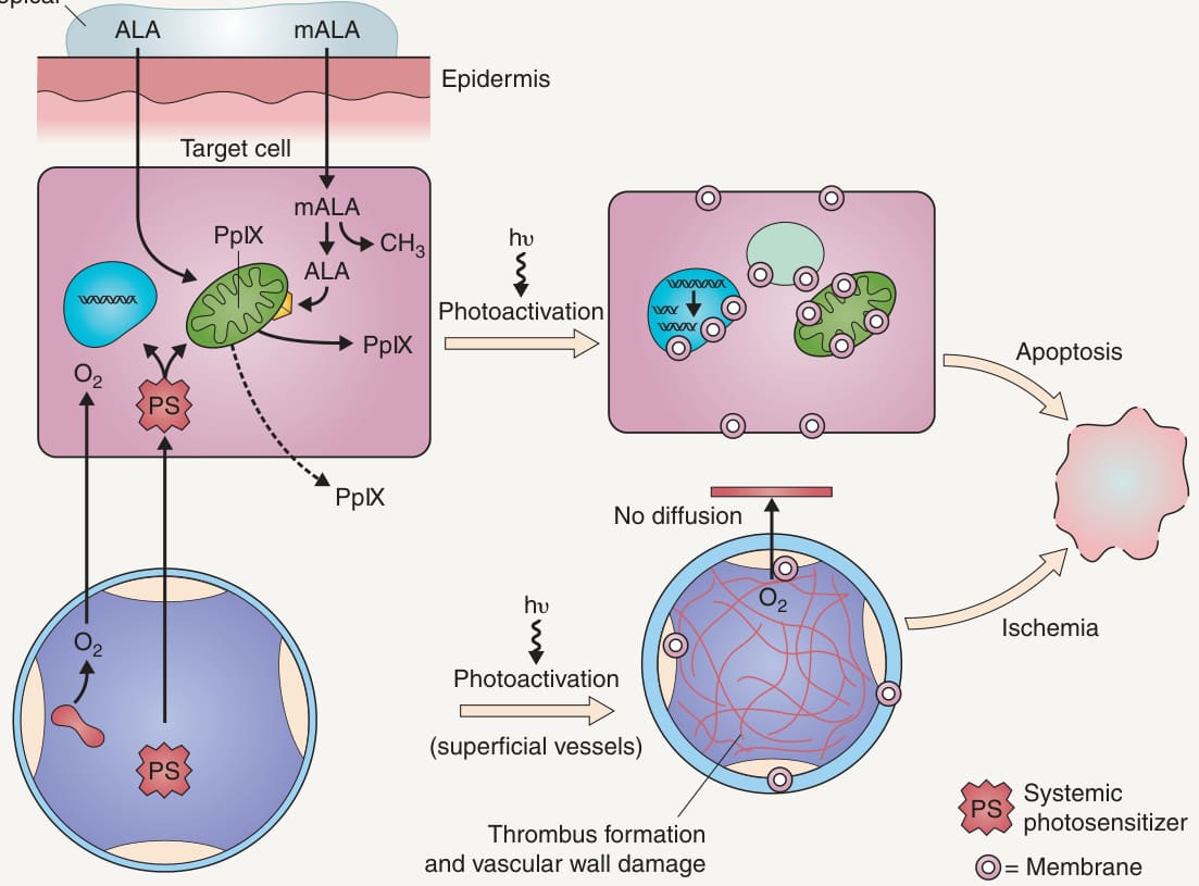
Fig. 135.1 Photosensitizer delivery and photodynamic effects on target cells and tissue vasculature.
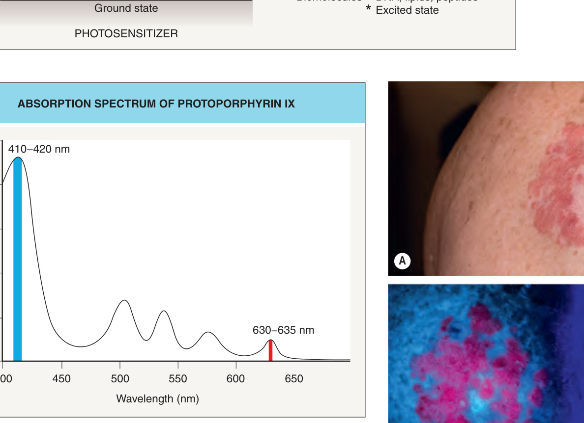
Fig. 135.3 Absorption spectrum of protoporphyrin IX (PpIX). The absorption spectrum depicts the efficiency of absorption of light as a function of wavelength. It reveals the major absorption (photoactivation) peaks of PpIX. Blue (410–420 nm) or red (630 nm) wavelengths are typically used to activate PpIX with artificial light; when daylight is used, the other PpIX absorption peaks are also activated.
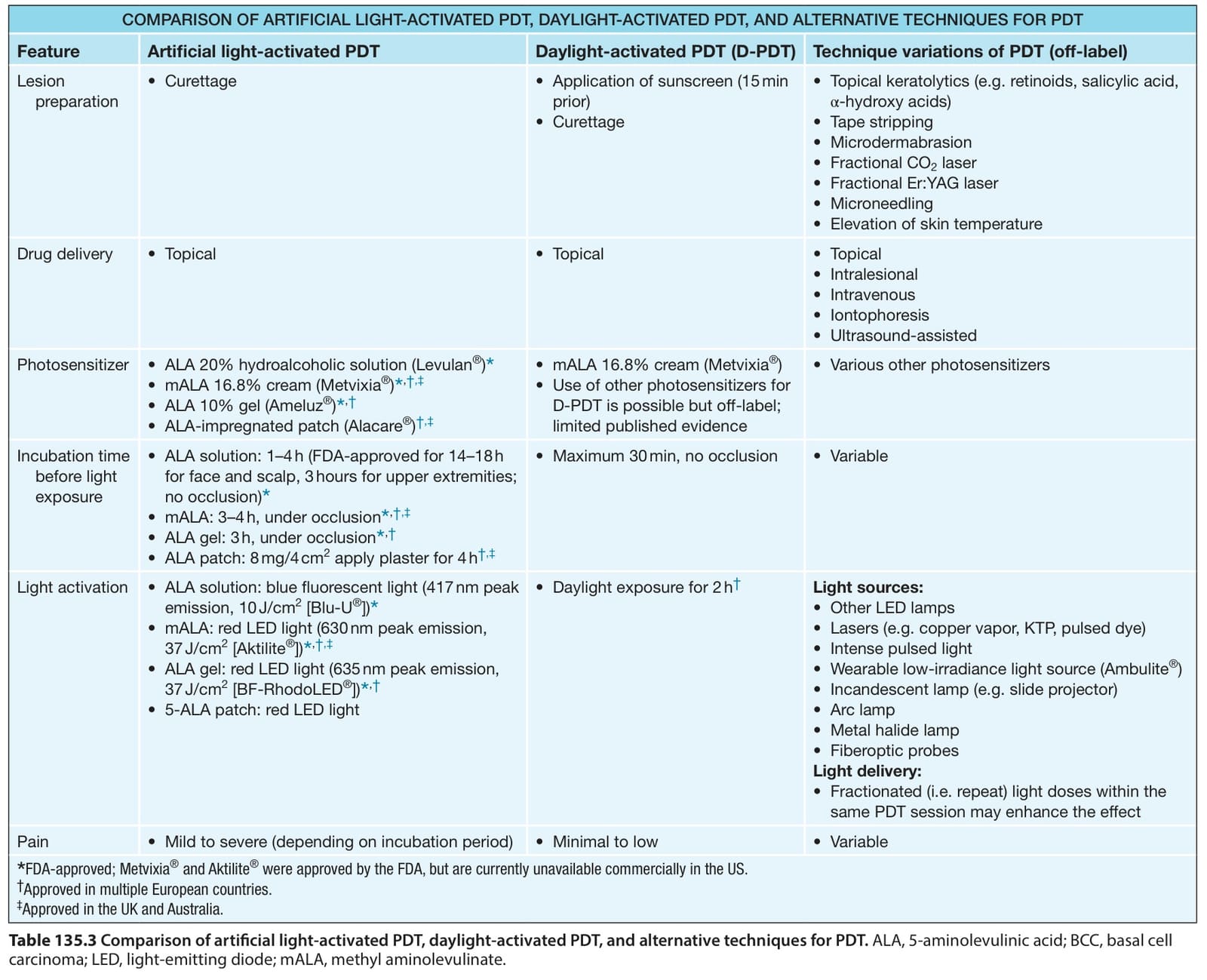
Table 135.3 Comparison of artificial light-activated PDT, daylight-activated PDT, and alternative techniques for PDT. ALA, 5-aminolevulinic acid; BCC, basal cell carcinoma; LED, light-emitting diode; mALA, methyl aminolevulinate.
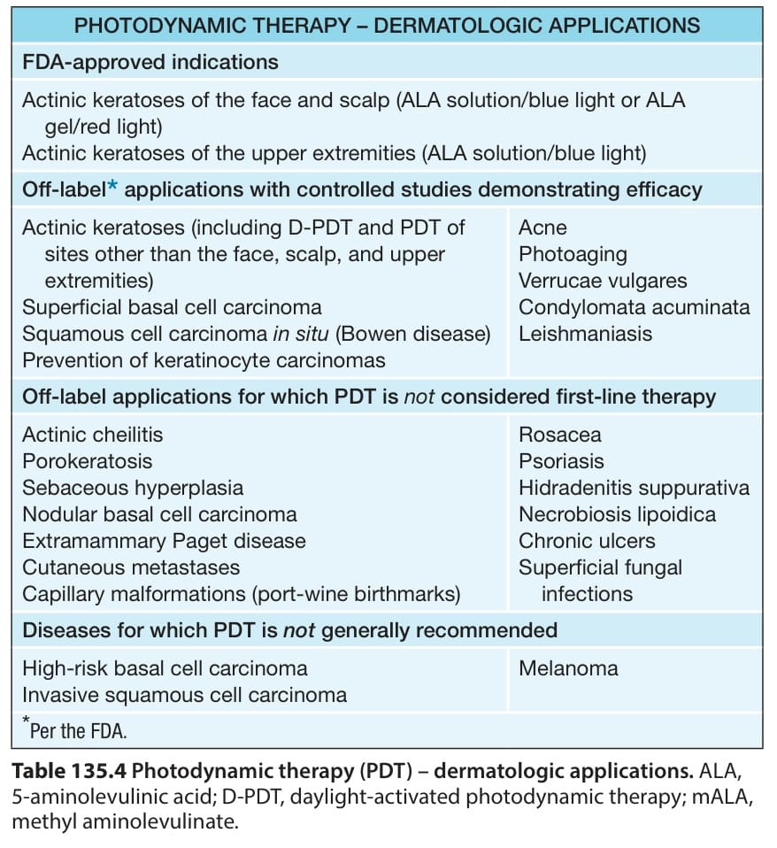
Table 135.4 Photodynamic therapy (PDT) – dermatologic applications. ALA, 5-aminolevulinic acid; D-PDT, daylight-activated photodynamic therapy; mALA, methyl aminolevulinate.