BASICS OF RADIOTHERAPY
Indications
In dermatology, the primary role of radiotherapy is to treat basal cell carcinoma (BCC) and cutaneous squamous cell carcinoma (SCC; see Ch. 108). It is also used to treat less common, but potentially aggressive, cutaneous malignancies and, occasionally, benign diseases. The former include Merkel cell carcinoma, Kaposi sarcoma (AIDS- and non-AIDS-related), angiosarcoma, adnexal carcinoma, lymphoma, and lentigo maligna.
While the majority of skin cancers are amenable to excision or other treatment modalities (e.g. electrodesiccation and curettage) which are often more cost-effective, there are clinical settings where radiotherapy is favored. For example, definitive radiotherapy is often recommended when the outcome (functional and/or cosmetic) is likely to be better with radiotherapy than with surgery, especially in the situation where the clinician is constrained by the site or size of the tumor. Likewise, elderly patients with advanced lesions for whom complex surgery is best avoided are also excellent candidates for radiotherapy. The aim of adjuvant radiotherapy is to reduce the risk of recurrence, whereas palliative radiotherapy plays an important role in patients with advanced and/or incurable disease.
Mechanism of Action
X-rays are a powerful form of ionizing electromagnetic radiation and the lethal damage they produce within double-stranded DNA represents the basis for radiation therapy. When X-rays (often referred to as photons) are absorbed by biological matter, an electron may be ejected from an atom with the local release of large amounts of energy resulting in DNA damage. Rapidly dividing cells, i.e. malignant cells, are more susceptible to lethal injury and mitotic death. Although normal adjacent tissues maintain mechanisms for repair following sublethal damage, there are limitations as to the dose of ionizing radiation normal tissue can tolerate, beyond which late reactions arise (see below). A therapeutic ratio is achieved when radiation is delivered in small divided doses (fractions), which allows time for repair of sublethal DNA damage in normal tissues without compromising tumor destruction.
Treatment Machines
In the modern era of radiation oncology, high-energy megavoltage (MV) photons (6–25 MV) are generated by a linear accelerator (LINAC) located within a heavily shielded concrete bunker. One of the advantages of megavoltage radiotherapy is its ability to treat deep-seated internal malignancies with relative skin sparing. However, in general, when treating cutaneous lesions, deep beam penetration and skin sparing are not desirable. Less penetrating modalities that avoid skin sparing, such as low-energy kilovoltage (kV) photons (50–300 kVp) supplied by a superficial/orthovoltage machine, superficial brachytherapy or electron beam therapy, are preferred modalities for cutaneous lesions. With these therapies, tissue penetration is measured in millimeters to centimeters and there is less normal tissue exposure. Radiotherapy modalities are summarized in Table 139.1.
Terminology
The International Unit of radiotherapy is a Gray (Gy). One Gy is equivalent to 100 cGy or 100 rads (in the older terminology). A fraction of radiotherapy refers to the dose delivered in one treatment. Treatments are typically delivered on a daily (Monday to Friday) basis. The daily outpatient treatments are only minutes in duration and treatment regimens usually consist of 10 to 30 weekday visits.
Radiotherapy Doses
Typical fractionation protocols for small SCCs and BCCs consist of 40 Gy in 10 (4 Gy) fractions or 50 Gy in 20 (2.5 Gy) fractions. Hypofractionation refers to the delivery of larger doses per fraction (4–7 Gy); typically a lower total dose is then delivered based upon the biological effective dose (BED; Table 139.2). The latter is calculated based upon a 2 Gy per fraction equivalent that is required to eradicate a tumor. This calculation provides an explanation for commonly prescribed schedules: 45 Gy in 15 (3 Gy) fractions, 40 Gy in 10 (4 Gy) fractions, and 35 Gy in 5 (7 Gy) fractions.
For BCCs and SCCs, there is not a marked dose–response such that local control is similar whether a patient receives 40 Gy in 10 fractions (47 Gy BED) or 50 Gy in 20 fractions (52 Gy BED). When the tumors are larger or more infiltrative, better local control is achieved with a BED of around 60 Gy. However, late effects increase as the total dose and dose per fraction increase and it is these late effects that will impact cosmesis and late tissue damage (e.g. fibrosis, atrophy, ulceration due to necrosis).
Radiation Modalities
Low-energy photons (superficial and orthovoltage)
Superficial and orthovoltage radiotherapy are two modalities that: (1) utilize low-energy photons; (2) lack skin-sparing properties; and
(3) have source-to-surface distances (SSD) ranging from 10 to 60 cm. The qualities and “hardness” of superficial/orthovoltage photon beams are determined by the placement of filters within the radiation beam. “Half value layer” (HVL) is an important concept in radiation physics and it refers to the thickness of an absorbent filter (often copper [Cu] or aluminum [Al]) that is required to reduce the beam intensity by 50%. These filters serve to remove the softer photon component and improve the penetration quality of the beam. The following are two examples of the influence of filters: a typical 100 kVp superficial photon beam with an HVL of 7 mm of Al delivers 100% of a prescribed dose to the surface, 85% at 5 mm, and 70% at 10 mm whereas a 250 kVp orthovoltage beam with an HVL of 2.5 mm of Cu delivers 95% of prescribed dose at 5 mm and 90% at 10 mm. In summary, different photon beam energies will have unique HVLs that result in varied beam qualities and dose penetration.
Office-based superficial radiotherapy was once the standard modality by which dermatologists treated skin cancer. Typical machines delivered photons of 30–150 kVp with a depth dose suitable for most patients with cutaneous malignancies <5 mm in depth. Over time, the high cost of radiotherapy machines and increased enthusiasm and training in dermatologic surgery led to a significant decline in office-based radiotherapy. More recently, introduction of superficial radiation therapy (SRT) machines has led to renewed interest in office-based radiotherapy for BCCs and cutaneous SCCs.
Orthovoltage units utilize higher energy photons of 150–300 kVp, and because of greater prescribed dose depths, are utilized for lesions >5 mm in depth. Some clinicians estimate tumor thickness and then choose an appropriate beam energy and HVL that will achieve 90%–95% of the prescribed dose at the depth of the tumor. However, this latter approach requires an accurate estimate of tumor thickness.
An important consideration when delivering orthovoltage radiotherapy is the generation of a photoelectric effect due to a higher energy range when compared to low-energy photons. A consequence of this photoelectric effect is the delivery of a higher dose of radiation to under-lying bone or cartilage. The latter does not occur with electrons (see below) or low-energy photons (see above). As a result, some clinicians use this as a justification for the use of electrons when treating lesions near bone or cartilage, although supportive clinical data are limited.
Electron beam therapy
A linear accelerator can generate electrons which can be utilized in a variety of treatment settings. Unlike X-rays, electrons are particulate radiation with a negative charge. Low-energy electrons (6–8 MeV [megaelectron volts]) offer an alternative to low-energy photons when treating cutaneous tumors, but the former do have some disadvantages. For example, electron beams are relatively skin-sparing (Fig. 139.1), and, as a result, a tissue equivalent bolus needs to be placed on the skin surface (Fig. 139.2). Also, utilizing electron beams for small treatment fields (<4 cm2) may result in tumor underdosing at the deepest extent of the tumor.
Electrons offer the advantage of a rapid linear decline in dose beyond a well-defined tissue depth that is a function of the selected beam energy
(see Fig. 139.1). For example, a 6 MeV electron beam provides some of its dose (~70%–75%) at the surface, but deposits 100% of the specified dose at a depth of ~1.5 cm. Using a higher-energy beam will result in deposition of a higher percentage of the dose at the skin surface (see Fig. 139.1), but with a higher dose to subcutaneous tissues as well as the skin surface (see Fig. 139.2A). On the other hand, if a tissue equivalent bolus of 1 cm is applied in conjunction with a 6 MeV beam, close to 100% of the desired dose will be obtained at the skin surface (see Fig. 139.2B). In other words, the dose will be brought 1 cm closer to the skin surface, further minimizing the dose to underlying structures.
Tumors treated with electrons require 10–20 mm margins due to a wider penumbra (dose drop-off at the field edge). Electrons also have a lower relative biological effectiveness (RBE) in comparison to photons, leading some clinicians to recommend a 10%–15% increase in total dose to compensate. It is important to appreciate the intricacies of using electron beam therapy, in particular the limitations of dose delivery with small fields. This requires careful attention to the margin and dose distribution at the edge of the radiation field when treating small cutaneous malignancies.
Brachytherapy
Radioisotope brachytherapy
Radioisotope brachytherapy represents radiotherapy that is delivered by the direct application of a radioactive source near or inside involved tissues. Most centers utilize a high dose-rate (HDR) system that delivers more than 12 Gy/hr, with Ir-192 being the most commonly used isotope. Treatments are delivered by the placement of surface applicators onto the skin at the site of the tumor. The radioactive source is often delivered to the applicators via a remotely controlled machine in a dedicated shielded HDR suite. Applicator selection varies depending on the location and surface contours of the target area, e.g. conical surface applicators for flat surfaces, flexible mesh style “flap” applicators for molding onto curved surfaces. When treating deeper tumors, interstitial catheters may be surgically placed within the target tissue.
The dose “fall-off” is steep with brachytherapy, providing an excellent opportunity to spare adjacent normal tissue which is not easily achieved with photon- or electron-based treatments. Per American Brachytherapy Society (ABS) consensus guidelines, HDR skin brachytherapy represents a standard of care option for appropriately selected patients with keratinocyte carcinomas. Because HDR delivery systems require Nuclear Regulatory Commission (NRC) oversight, special shielded vaults, and careful security and safety protocols, radioisotope brachytherapy is performed by a radiation oncologist in a controlled setting.
Electronic brachytherapy
A newer, non-isotope-generated form of brachytherapy, referred to as electronic (surface) brachytherapy, utilizes a miniature electronic X-ray source instead of a radioactive source. The currently available systems operate in the 50–70 kVp energy range, require little shielding, and are considered more portable. Electronic brachytherapy systems are not subject to NRC oversight, but may need to adhere to local regulations. Similar to radioisotope brachytherapy, a steep dose fall-off allows for sparing of adjacent organs and tissues. However, there are some concerns as to tissue dose calculations as well as the potential for different radiobiological effectiveness due to the photons’ low energy. Therefore, consensus guidelines for calculating tissue dosages are yet to be finalized.
As with radioisotope brachytherapy, suitable lesions need to be super-ficial (<3–4 mm in depth) and well-defined. While data are emerging regarding the safety and efficacy of electronic brachytherapy for the treatment of keratinocyte carcinomas, given the lack of both long-term follow-up and a consensus regarding dosimetric properties of this modality, the ABS does not advise its use outside of prospective or registry trials.
Advantages of Radiotherapy
Radiotherapy offers the advantage of a non-surgical approach, thereby avoiding surgical morbidity, scarring, and the need for reconstruction. In situations where reconstruction with grafts or flaps is required, improved cosmesis may result from radiotherapy. Radiotherapy fields can also be tailored with generous margins in order to encompass “at-risk” areas
(e.g. possible sites of clinically inapparent tumor extensions) that, if surgically approached, would require involved and complex surgery. In addition, standard oncologic excision margins in certain sites are often difficult to achieve without causing surgical morbidity; such sites include the midface triangle, i.e. the periorbital region (especially the medial canthus), the lower eyelid, nose (especially the ala nasi and nasal tip), nasolabial fold, lip, and chin.
Disadvantages of Radiotherapy
Radiotherapy does have its disadvantages; in particular, most patients require a protracted course of treatment compared to what is often a simple outpatient excision. Patients should also be informed that because of the risk of serious late effects such as soft tissue and cartilage necrosis, a second course of definitive radiotherapy cannot be delivered to the same site. In addition, the reduced “quality” of irradiated skin may make subsequent surgery difficult if the tumor recurs or persists following radiation therapy. Over time, irradiated skin may also develop dyspigmentation, telangiectasias, and a shiny appearance. Lastly, individuals with xeroderma pigmentosum or basal cell nevus syndrome should not be irradiated owing to their marked predisposition for developing multiple and recurrent skin cancers, often at a young age.
Tumors that have deeply invaded cartilage or bone often do better with a combination of excision plus adjuvant radiotherapy, although definitive radiotherapy still remains an option. Radiotherapy of lesions located on the foot, anterior lower leg, or dorsum of the hand, although not contraindicated, should be avoided if possible; lesions in such sites are best treated with excision. Unfortunately, radiotherapy to poorly vascularized tissue is often associated with poor healing, thus irradiation of skin cancers arising in sites of chronic ulceration, trauma, or thermal burns should be avoided, if possible. In addition, patients with systemic sclerosis and other causes of cutaneous fibrosis may experience severe fibrotic reactions following radiotherapy.
Radiotherapy has a very small (~1 in 1000) risk of causing a radiation-induced in-field malignancy (often soft tissue/bone sarcoma) 10–15 or more years after exposure. However, these data were obtained from studies involving many different malignancies treated by various techniques and dosing schedules. It is likely that with modern radiotherapeutic techniques as well as the smaller volumes being irradiated when treating skin cancers, a figure of 1 in 1000 represents an overestimate of the real risk. Nonetheless, for younger patients (<50–60 years of age), this is an important issue and patients need to be informed of the potential 5-10X greater risk of a second malignancy within the treatment field when compared to individuals not receiving radiotherapy12a.
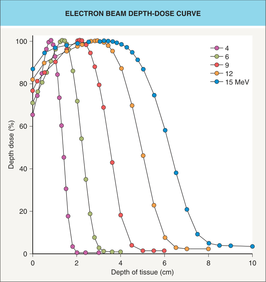
Fig. 139.1 Electron beam depth-dose curve. Electrons offer the advantage of a rapid linear decline in dose beyond a well-defined tissue depth that is a function of the selected beam energy. Courtesy Lynn Wilson, MD.
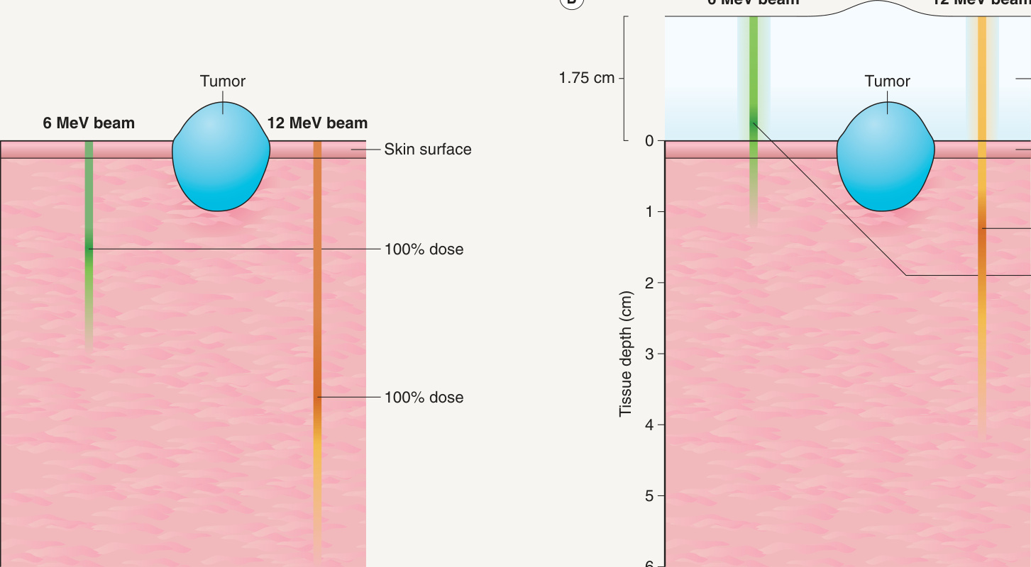
Fig. 139.2 Depth dose of electron beams – with and without a bolus.A Beams with a dose of 6 MeV and 12 MeV. B Demonstration of the bolus effect whereby a more appropriate dose of radiation is obtained at the skin surface. Bolus material needs to be tissue equivalent and maintain a constant thickness; its composition varies from paraffin wax to thermoplastics (e.g. Aquaplast®). Courtesy Lynn Wilson, MD.
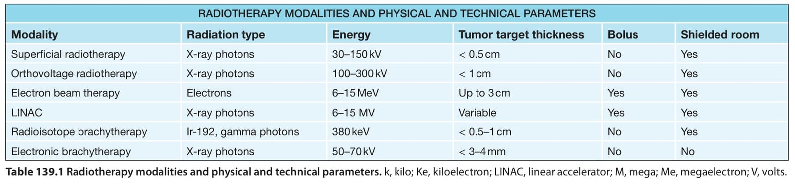
Table 139.1 Radiotherapy modalities and physical and technical parameters. k, kilo; Ke, kiloelectron; LINAC, linear accelerator; M, mega; Me, megaelectron; V, volts.
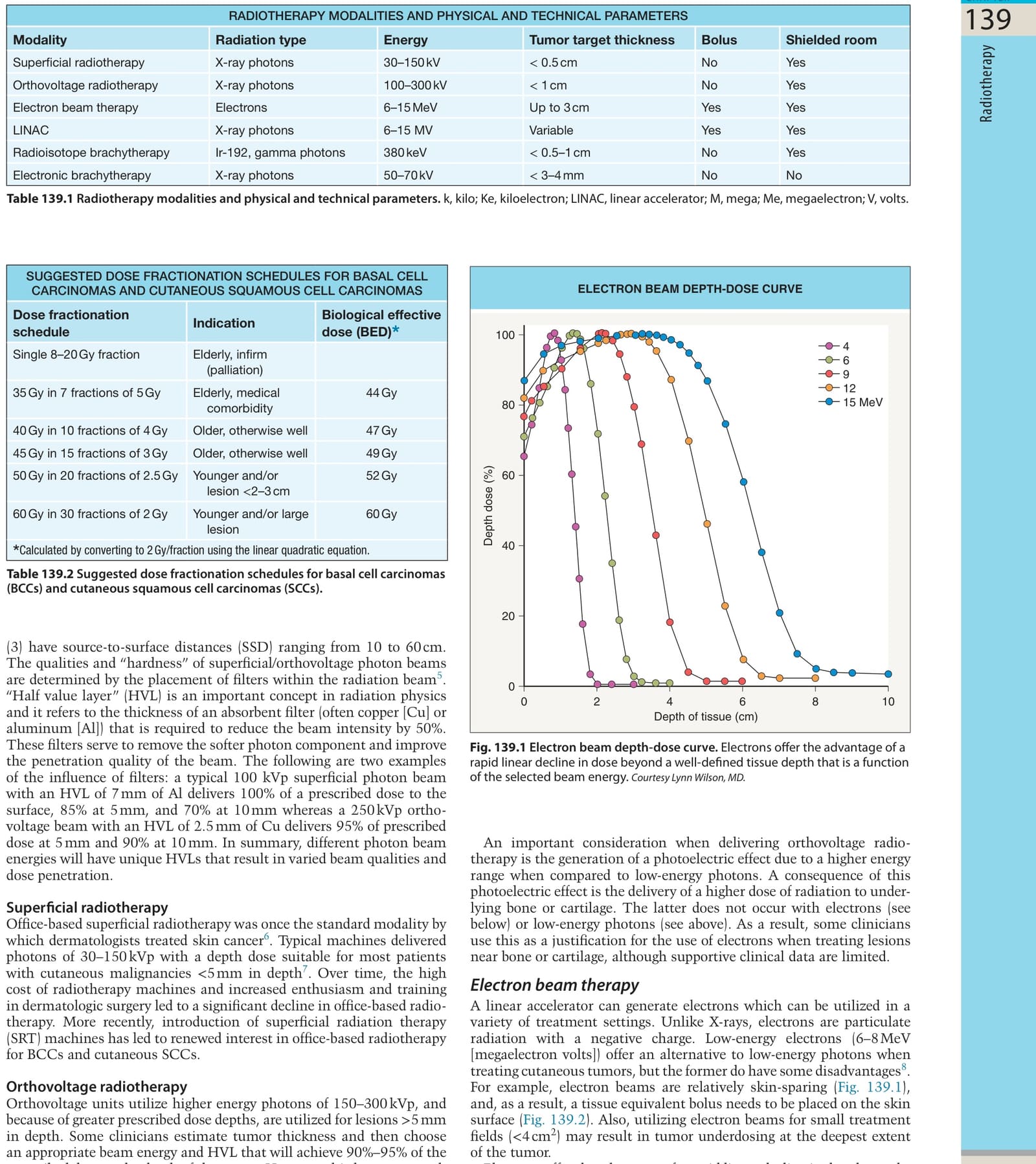
Table 139.2 Suggested dose fractionation schedules for basal cell carcinomas (BCCs) and cutaneous squamous cell carcinomas (SCCs).