NEUTROPHIL BIOLOGY
Granulocytes (neutrophils, eosinophils, and basophils) are cells essential to our defense against microbes as well as other inflammatory responses. The neutrophil is a terminally differentiated, non-dividing cell which is packed with granules whose contents kill and degrade target microorganisms. Recent advances in our understanding of neutrophil biology include the molecular and cellular mechanisms responsible for their production and release from the bone marrow (e.g. chemokine CXC receptor 4 [CXCR-4]; see Ch. 60); their recruitment, priming, and activation within inflamed tissues (Table 25.1); and the events resulting in their removal.
Granulocytes, including neutrophils, originate in the bone marrow from pluripotent cells where granulocyte colony-stimulating factor (G-CSF) plays a critical role in regulation of neutrophil development. To supply sufficient numbers of circulating cells, neutrophils are produced within the bone marrow at a prodigious baseline rate (>5–10 × 10 neutrophils daily). The bone marrow also has the capacity to upregulate granulocyte production sharply in response to a number of stresses such as infection. Mature neutrophils appear to circulate in the peripheral bloodstream anywhere from 6 hours to up to 6 days before migrating into tissues; within such tissues the neutrophils survive for an additional 2 to 3 days. Senescent neutrophils, unlike platelets and erythrocytes, appear to be removed from the circulation following a return to the bone marrow where they undergo apoptosis.
Transcriptional profiling studies suggest that granulocytes arise via the selective expression of a subset of transcription factors (e.g. STAT3), granulocyte proteins (e.g. neutrophil elastase), and receptors (e.g. N-formyl-methionyl-leucyl-phenylalanine [fMLP]). Differentiation from pluripotent stem cells requires 7–10 days, and during this period, under the influence of cytokines, neutrophils acquire their characteristic appearance and granules (primary, secondary, and tertiary). The following stages of myeloid maturation are recognized: myeloblast, promyelocyte, myelocyte, metamyelocyte, band, and, finally, the segmented neutrophil. The progressive gain of differentiated characteristics is accompanied by a loss in the potential to proliferate, i.e. beyond the myelocyte stage, the cells are non-dividing. The intracellular granules acquired during maturation contain enzymes that mediate the oxidative and non-oxidative killing functions of the neutrophil: 1.primary (azurophilic) granules – acquired at the promyelocyte stage and their contents include myeloperoxidase, lysozyme, neutrophil elastase, defensins, proteinase 3, cathepsin 3, and bactericidal/ permeability-increasing protein 2.secondary granules – acquired at the transition to the myelocyte stage and their contents include lactoferrin, neutrophil collagenase, neutrophil gelatinase-associated lipocalin, and lysozyme 3.tertiary granules – acquired during later stages of neutrophil maturation and contain gelatinase B (matrix metalloproteinase 9) and leukolysin. While much remains to be discovered about the pathophysiology of neutrophilic dermatoses, contributing factors include genetic predisposition, aberrant cell signaling and effector molecules, and ultimately abnormal neutrophil activity. Proinflammatory cytokines that play a key role in neutrophilic inflammation include IL-1β, TNF, and IL-172a. The latter upregulates G-CSF activity, which is important for neutrophil differentiation and release, and it enhances production of IL-8. In addition to being a potent neutrophil chemoattractant, IL-8 has synergistic effects when combined with TNF. Under normal conditions, phagocytosis of neutrophils limits IL-23 secretion, and this in turn limits IL-17 activity2b. The response of neutrophilic dermatoses to inhibitors of IL-17, TNF, and IL-1 lends support to this proposed pathogenesis.
Inflammation
A critical role of inflammation is to deliver neutrophils and other leukocytes to a site of injury and then activate these cells to perform their function of protecting the host against infection. Neutrophils are among the first cells to arrive at sites of inflammation. Reasons for this include their abundance in the bloodstream and their rapid response to chemokines. When activated, neutrophils move at speeds up to 30 microns/ min – the fastest cell in the body.
The motile responses of neutrophils to microbial infection include emigration out of the vasculature and movement toward the source of the inflammatory chemoattractant. This culminates in the phagocytic ingestion of opsonized microbes. In order to arrive at the site of infection, leukocytes must migrate out of the vasculature via margination, rolling, activation, and tight adhesion (see Ch. 102). They then move toward the site of injury or infection (see Table 25.1) and eventually undergo degranulation then apoptosis.
The price to be paid for the defensive potency of neutrophils for destroying microbes and necrotic tissues is that they can injure normal tissue. During activation and phagocytosis, neutrophils release products (e.g. lysosomal enzymes, reactive oxygen intermediates, products of arachidonic acid metabolism [prostaglandins and leukotrienes]) not just within
the phagolysosome, but also into the extracellular space. Endothelial injury and tissue damage ensue, thus contributing to a number of acute and chronic diseases that affect the skin as well as other organs.
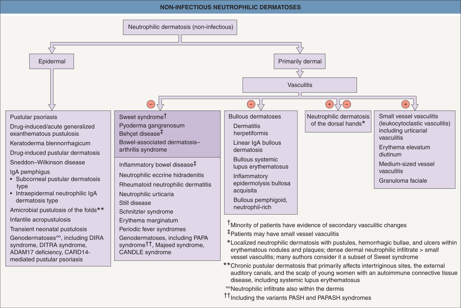
Fig. 25.1 Non-infectious neutrophilic dermatoses. Entities in the darker box are discussed in this chapter. CANDLE, chronic atypical neutrophilic dermatosis with lipodystrophy and elevated temperature; DIRA, deficiency of the interleukin-1 receptor antagonist; DITRA, deficiency of the interleukin-36 receptor antagonist; PAPA, pyogenic arthritis, pyoderma gangrenosum, and acne; PAPASH, pyogenic arthritis, pyoderma gangrenosum, acne, and suppurative hidradenitis; PASH, pyoderma gangrenosum, acne, and suppurative hidradenitis.
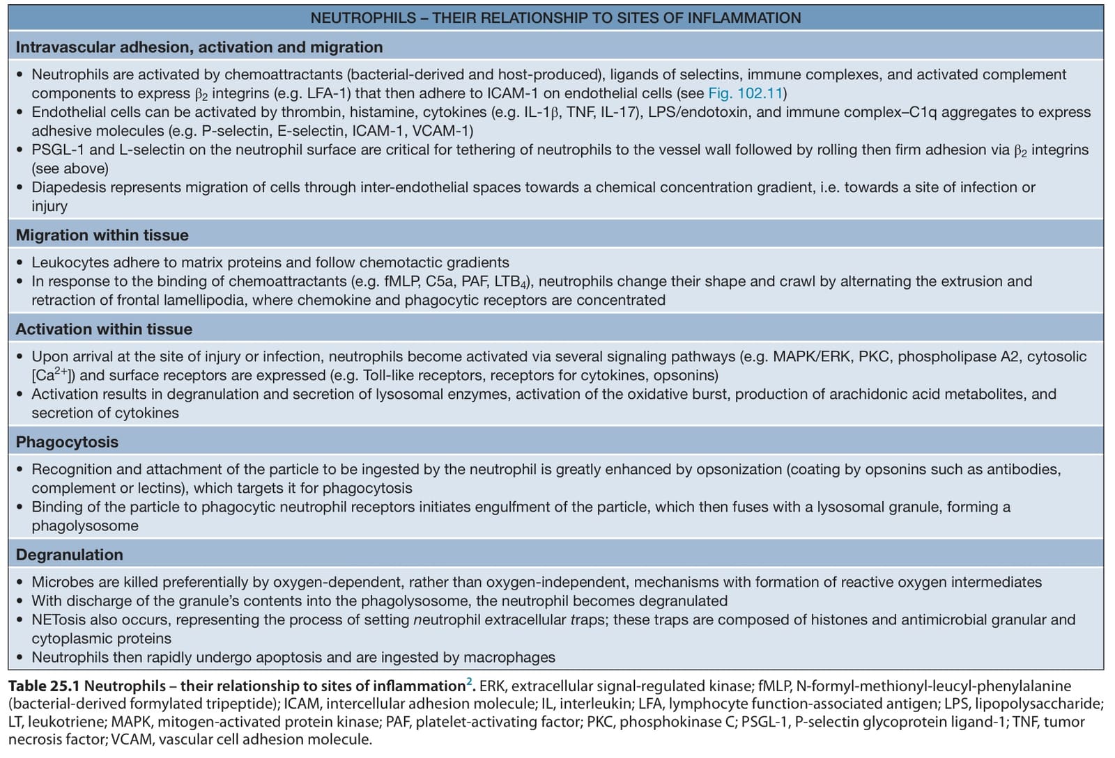
Table 25.1 Neutrophils – their relationship to sites of inflammation. ERK, extracellular signal-regulated kinase; fMLP, N-formyl-methionyl-leucyl-phenylalanine (bacterial-derived formylated tripeptide); ICAM, intercellular adhesion molecule; IL, interleukin; LFA, lymphocyte function-associated antigen; LPS, lipopolysaccharide; LT, leukotriene; MAPK, mitogen-activated protein kinase; PAF, platelet-activating factor; PKC, phosphokinase C; PSGL-1, P-selectin glycoprotein ligand-1; TNF, tumor necrosis factor; VCAM, vascular cell adhesion molecule.