PATHOGENESIS
Pathogenic Autoantibodies in Pemphigus
The hallmark of pemphigus is the finding of IgG autoantibodies against the cell surface of keratinocytes (see Fig. 29.1). The pemphigus autoantibodies found in patients’ sera play a primary pathogenic role in inducing the loss of cell adhesions between keratinocytes, and subsequent blister formation. Neonates of mothers with pemphigus vulgaris may have a transient disease caused by maternal IgG that crosses the placenta. As maternal antibody is catabolized, the disease subsides. IgG fractions from patients can induce blister formation in the absence of complement or inflammatory cells in a skin organ-culture system. Furthermore, passive transfer of patients’ IgG to neonatal mice results in blisters in the mice with typical histologic findings. Even monovalent Fab′ fragments of IgG from patients with pemphigus foliaceus are sufficient to cause blisters in neonatal mice, indicating complement activation and surface cross-linking may not be essential in keratinocyte detachment.
Desmogleins as Pemphigus Antigens
Immunoelectron microscopy localized both pemphigus vulgaris and pemphigus foliaceus antigens to the desmosomes, the most prominent cell–cell adhesion junctions in stratified squamous epithelia. Immunochemical characterization of pemphigus antigens by immuno precipitation or immunoblotting with extracts from cultured keratinocytes or epidermis demonstrated that the pemphigus vulgaris and foliaceus antigens were 130 kDa and 160 kDa transmembrane glycoproteins, respectively (Table 29.2). By comparative immunochemical studies using anti-desmoglein 1 (Dsg1) monoclonal and polyclonal antibodies, the 160 kDa protein recognized by pemphigus foliaceus sera was subsequently shown to be identical to Dsg1. An 85 kDa plaque protein, plakoglobin, was co-immunoprecipitated with the 130 kDa and 160 kDa pemphigus antigens, demonstrating that plakoglobin forms a molecular complex with pemphigus vulgaris and foliaceus antigens.
Molecular cloning of cDNA encoding Dsg1 and pemphigus vulgaris antigens indicated that both molecules were members of the cadherin supergene family. Thus, pemphigus was discovered to be an anti-cadherin autoimmune disease. The pemphigus vulgaris antigen was termed desmoglein 3 (Dsg3). The basic pathophysiology of pemphigus is as follows: autoantibodies inhibit the adhesive function of desmogleins and lead to the loss of the cell–cell adhesion of keratinocytes, resulting in blister formation.
Cadherins are a family of calcium-dependent cell–cell adhesion molecules that play an important role in the formation and maintenance of complex tissue integrity. Based on sequence similarity, cadherins have two major subgroups: classic cadherins (e.g. E-, P-, N-cadherins) and desmosomal cadherins (desmogleins and desmocollins). All members of the cadherin family contain conserved repeated amino acid sequences (cadherin repeats) with calcium-binding motifs in their extracellular domains (see Fig. 29.2). When classic cadherins were introduced by gene transfection into non-adhesive mouse fibroblast L cells, the cells acquired strong cell adhesion activity mediated by a homophilic type of
interaction. Cadherins require their well-conserved cytoplasmic domains in order to associate with the plaque proteins, α-catenin, β-catenin and plakoglobin, which mediate and regulate binding to the cytoskeleton network (Fig. 29.3). As a consequence of these interactions, cadherins produce strong cellular adhesion and morphologic changes in the cells. Cadherin molecules form dimers as their functional unit, with the distal extracellular domain (EC1) of the cadherin from one cell binding to the same region of a second cadherin from an opposing cell.
Two major types of adhering junctions of epithelial cells are commonly distinguished: adherens junctions and desmosomes (see Fig. 29.3). The adherens junction anchors bundles of actin microfilaments and contains classic cadherins as its transmembrane components and α-catenin, β-catenin, and plakoglobin as its cytoplasmic components. In contrast, the desmosome anchors intermediate filaments like keratins and contains desmosomal cadherins as its transmembrane components and plakoglobin, plakophilin, and desmoplakin
as its cytoplasmic components. In general, adherens junctions mediate quick but weak cellular adhesion, whereas desmosomes mediate slow but strong cellular adhesion.
Desmogleins have four cadherin repeats in their extracellular domain, as do classic cadherins (see Fig. 29.2). Desmogleins have four isoforms (Dsg1–4). Expression of Dsg1 and Dsg3 is basically restricted to stratified squamous epithelia, where blisters are formed in pemphigus, while Dsg2 is expressed in all desmosome-possessing tissues, including simple epithelia and myocardium. Dsg4 plays an important adhesive role primarily in hair follicles, and mutations in DSG4 can lead to abnormal hair development (e.g. localized autosomal recessive hypotrichosis).
Desmocollins are another group of transmembrane glycoproteins within desmosomes and they have three isoforms (Dsc1–3). Each isoform has two products derived from alternatively spliced mRNA of a single gene. Desmosomes always have desmoglein and desmocollin as a pair, with Dsg:Dsc heterodimers representing the fundamental adhesive unit of desmosomes and thus providing a structural framework for understanding desmosome assembly.
Plakoglobin and plakophilin, together with β-catenin, are members of the armadillo family of nuclear and junctional proteins, which are not only simple anchoring molecules but also dynamic regulators of cellular adhesion and proliferation. Desmoplakin is a dumbbell-shaped molecule composed of three domains: a central α-helical coiled-coil rod, flanked by globular carboxy- and amino-terminal domains that interact with intermediate filaments and armadillo family members, respectively (see Fig. 29.3). Desmoplakin has two products derived from alternatively spliced mRNA of a single gene: desmoplakin I (250 kDa) and II (210 kDa) (see Table 29.2). Desmoplakin, a member of the plakin family, plays an important role in the anchorage of the cytoskeleton to filament attachment sites on desmosomes.
Compelling evidence has accumulated that IgG autoantibodies against Dsg1 and Dsg3 are pathogenic and play a primary role in inducing the blister formation in pemphigus. Essentially, all patients with pemphigus have IgG autoantibodies against Dsg1 and/or Dsg3, depending on the subtype of pemphigus. When anti-desmoglein IgG autoantibodies are removed from the sera of patients with pemphigus vulgaris, pemphigus foliaceus or paraneoplastic pemphigus (by immunoadsorption with recombinant desmoglein proteins), the sera are no longer pathogenic in inducing blister formation. Furthermore, anti-desmoglein IgG autoantibodies that have been affinity-purified from pemphigus sera via desmoglein recombinant proteins can cause blisters when injected into neonatal mice. Some pemphigus sera react with Dsg4 due to cross-reactivity of a subset of anti-Dsg1 IgG, although Dsg4/Dsg1-cross-reacting IgG has no demonstrable pathogenic effect. IgG autoantibodies against non-desmoglein molecules, such as acetylcholine receptors or annexin-like molecules, have also been detected, but their pathogenic relevance in pemphigus remains to be determined.
Desmoglein Compensation Theory as Explanation for Localization of Blisters
The sites of blisters in pemphigus vulgaris and foliaceus are explained logically by the desmoglein compensation theory: Dsg1 and Dsg3 compensate for each other when they are coexpressed in the same cell (Fig. 29.4). While patients with pemphigus foliaceus have only anti-Dsg1 IgG autoantibodies, individuals with the mucosal-dominant type of pemphigus vulgaris have only anti-Dsg3 IgG autoantibodies. Those with the mucocutaneous type of pemphigus vulgaris have both anti-Dsg3 and anti-Dsg1 IgG autoantibodies. Of note, the intraepithelial expression pattern of Dsg1 and Dsg3 differs between the skin and mucous membranes. In the skin, Dsg1 is expressed throughout the epidermis, but more intensely in the superficial layers (see Fig. 29.1C), whereas Dsg3 is expressed in the lower portion of the epidermis, primarily in the basal and parabasal layers (see Fig. 29.1A). In contrast to the skin, Dsg1 and Dsg3 are expressed throughout the squamous layer of mucosa, but Dsg1 is expressed at a much lower level than Dsg3 (see Fig. 29.4B).
When sera contain only anti-Dsg1 IgG (which interferes with the function of Dsg1), blisters appear only in the superficial epidermis of the skin because that is the only area in which Dsg1 is present without coexpression of Dsg3. The splits occur between the second (SG2) and the third (SG3) layers of the stratum granulosum (SG) because IgG does not go beyond the tight junctions present between SG2 cells. In the unaffected deep epidermis, the presence of Dsg3 compensates for the loss of function of Dsg1. Although the anti-Dsg1 IgG binds to mucosa, no blisters are formed, because of the coexpression of Dsg3.
Thus, sera containing only anti-Dsg1 IgG cause superficial blisters in the skin without mucosal involvement, as is seen in patients with pemphigus foliaceus (see Fig. 29.4).
When sera contain only anti-Dsg3 IgG, they are inefficient in producing cutaneous blisters because coexpressed Dsg1 compensates for the impaired function of Dsg3, resulting in no, or only limited, skin lesions. However, in the mucous membranes, Dsg1 cannot compensate for the impaired Dsg3 function because of its low expression. Therefore, sera containing only anti-Dsg3 IgG cause oral erosions without apparent skin involvement, as is seen in patients with the mucosal-dominant type of pemphigus vulgaris (see Fig. 29.4).
When sera contain both anti-Dsg1 and anti-Dsg3 IgG, they inter-fere with the function of both Dsg1 and Dsg3, resulting in extensive blisters and erosions of the skin as well as the mucous membranes, as is seen in patients with the mucocutaneous type of pemphigus vulgaris. Possible explanations for why splits appear just above the basal layer, rather than the whole epithelium falling apart, include: (1) cell–cell adhesion in the basal and parabasal layers may be weaker than in other parts of the epithelium; and (2) autoantibodies, which penetrate from the dermis, might have better access to the lower part of the epithelia.
In pregnant women with pemphigus, autoantibodies cross the placenta and bind to the fetal epidermis. However, neonates develop blisters if the mother has pemphigus vulgaris, but very rarely if she has pemphigus foliaceus. This confusing observation is also explained by the desmoglein compensation theory. The distribution of Dsg3 within neonatal epidermis is unlike that in adult epidermis; it is found on the surface of keratinocytes throughout the epidermis, which is similar to its distribution in mucous membranes (remember, neonatal skin is bathed
in amniotic fluid). Therefore, pemphigus foliaceus sera containing only anti-Dsg1 IgG cannot efficiently induce blisters in neonatal skin.
As an extension of this compensation theory, exfoliative toxins, which are produced by Staphylococcus aureus and lead to bullous impetigo as well as staphylococcal scalded skin syndrome, specifically cleave Dsg1. Inactivation of Dsg1 by this toxin induces superficial blisters in the epidermis that are clinically and histologically similar to those seen in pemphigus foliaceus.
In pemphigus, the disruption of cell–cell adhesion is currently thought to be mediated via the combined effects of direct inhibition by antibodies plus subsequent signal transduction induced by antibody binding. The direct inhibition is mediated by steric hindrance, i.e. the binding of autoantibodies to desmogleins spatially interferes with the adhesive interaction of desmogleins between cells. This proposed pathogenesis is supported by the following observations: (1) in pemphigus vulgaris and foliaceus, dominant epitopes are localized to the functionally important N-terminal regions of desmogleins; and (2) pathogenic anti-Dsg3 mouse or human monoclonal antibody, but not non-pathogenic monoclonal antibodies, recognizes the N-terminal adhesive surface of Dsg3 (see Fig. 29.3B). The phenotype of the Dsg3 null mouse (whose Dsg3 gene is genetically deleted) closely resembles that of human pemphigus vulgaris patients. The role of signal transduction induced by antibody binding is supported by the in vitro observation that IgG from pemphigus vulgaris sera (when added to the media of cultured keratinocytes) causes a transient increase in intracellular calcium and/or inositol 1,4,5-triphosphate, activation of protein kinase C, or phosphorylation of Dsg3. Furthermore, with desmosomal disassembly, Dsg3 is internalized or endocytosed from the cell surface in association with keratin retraction.
Humoral and Cellular Autoimmunity in Paraneoplastic Pemphigus
Patients with paraneoplastic pemphigus develop characteristic IgG autoantibodies against multiple antigens, including Dsg3 and/or Dsg1, multiple members of the plakin family (plectin, epiplakin, desmoplakins I and II, bullous pemphigoid antigen 1, envoplakin, and periplakin), the protease inhibitor alpha-2-macroglobulin-like-1, and transglutaminase 138a (see Table 29.2). Anti-desmoglein antibodies play a role in inducing the loss of cell adhesion of keratinocytes and initiate blister formation, while the pathophysiologic relevance of the anti-plakin autoantibodies is unclear, in that plakin molecules are intracellular and IgG cannot penetrate cell membranes. In addition to humoral autoimmunity, cell-mediated cytotoxicity is involved in the pathogenesis of paraneoplastic pemphigus, in which more severe and refractory oral erosions and stomatitis as well as more polymorphic skin eruptions are seen, in comparison with classic forms of pemphigus. It was demonstrated in mice that Dsg3-specific T cells not only help B cells produce anti-Dsg3 IgG (which causes acantholysis), but also directly infiltrate into the epidermis and induce an interface dermatitis. Clarification of the exact roles of autoimmune T cells should provide valuable insights into the pathophysiology of paraneoplastic pemphigus.
Immunologic Mechanism of Pathogenic Autoantibody Production in Pemphigus
In contrast to the significant progress since the late 1980s in under-standing the pathophysiologic mechanisms of blister formation in pemphigus, it is still unclear why patients with pemphigus begin to produce the pathogenic autoantibodies.
Pemphigus autoantibodies are composed of IgG isotypes, which may be produced after isotype switching, and they have a high affinity towards the antigen, which may be a result of affinity maturation of the antibodies. In addition, pemphigus sera recognize several distinct epitopes on desmogleins, and the presence of autoantibodies is associated with specific HLA class II alleles, including DRB10402, DRB11401, and DQB10302 in White individuals and DRB114 and DQB10503 in Japanese. All of these features suggest that autoantibody production in pemphigus is T cell-dependent. More recently, T cells reactive against Dsg3 were shown to be present in peripheral blood from patients with pemphigus vulgaris as well as healthy individuals. Certain peptides from Dsg3, predicted to fit into the DRB10402 pocket, were able to stimulate T cells from the pemphigus patients.
Another advance that will allow the study of T cells and B cells is the development of an active disease mouse model for pemphigus vulgaris. This model is valuable not only for dissecting the cellular and molecular mechanisms involved in antibody production but also for developing novel therapeutic strategies.
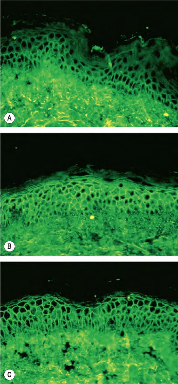
Fig. 29.1 Indirect immunofluorescence of pemphigus sera with normal human epidermis as a substrate. The hallmark of pemphigus is the finding of IgG autoantibodies directed against the cell surface of keratinocytes. A Pemphigus vulgaris sera containing anti-desmoglein 3 (anti- Dsg3) IgG alone stain predominantly the cell surfaces in the lower epidermis. B Pemphigus vulgaris sera containing both anti-Dsg3 IgG and anti-Dsg1 IgG stain the cell surfaces throughout the epidermis. C Pemphigus foliaceus sera, which contain only anti-Dsg1 IgG, stain the cell surfaces throughout the epidermis, but more intensely in the superficial layers.
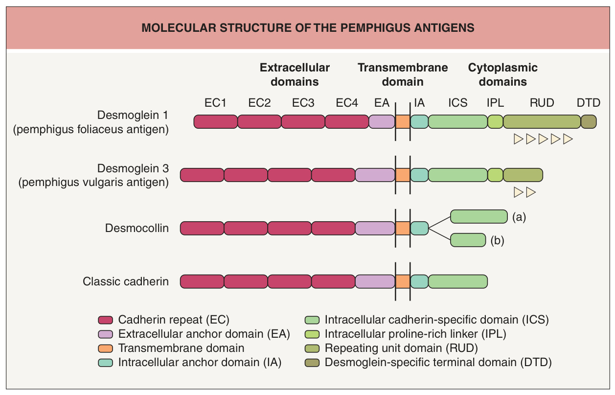
Fig. 29.2 Molecular structure of the pemphigus antigens. The extracellular (EC) region has four cadherin repeats, which have calcium-binding motifs. The intracellular cadherin-specific (ICS) domain is well conserved among cadherins and it is responsible for interactions with β-catenin or plakoglobin (see Fig. 29.3). Desmogleins have their own unique sequences (repeating unit domain [RUD]) of repeats of 29 ± 1 residues (arrowheads). Each desmocollin isotype has two products (a and b) derived from alternatively spliced transcripts of a single gene.
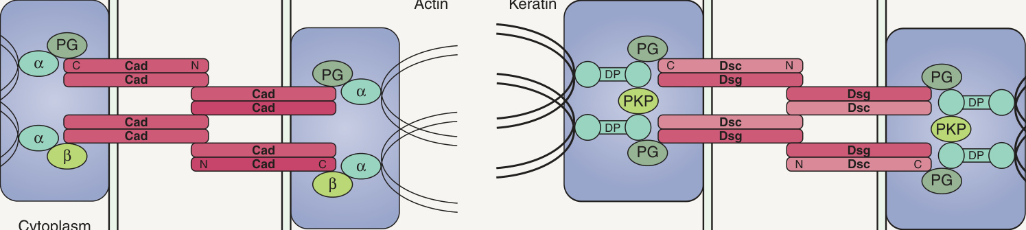
Fig. 29.3 The adherens junction and desmosome.A The adherens junction complex contains classic cadherins as transmembrane constituents and α-catenins, β-catenins, and plakoglobin as cytoplasmic constituents. A classic cadherin is directly coupled through its cytoplasmic tail to β-catenin or plakoglobin, which in turn is linked to α-catenin, which binds to actin. B The desmosome complex includes desmogleins and desmocollins as transmembrane constituents, and plakoglobin, plakophilin, and desmoplakin as cytoplasmic constituents. Desmogleins and desmocollins form heterodimers as a functional adhesive unit and associate with plakoglobin, which in turn binds to desmoplakin which links keratin to the membrane. C, carboxy terminus; N, amino terminus.
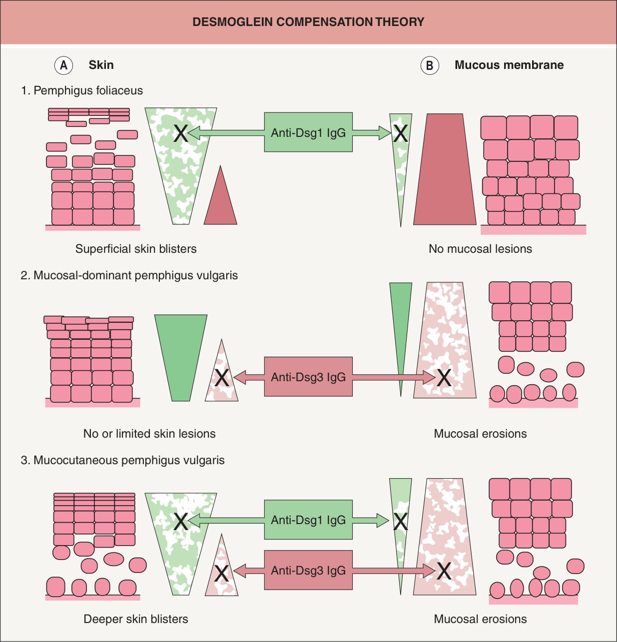
Fig. 29.4 Logical explanation for the localization of blister formation in classic pemphigus by desmoglein compensation theory. The colored triangles represent the distribution of desmoglein 1 (Dsg1, green) and desmoglein 3 (Dsg3, pink) in the skin (A) and mucous membranes (B). Pemphigus foliaceus sera contain only anti-Dsg1 IgG, which causes superficial blisters in the skin because Dsg3 functionally compensates for the impaired Dsg1 in the lower part of the epidermis (A1), whereas those antibodies do not cause blisters in the mucous membranes because cell–cell adhesion is mainly mediated by Dsg3 (B1). Sera containing only anti-Dsg3 IgG cause no or only limited blisters in the skin because Dsg1 compensates for the loss of Dsg3- mediated adhesion (A2); however, these sera induce separation in the mucous membranes, where the low expression of Dsg1 will not compensate for the loss of Dsg3-mediated adhesion (B2). When sera contain both anti- Dsg1 and anti-Dsg3 IgG, the function of both Dsgs is compromised and blisters occur in both the skin and mucous membranes (A3, B3). In neonatal skin, the situation is similar to that shown here for mucous membranes.
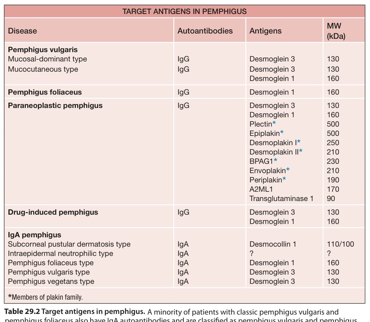
Table 29.2 Target antigens in pemphigus. A minority of patients with classic pemphigus vulgaris and pemphigus foliaceus also have IgA autoantibodies and are classified as pemphigus vulgaris and pemphigus foliaceus. A2ML1, alpha-2-macroglobulin-like-1 protease inhibitor; BPAG1, bullous pemphigoid antigen 1.