PATHOGENESIS
Papillomavirus Evolution
Historically, papillomaviruses were grouped together with polyomaviruses, but their fundamentally different biology and genetic organization resulted in their reclassification as a separate family in 2000. The National Center for Biotechnology Information (NCBI) nucleotide sequence database (www.ncbi.nlm.nih.gov) and the Karolinska Institutet International HPV Reference Center (www.hpvcenter.se) currently list more than 200 different HPV types as well as numerous incompletely characterized putative newer HPV types. By definition, a novel type has more than 10% dissimilarity from any previously characterized type in the DNA sequences of three HPV genes (E6, E7, and L1). Dissimilarity of 2%–10% is defined as a subtype, and <2% as a variant.
By comparing DNA sequences in conserved portions of the papillomavirus genomes across different types, a phylogenetic tree establishing the degree of relatedness among these types has been generated. Two general, the α- and β-papillomaviruses, encompass almost all known HPV types, with the genus α containing mucosal and cutaneous HPV types that are pathogenic in normal hosts and the genus β containing the types associated with EV; γ, μ, and ν genera contain additional cutaneous types. Within each genus, HPV types are grouped according to sequence homology into species, which often share similar biologic and pathologic properties (e.g. HPV-16 and -31). Present models postulate that HPVs co-evolved with our human ancestors and novel HPV types or variants were generated through either genetic drift or natural selection via adoption of specialized ecologic niches. Genetic recombination between HPV types does not occur.
Virology
Papillomaviruses are non-enveloped, double-stranded DNA viruses ~55–60 nm in diameter. The spherical capsid is composed of two virally encoded proteins: the major structural protein L1 and the minor structural protein L2. During virion assembly within cells, L1 proteins form pentamers called capsomeres, and 72 capsomeres multimerize to form the viral capsid with an icosahedral symmetry arranged on a T = 7 surface lattice. The capsid surrounds the viral DNA, thereby protecting it from degradation, and it also enables the virus to bind efficiently to target cells. Packaged viral DNA is associated with L2 and cellular histones, forming a mini chromosome. In contrast, the early (E) viral proteins are not incorporated into infectious virions.
When expressed in cell culture, the L1 major capsid proteins of papillomaviruses self-assemble into VLPs that are morphologically similar to native virions (Fig. 79.1). Papillomavirus VLPs display type-restricted and neutralization epitopes that are used to detect serum antibodies and as the basis for prophylactic vaccines (see below).
The HPV genome, ~8 kilobases (kb) in length, is composed of three domains: the upstream regulatory region (URR), the early region, and the late region (Fig. 79.2). The URR, ~1 kb in length, lacks open reading frames (ORFs) and contains the origin of replication and many of the control elements for transcription and replication. The early region, ~4 kb in length, contains the ORFs for genes expressed early in the papillomavirus life cycle. The late region, ~3 kb in length, encodes the capsid proteins (see above).
Papillomavirus Life Cycle
The papillomaviruses are highly species-specific, and productive infection has not been observed outside natural host tissue. The papillomavirus life cycle is completed only in fully differentiated squamous epithelia. This has impeded its study in monolayer tissue culture cells, where late gene expression and virion production do not occur. Only
a small number of HPV types have been successfully propagated in mouse xenograft or raft culture systems, and limited amounts of infectious virions have been generated.
Productive infection and induction of hyperproliferation are initiated when the virus enters proliferating basal epithelial cells (Fig. 79.3). This layer of cells is not normally accessible to the virus because of the mechanical barrier provided by the overlying differentiated cell layers. Therefore, it is likely that infection requires an abrasion or other trauma to the epithelium to expose the basal cells to the virus. Our knowledge about early steps of infection such as viral entry and uncoating is limited. The receptor(s) that mediate virus binding to epithelial cells have not been definitively identified, but binding appears to depend on the L1 major capsid protein, and cell surface and/or basement membrane heparan sulfate is necessary for efficient infection in vitro. The amino terminus of the L2 minor capsid protein is then cleaved by furin (a proprotein convertase), which exposes both a cross-neutralization epitope on L2 and a binding site (thought to be on L1) that facilitates viral entry via a putative secondary receptor on keratinocytes. Upon infection, the covalently closed circular DNA genome becomes established as a low-copy autonomous replicon in the nuclei of basal cells, thereby creating a long-term reservoir of viral DNA.
The program of viral gene expression is intimately linked to the differentiation state of the infected cells. The E genes are transcribed at low levels in the basal cell layer and spinous layer. The first genes to be expressed following infection are the E1 and E2 genes, which are responsible for controlling the transcription of other viral genes and replication of the viral genome. HPVs do not encode the enzymes required for transcription or replication of viral DNA. Consequently, HPV is entirely dependent on the co-opting of cellular machinery for these functions. One major effect of HPV proteins E5, E6, and E7 is that the epidermal cell cycle, which is normally blocked for cells that are suprabasal, continues so that HPV genome copy number can be amplified to high levels during vegetative viral replication for assembly into virions (see Fig. 79.3). The E6 and E7 proteins of the high-risk mucosal HPV types act as viral oncoproteins, but no such functions are associated with the corresponding proteins of the low-risk mucosal and EV types. There is limited understanding of possible differences between oncogenic (e.g. HPV-5, -8) and the non-oncogenic β HPV types.
In combination with other cellular proteins, E6 from high-risk mucosal HPV causes the ubiquitin-mediated degradation of the cellular protein p53. Elevated levels of p53 arrest cells in the G1 phase of the cell cycle or induce apoptotic cell death, so that E6-facilitated destruction of p53 removes a brake on suprabasal cell cycling. One of the principal effects of high-risk mucosal HPV E7 is to bind to the underphosphorylated form of RB (the retinoblastoma tumor suppressor protein). A physiologic role of underphosphorylated RB is to bind to and inhibit the function of the E2F transcription factor (see Fig. 107.4). When HPV E7 binds RB, E2F is liberated from this inhibition and is thereby able to induce the expression of genes required for DNA replication. Both E6 and E7 are multifunctional proteins, and while their effects on p53 and RB are critical ones, additional targets are also important to the oncogenic potential of the virus. These include activation of telomerase by E6, binding of histone deacetylases by E7, and synergistic effects of both proteins that lead to centrosome abnormalities and chromosomal instability.
The genes for the virion structural proteins, i.e. the late (or L) genes, are expressed in the terminally differentiated superficial layers of the epithelium. The more superficial epithelial layers also have higher levels of expression of E1 and E2 and amplification of viral DNA. The amplified genomes are encapsulated by the L1 and L2 capsid proteins to generate infectious virions, and virus particles are observed in the granular layer of the epithelium and above. Virus assembly is not believed to lyse the cells; instead, the virus is shed with the cornified layer as cells slough from the epithelial surface (see Fig. 79.3). E4 proteins are hypothesized to disrupt the intracellular filamentous network of keratinocytes, which may facilitate virus release from the corneocytes.
Host Immune Response
Persistent papillomavirus infections are common, indicating that HPVs have evolved mechanisms to evade immune surveillance. There is no viremic phase during the life cycle, so a systemic immune response is avoided. In addition, low levels of viral proteins are expressed in the basal and spinous cell layers of the epidermis, where they would be most likely to be recognized by Langerhans cells and infiltrating lymphocytes. Only in the more immunologically privileged terminally differentiated layers is there extensive production of virion proteins, with shedding of infectious virus exclusively from the external epithelial surface. Despite the relative success of papillomaviruses in evading immune responses, up to two-thirds of cutaneous warts spontaneously regress within 2 years, and lesions of multifocal infections often regress concomitantly. There is also an increased prevalence of warts in patients with suppressed cell-mediated immunity. In contrast, patients with underlying defects in humoral immunity do not seem to be predisposed to HPV infections, suggesting a primary role of cell-mediated responses.
Although low amounts of virion capsid proteins are produced in the lower epidermal cell layers, serum antibodies specific to conformational virion epitopes of high-risk HPV-16 can be detected in more than half of women with low-grade or asymptomatic genital HPV-16 infections. It is unknown if these antibodies play a role in limiting reinfection with the same type from autoinoculation or from an infected sexual partner.
Latent HPV infections are defined as persistence of HPV DNA in clinically and cytologically normal epithelium. However, it is difficult to distinguish true viral latency from persistent infection with a low level of DNA replication. The mechanisms by which HPV enters into and is activated out of latency are unknown.
Oncogenic Potential of HPV
Different HPV types have markedly different oncogenic potentials. The vast majority of cervical cancers, for example, contain the sequences of one of five HPV types: 16, 18, 31, 33, or 45. In contrast, infection with HPV-6 or -11 occurs commonly in benign or low-grade intraepithelial lesions but is very rarely associated with the development of anogenital malignancies. HPV-5 and -8 DNA sequences are frequently detected in SCCs associated with EV.
Progression of HPV-associated cervical lesions to invasive cancer typically requires several decades. Both persistent HPV infection and the accumulation of additional genetic mutations are required in the current model of multistage carcinogenesis. Integration of the viral genome randomly into the host DNA is a common event, invariably leading to loss of E1 and E2 expression. In HPV-16 and -18, E2 functions to repress the transcription of E6 and E7 genes. Therefore, integration is thought to increase expression of E6 and E7, which are selectively retained and expressed at elevated levels in carcinomas, suggesting a critical role for oncogenic progression (see above).
Differences in the activities of E6 and E7 genes between oncogenic and non-oncogenic HPV types principally account for the different risk of progression. E6 and E7 proteins derived from high-risk HPV types (e.g. 16 and 18) degrade p53 and interact with RB (respectively) with greater efficiency than do E6 and E7 proteins from low-risk types (e.g. 6 and 11). Degradation of p53 by E6, which eliminates from infected cells the principal “guardian of the genome” (the protein responsible for cell cycle arrest in the presence of damage to cellular DNA), can lead to the accumulation of genetic mutations in infected cells. Thus, carcinogenic progression appears to be promoted or facilitated by increased E6 and E7 expression as well as by activity of external carcinogens, resulting in genomic instability.
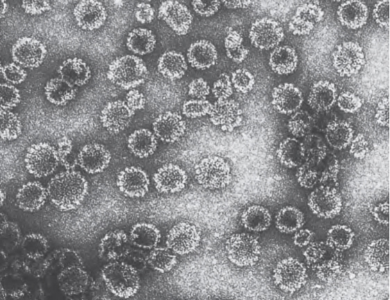
Fig. 79.1 Transmission electron photomicrograph of purified HPV-16 virus- like particles (VLPs). A prophylactic vaccine that contains HPV-6, -11, -16, -18, -31, -33, -45, -52, and -58 VLPs has been approved for the prevention of anogenital warts and cancers. Following expression in cell culture, the L1 capsid protein self-assembles into VLPs (empty capsids ~50 nm in diameter devoid of HPV DNA) that display type-restricted and neutralization surface epitopes similar to native virions. Courtesy Saeed Shafti-Keramat.
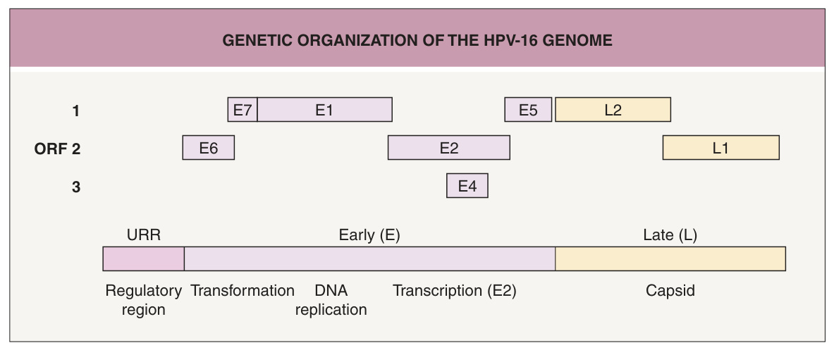
Fig. 79.2 Genetic organization of the HPV-16 genome. This is a linearized map of the 7.9 kb circular double-stranded genome. All open reading frames (ORFs) reside on one strand. Early (E) and late (L) regions are indicated. The upstream regulatory region (URR) contains the origin of replication and control elements for transcription and replication.
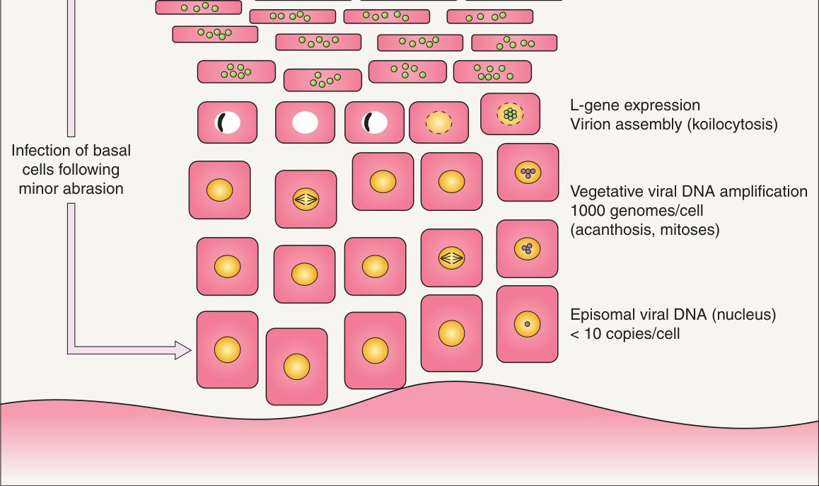
Fig. 79.3 The papillomavirus life cycle. The figure depicts a productive (virion-producing) benign wart. Adapted from Orth G. Epidermodysplasia verruciformis. In: Salzman NP, Howley PM (eds). The Papovaviridae: Volume 2 The Papillomaviruses. New York: Plenum Press; 1987:199–243.