UV INDUCTION OF DNA DAMAGE
Different wavelengths of UV light induce different types of DNA damage. UVC and UVB, but much less so UVA, are capable of exciting the DNA molecule directly and subsequently generating DNA photoproducts. Indeed, DNA is regarded as the chromophore for most of the biologic effects of UVB and UVC, including erythema, tanning, immunosuppression, and mutagenesis. DNA photoproducts are dimers, formed by covalently binding two adjacent pyrimidines in the same polynucleotide chain. The two major types of pyrimidine dimers are cyclobutane–pyrimidine dimers and 6,4-photoproducts.
Cyclobutane–pyrimidine dimers (CPDs) are the most common DNA photoproducts formed by UV irradiation. They are generated upon saturation of the 5,6 double bonds and formation of a four-membered cyclobutyl ring (Fig. 86.8). CPDs are observed at all possible dipyrimidine sites, with the thymine–thymine dimer (T–T) being the most common, followed by C–T and T–C dimers. C–C dimers are the least common. The formation of CPDs is not a random phenomenon: it is influenced by the sequence and conformational context of the affected DNA sequence. More recently, an additional molecular mechanism for the formation of CPDs was described, consisting of photoexcitation of melanin and formation of peroxynitrite. Due to the long half-life of these reactive molecules, this mechanism adds to CPD formation for hours after UV irradiation, but only in melanin-containing cells. These CPDs have been termed “dark CPDs”.
The 6,4-photoproduct is a non-cyclobutane dipyrimidine photoproduct, which is formed upon covalent linkage between the C-6 position of one pyrimidine and the C-4 position of the 3′ adjacent pyrimidine (Fig. 86.9). The T–C (6–4) dimer is the most common dimer of this type, but C–C and T–T dimers are also observed after UV irradiation. Upon further irradiation with UV wavelengths between 280 and 360 nm, the normal isomers of 6,4-photoproducts can be converted
to their Dewar valence isomers, which are less mutagenic than the normal isomers but may still contribute to solar mutagenesis. Although eightfold fewer in number than CPDs, 6,4-photoproducts can activate the ATR-CHK1 DNA damage response pathway and may be the major trigger for UV-induced DNA damage responses. A few other rare DNA photoproducts have been described, such as complex purine lesions and pyrimidine hydrates, but their physiologic significance in cutaneous photobiology is unknown.
The absorption maximum of DNA is at 260 nm. This makes UVC the most effective wavelength for the induction of DNA photoproducts in naked DNA. However, in vivo, due to the absorption of shorter wavelengths in upper layers of the epidermis, 300 nm (UVB) is the most effective wavelength for inducing DNA photoproducts in the basal layer of the epidermis (Fig. 86.10). It is now well recognized that when solar-available doses are compared, UVA can also generate pyrimidine dimers, albeit at a lower frequency than UVB.
While formation of UVB-induced pyrimidine dimers results from the direct absorption of photons by bases of DNA, UV irradiation can also damage DNA indirectly. After absorption of photons by chromophores other than DNA, energy can be transferred either to DNA (type I photosensitized reaction) or to molecular oxygen, with reactive oxygen species in turn being able to damage DNA (type II photosensitized reaction). Many of the biologic properties of UVA, including its toxicity to cells, are dependent upon the presence of molecular oxygen, which points to a prominent role for reactive oxygen species. Although these reactive
A pyrimidine–pyrimidone (6–4) photoproduct is formed by covalent linkage between C-6 and C-4 of two adjacent pyrimidines (here a thymine and a cytosine base), following excitation of the bases by UV light.
oxygen species are also formed by UVB, UVA is responsible for almost all oxidative DNA damage after exposure of cells to natural sunlight.
UV-induced reactive oxygen species include singlet oxygen and probably others such as hydrogen peroxide and the superoxide radical. Even the highly reactive hydroxyl radical may be formed by a reaction of hydrogen peroxide with nuclear metals through a Fenton reaction. This oxidative stress affects not only DNA, but also membranes and proteins. Singlet oxygen and other reactive oxygen species react predominantly with guanine and generate several DNA changes, including the mutagenic and well-studied 7,8-dihydro-8-oxoguanosine (8-oxoG; Fig. 86.11).
Kielbassa et al. published a UV action spectrum for the formation of dimers and oxidative guanine base modifications in mammalian cells (Fig. 86.12). Differences between the DNA-damaging properties of
UVA, UVB, and UVC are gradual, and, as noted previously, only a few pyrimidine dimers are generated by UVA. There is mounting evidence that pyrimidine dimers are the most important pre-mutagenic DNA lesions, not only in UVB-induced, but also in UVA-induced, mutation formation, with oxidative DNA damage playing only a minor role in mutagenesis. Lastly, DNA double-strand breaks are probably not formed by either UVB or UVA.
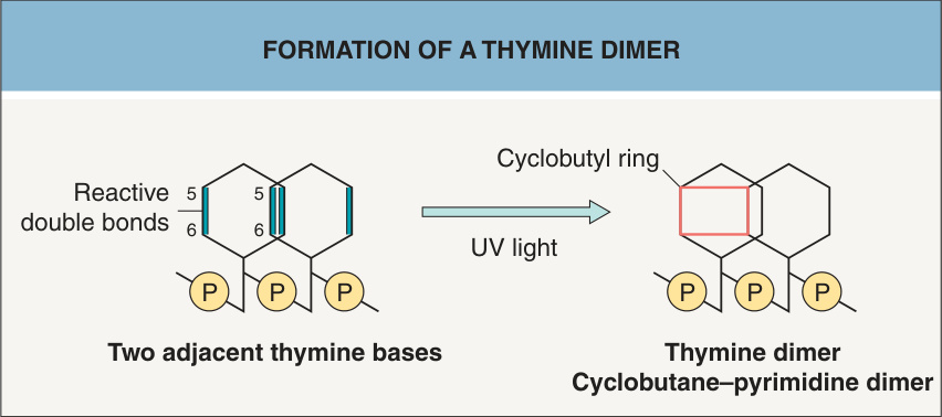
Fig. 86.8 Formation of a thymine dimer. Following excitation of the bases by UV light, a cyclobutane–pyrimidine dimer is formed by covalent linkage between two adjacent pyrimidines (here two thymine bases) and formation of a cyclobutyl ring.
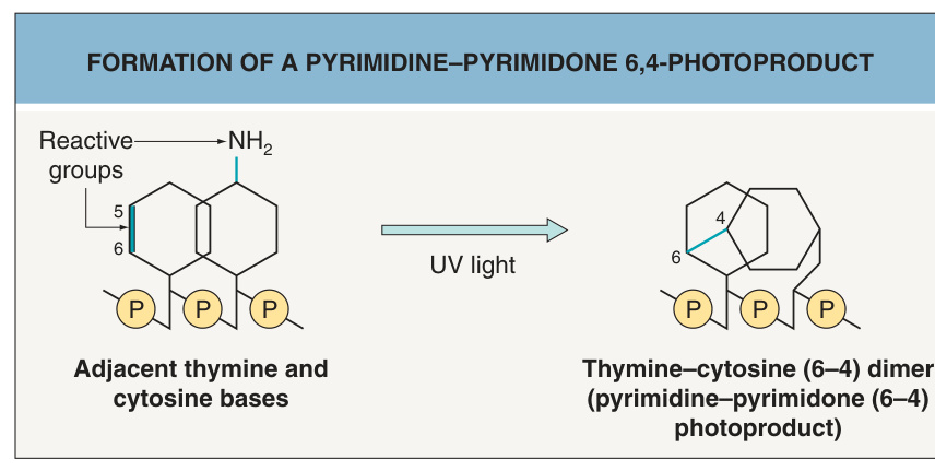
Fig. 86.9 Formation of a pyrimidine–pyrimidone 6,4-photoproduct.
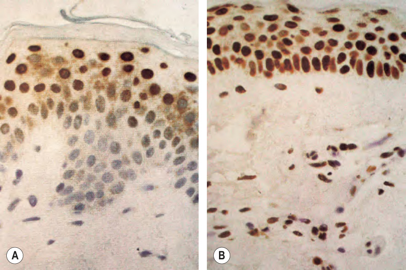
Fig. 86.10 A wavelength of 300 nm is more effective than one of 290 nm in inducing thymine dimers in the basal layer of the human epidermis.A After irradiation of human skin with monochromatic 290 nm UVB (2 MED) and staining with anti-thymine dimer antibodies, most cells in the basal layer show only blue counterstaining, while suprabasal layers demonstrate pronounced reactivity. B In contrast, with 2 MED of monochromatic 300 nm UVB, a pronounced immunostaining is also evident in the basal layer of the epidermis (as well as in cells within the dermis). Reproduced with permission from Young AR, Chadwick CA, Harrison GI, et al. The similarity of action spectra for thymine dimers in human epidermis and erythema suggests that DNA is the chromophore for erythema. J Invest Dermatol 1998;111:982–8.
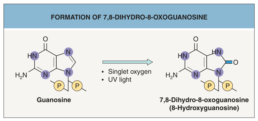
Fig. 86.11 Formation of 7,8-dihydro-8-oxoguanosine. 7,8-Dihydro-8- oxoguanosine (tautomer: 8-hydroxyguanosine) is an oxidation product of guanosine. It is formed by singlet oxygen, which is generated through a photosensitized reaction after excitation of a cellular chromophore by UV light.
Fig. 86.12 Action spectrum for the induction of cyclobutane dimers and oxidative guanine modifications in Chinese hamster ovary cells. The number of DNA lesions was assessed by the ability of repair enzymes to incise DNA from cells irradiated with different wavelengths from a monochromator. The ability of UV light to induce cyclobutane dimers and oxidative DNA damage rapidly declines with increasing wavelengths, e.g. 320 nm UV light is approximately 1000-fold less capable of inducing cyclobutane dimers than is 290 nm UV light. This decline parallels well with the decline of skin cancer formation in mice from 300 to 340 nm (Utrecht–Philadelphia skin cancer action spectrum). This decline does not mean that longer wavelengths do not contribute to photocarcinogenesis, because longer-wavelength UVA is much more abundant in natural sunlight than is UVB, which offsets at least some of the weaker effects of UVA. The second peak of oxidative base damage formation in the UVA range parallels with a second peak of skin cancer formation at 380 nm. Since there is no second peak of cyclobutane dimer formation with UVA, this has been interpreted as evidence that oxidative base damage contributes to mutagenesis and skin cancer formation. However, more recent data indicate that pyrimidine dimers are the most important pre-mutagenic DNA lesion not only for UVB, but also for UVA.