SKIN STEM CELLS
Stem cells are present in each self-renewing tissue and are responsible for maintaining and repairing the tissue in which they reside. These somatic stem cells have two defining properties: (1) the ability to produce daughter cells that can differentiate to renew or repair the tissue; and (2) the ability to produce a new stem cell (self-renewal). The epidermis, dermis, appendages, and melanocytes are each maintained by different lineage-specific stem cells. Stem cells often reside in a protective niche microenvironment that provides necessary signaling factors. During periods of homeostasis, stem cells generally divide very infrequently, a feature believed to protect them from acquiring mutations during cell cycle progression. After an injury, however, stem cells can divide more rapidly as part of wound healing.
One distinguishing characteristic of stem cells is that they can be maintained in culture virtually indefinitely. For example, a subset of epidermal basal cells forms highly proliferative colonies that can be passaged long-term in vitro; referred to as holoclones, they presumably represent stem cells. Holoclones can reconstitute epidermis in vitro, and they have been utilized as skin grafts for the treatment of burns and inherited skin disorders (see below). Within the skin, stem cells that maintain the epidermis and hair follicles have been most extensively studied. The properties of different cutaneous stem cells, including their molecular markers, are summarized in Table 2.2.
Epidermal Stem Cells
The human epidermis renews itself every 40–56 days. This constant turnover is mediated by epidermal stem cells, which reside in the basal layer. Stem cells comprise a small percentage of the basal epidermis, although their precise location and number is unclear. There is conflicting evidence regarding whether palmoplantar epidermal stem cells are found at the bottom or top of rete ridges, with more protection from environmental stress in the former location. Similarly, there are two competing models – hierarchical and stochastic – to account for proliferative, undifferentiated cells in the basal epidermis (Fig. 2.5).
Hair Follicle Stem Cells
The hair follicle bulge, which is located in the outer root sheath just deep to the sebaceous gland and adjacent to the arrector pili muscle attachment site, is thought to harbor hair follicle stem cells (Fig. 2.6).
The bulge represents the lowest portion of the hair follicle that does not regress during the catagen phase, and it contains morphologically undifferentiated cells. Hair follicle bulge cells are slow-cycling and have a high clonogenic potential in culture, as well as being self-renewing and multipotent. In vivo, they typically proliferate only when the hair follicle re-enters the anagen phase, in order to regenerate the bottom two-thirds of the follicle. In addition, appendageal stem cells of the bulge and eccrine duct provide an important ectodermal reserve, allowing reconstitution of the surface epidermis after wounding.
The bulb region at the base of the anagen follicle contains self-renewing, rapidly dividing, clonogenic matrix cells that produce the hair shaft and inner root sheath. These cells have the properties of stem cells, except they regress along with the rest of the lower follicle during catagen. Whether bulb cells are best described as transit amplifying cells or committed progenitor cells is a matter of debate.
Stem Cell Plasticity
Under homeostatic conditions, epidermal and hair follicle stem cells only contribute to their respective tissues of origin. However, bulge-derived keratinocytes can also contribute to the repair of the interfollicular epidermis in response to injury. Although most bulge-derived cells survive only transiently within the healing epidermis, a subpopulation of bulge cells can transdifferentiate into long-living epidermal stem cells. Similarly, epidermal stem cells can contribute to new hair follicle formation in the center of large wounds.
Like many organs, the skin and subcutaneous fat contain rare cells that can produce sphere-like colonies when provided with multiple growth factors in three-dimensional culture. These sphere cells represent pluripotent stem cells that can be directed to produce many tissue types, and their use for autologous stem cell therapies is being explored. However, the in situ location and function of these cells in the skin remains unknown.
Along the same lines, cells in adult mammals are far more “flexible” in their potential to form other cell types than previously thought. Some cell types, particularly bone marrow cells, have the potential to incorporate themselves into diverse tissues (e.g. epidermis, heart) and adopt the fates of resident cells, contributing progeny to the tissue. Induced pluripotent stem (iPS) cells are somatic cells
(e.g. fibroblasts) that have been reprogrammed into an embryonic stem cell-like state; these cells can be stimulated to differentiate into a variety of cell types, including keratinocytes. Thus, adult cells, including different skin cell types, may be capable of dedifferentiating into pluripotent stem cells.
Stem Cell-Based Therapy for Genetic Skin Disease
Stem cells have been utilized in several strategies for gene therapy of heritable skin diseases (Fig. 2.7). Treatment via delivery of gene-corrected stem cells has the advantage of continuous protein synthesis in the skin, but it may carry a risk of oncogenesis, especially with retroviral vectors. Laminin-β3-deficient skin from patients with junctional EB has been corrected by transplantation of stem cell-enriched epidermal grafts transduced ex vivo with a retroviral vector encoding normal LAMB3 cDNA. The treatment resulted in expression of functional laminin 332 and an absence of blistering in the corrected epidermis, both sustained over years of follow-up. In patients with severe recessive dystrophic EB (RDEB), similar genecorrected autologous keratinocyte grafts as well as intradermal and intravenous injections of allogeneic mesenchymal stromal/stem cells have also led to improved wound healing. In clinical trials investigating the use of allogeneic bone marrow-derived stem cell transplantation in children with severe RDEB, collagen VII deposition at the dermal–epidermal junction and variably decreased blistering were observed, with associated morbidity and mortality partially ameliorated by using reduced-intensity conditioning regimens. Other investigators are studying alternative sources of stem cells for the treatment of EB, including autologous iPS cells derived from revertant keratinocytes.
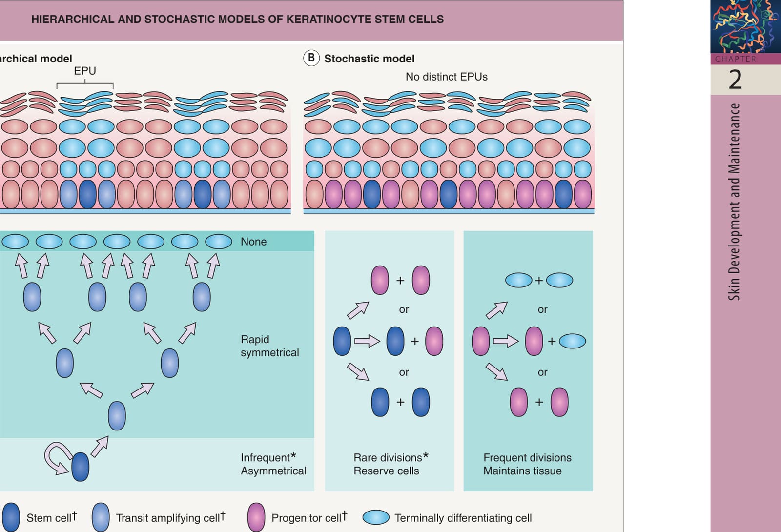
Fig. 2.5 Hierarchical and stochastic models of keratinocyte stem cells. These two models have been utilized to explain the behavior of stem cells and other proliferative cells in the basal epidermis. A In the hierarchical model, stem cells undergo infrequent asymmetric division, generating one new stem cell and one daughter transit amplifying (TA) cell. Unlike stem cells, TA cells divide frequently and symmetrically, producing two TA cells; in this model, they constitute the majority of basal keratinocytes. After a few rounds of division, TA cells withdraw from the cell cycle and move suprabasally, initiating the keratinocyte terminal differentiation program. Each epidermal stem cell and its progeny form a vertical column of progressively differentiating cells, which is known as an epidermal proliferation unit (EPU). B In the stochastic model, epidermal stem cell divisions are rare and usually asymmetrical, generating a new stem cell and a committed progenitor cell; however, random symmetric divisions resulting in either two stem cells or two progenitor cells also occasionally occur. In this model, committed progenitor cells make up the bulk of the basal layer, where they divide frequently to maintain the epidermis by producing either a new progenitor and a cell that leaves the basal layer to undergo terminal differentiation or two cells of either fate. It is possible that aspects of both stem cell models occur in the skin, depending on location or external stimuli, explaining why experimental evidence exists to support both models. Retention of a DNA label (e.g. 5-bromo-2-deoxyuridine or tritiated thymidine) is utilized as an in vivo marker of stem cells based on their slowly cycling nature.
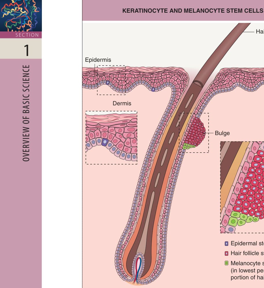
Fig. 2.6 Keratinocyte and melanocyte stem cells. Note that the bulge is continuous with the outer root sheath.
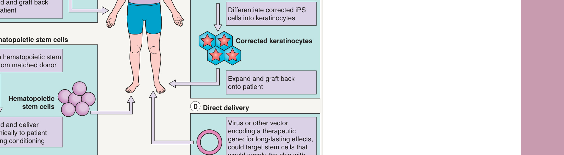
Fig. 2.7 Strategies for stem cell therapy of genetic skin disorders.A One strategy is to isolate epidermal stem cells from a patient, correct the genetic defect in vitro (e.g. using a viral vector), and graft epithelial sheets containing the corrected stem cells back onto the patient. B Gene therapy could also potentially utilize induced pluripotent stem (iPS) cells, which are generated from somatic cells (e.g. fibroblasts). The genetic defect could be corrected via techniques such as CRISPR/ Cas9-based targeted genome editing in these cells (see Fig. 3.13), which would then be differentiated into keratinocytes and grafted onto the patient. C A third strategy involves the use of hematopoietic stem cells (HSC). Upon systemic administration (following conditioning), allogeneic HSC cells have the capacity to home to the skin (e.g. to sites of injury in patients with epidermolysis bullosa) and produce differentiated progeny cells that provide the needed skin protein (e.g. collagen VII). D Finally, therapeutic genes could be directly administered to patients via a virus or another vector. To achieve permanent correction, the vector must target stem cells that could supply the protein to the skin. Courtesy Maranke I. Koster, PhD.
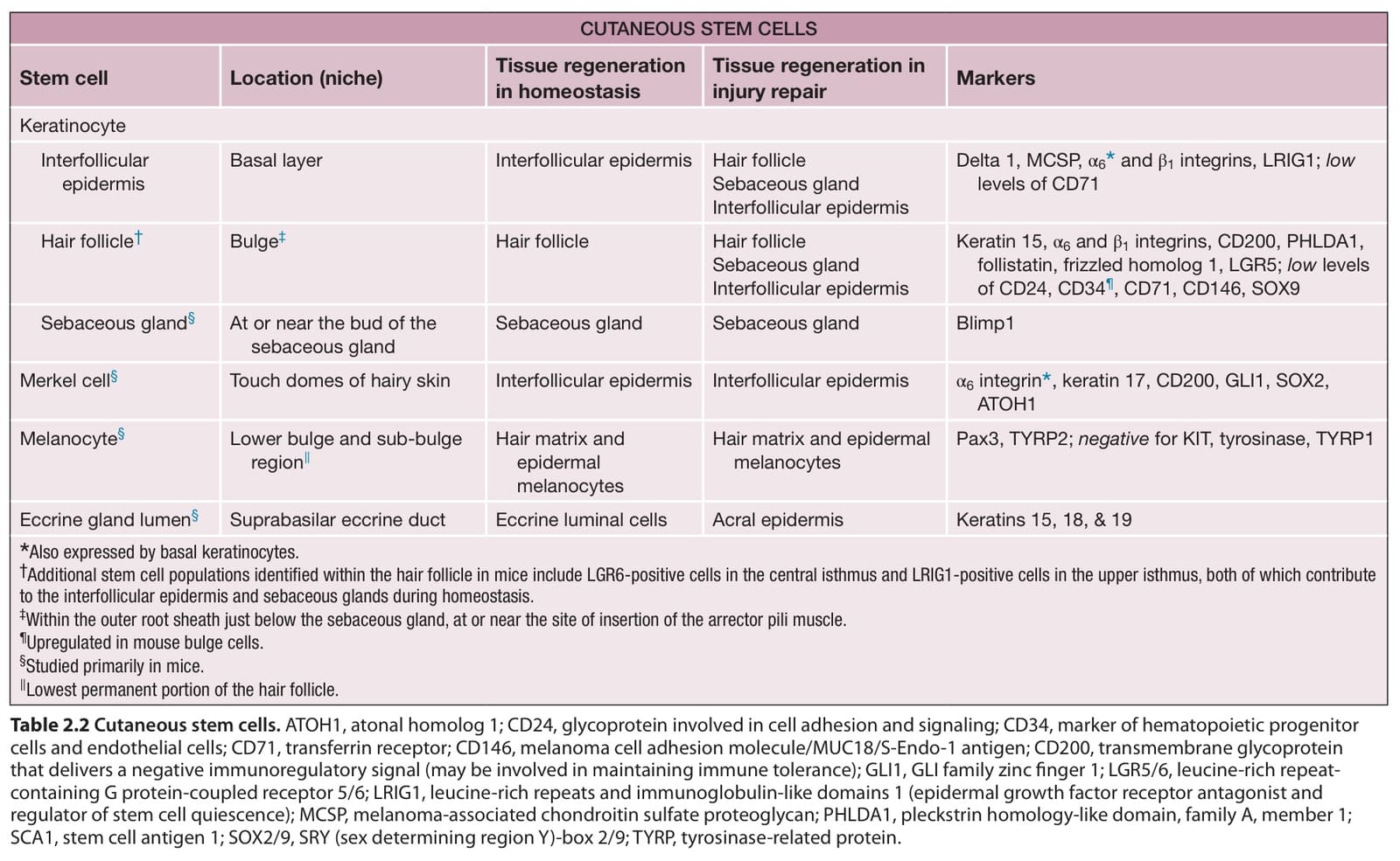
Table 2.2 Cutaneous stem cells. ATOH1, atonal homolog 1; CD24, glycoprotein involved in cell adhesion and signaling; CD34, marker of hematopoietic progenitor cells and endothelial cells; CD71, transferrin receptor; CD146, melanoma cell adhesion molecule/MUC18/S-Endo-1 antigen; CD200, transmembrane glycoprotein that delivers a negative immunoregulatory signal (may be involved in maintaining immune tolerance); GLI1, GLI family zinc finger 1; LGR5/6, leucine-rich repeatcontaining G protein-coupled receptor 5/6; LRIG1, leucine-rich repeats and immunoglobulin-like domains 1 (epidermal growth factor receptor antagonist and regulator of stem cell quiescence); MCSP, melanoma-associated chondroitin sulfate proteoglycan; PHLDA1, pleckstrin homology-like domain, family A, member 1; SCA1, stem cell antigen 1; SOX2/9, SRY (sex determining region Y)-box 2/9; TYRP, tyrosinase-related protein.