PATHOGENESIS
IHs represent localized or regional areas of abnormal vascular development and proliferation. Their development involves de novo formation of vessels from multipotent hemangioma-derived stem cells (HemSCs) and endothelial progenitor cells (vasculogenesis) as well as formation of new vessels from existing ones (angiogenesis). Other cell types in the hemangioma’s environment, including monocytes, fibroblasts, pericytes, mesenchymal cells, adipocytes and mast cells, can influence endothelial cell proliferation and may have a role in hemangioma formation and/or involution.
The overlapping immunohistochemical phenotype of hemangioma cells and human placental endothelium has led to the suggestion that hemangioma cells may be of placental origin or undergo differentiation toward a placental microvascular phenotype. North and co-workers reported that glucose transporter protein-1 (GLUT1) is expressed by IHs during all phases of their development (proliferating, involuting, involuted) as well as by the placenta, but not by other vascular tumors or malformations. Additional placenta-associated vascular antigens, including merosin, FcγRII, and Lewis Y antigen, are present in hemangioma specimens and placental chorionic villi but absent in microvessels of the normal skin and subcutis. However, hemangiomas do not express placental trophoblastic markers (e.g. human placental lactogen) and are not thought to represent placental emboli.
Role of Hypoxia
The role of hypoxia in the pathogenesis of IHs is supported by their association with hypoxic placental changes, prematurity/low birth weight (often caused by placental insufficiency), retinopathy of prematurity (GLUT1-positive, hypoxia-induced neovascularization), and regional arterial insufficiency in PHACE(S) and LUMBAR syndromes (see below). Hypoxia upregulates expression of GLUT1 and vascular endothelial growth factor (VEGF), leading to mobilization of endothelial progenitor cells. Moreover, the combination of hypoxia and estrogen has a synergistic effect on hemangioma endothelial cell proliferation in vitro, potentially explaining the predilection of IHs for female infants.
Investigators have also found that hypoxia inducible factor (HIF)-1α and its downstream effectors are upregulated in IHs during their proliferative phase.
Signaling Pathways
Several signaling pathways play a role in IH development. Studies have confirmed the importance of VEGF (also known as VEGF-A) signaling, with a shift from expression of VEGF receptor 1 (VEGFR-1), which binds VEGF with high affinity but transmits a very weak signal, to VEGFR-2, which strongly stimulates endothelial cell proliferation upon VEGF binding. VEGFR-2 signaling activates the phosphatidylinositol 3-kinase (PI3K)-mechanistic target of rapamycin (mTOR) pathway, leading to upregulation of HIF-1α and increased levels of VEGF. Accordingly, treatment of hemangioma endothelial cells in vitro with the mTOR inhibitor sirolimus (rapamycin) results in reduced proliferation.
Hemangiomas also have increased expression of the endothelial cellspecific Tie-2 tyrosine kinase receptor and dysregulated production of angiopoietin-2, which may promote angiogenesis and inhibit vessel maturation (see Ch. 102). The Notch pathway is thought to mediate differentiation of HemSCs into pericytes/vascular smooth muscle cells, and the latter appear to have pro-angiogenic properties and support IH development. The renin–angiotensin system (RAS) has also been implicated in IH proliferation via activation of angiotensin II receptor 2, and treatment of IH with β-blockers reduces plasma renin levels.
Genetic Factors
A variety of genetic factors may be involved in the development of IH. There have been reports of somatic mutations in genes that encode proteins involved in VEGF signaling (e.g. VEGFRs) and other pathways that affect vascular development within hemangioma tissue. Heterozygous germline mutations in VEGFR2 and ANTXR1 (anthrax toxin receptor 1), which encodes an integrin-like endothelial cell receptor, have been identified in a small subset of IH patients and may represent hemangioma predisposition factors.
Factors Influencing Natural History
Hemangiomas have been studied during different phases of their clinical course to gain insights into the mechanisms that govern their growth and involution. In the proliferative phase, hemangiomas express markers of proliferation such as proliferating cell nuclear antigen (PCNA) and increased levels of pro-angiogenic molecules such as VEGF and basic fibroblast growth factor (bFGF). In addition, other angiogenesis mediators, proteins found within the perivascular extracellular matrix, and enzymes involved in matrix remodeling are produced at higher levels during proliferation (Table 103.3).
The factors that govern the transition of hemangiomas from proliferation to involution and the mechanisms of the associated decrease in
pro-angiogenic factors are not fully understood. Increased expression of angiogenesis inhibitors and apoptosis promoters has been observed in involuting hemangiomas (see Table 103.3). Depending on the extracellular environment, mesenchymal stem cells may contribute to endothelial cell proliferation during the hemangioma growth phase or differentiate into adipocytes contributing to fibrofatty residua in involuting hemangiomas.
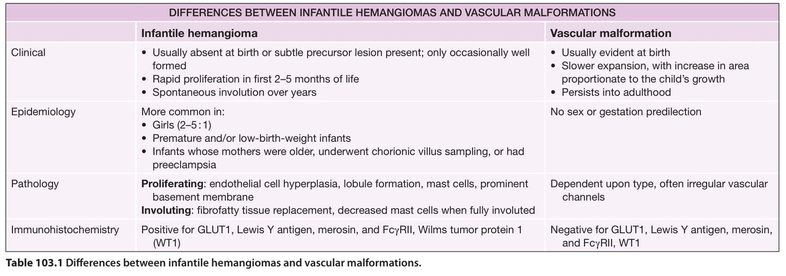
Table 103.1 Differences between infantile hemangiomas and vascular malformations.
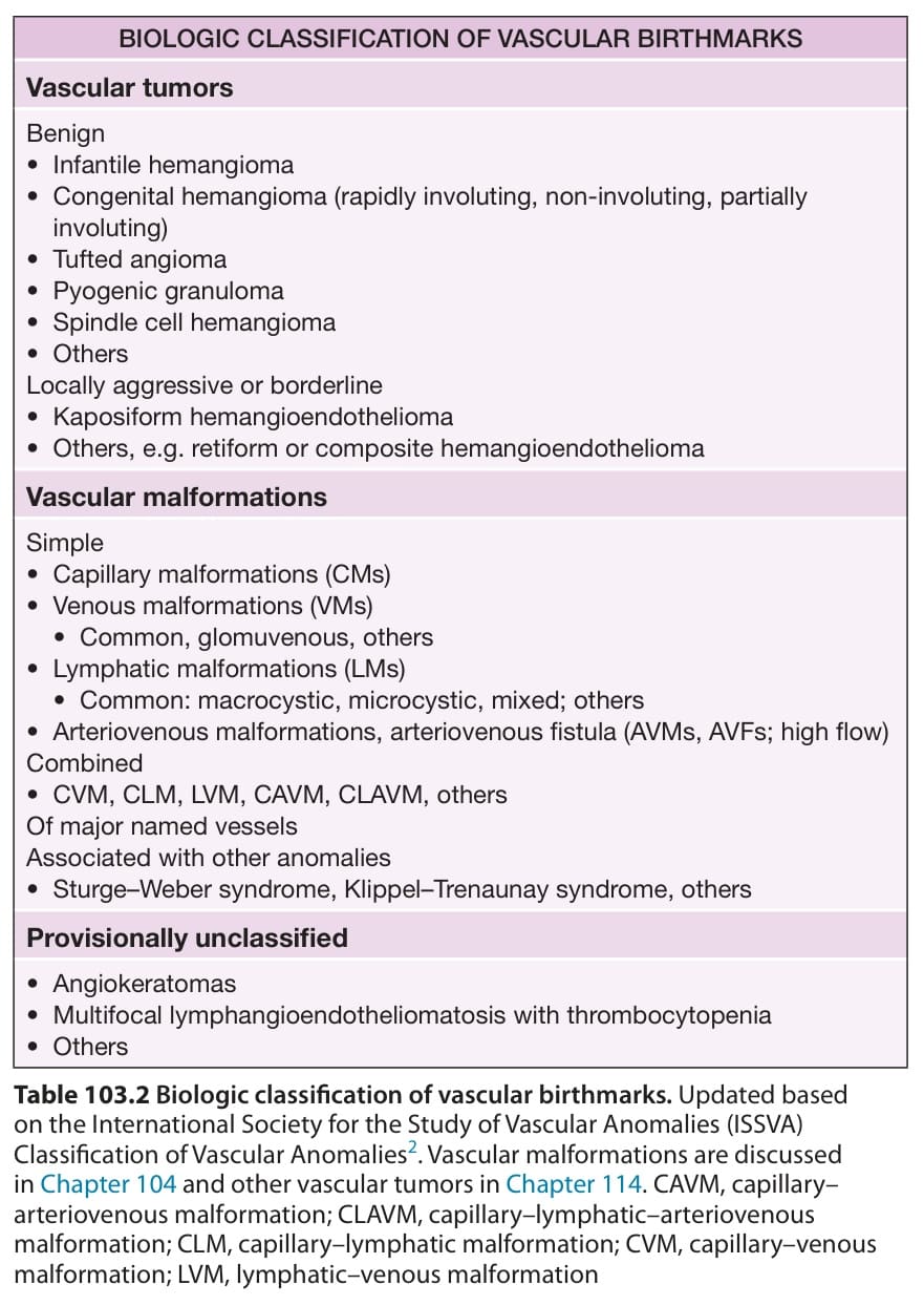
Table 103.2 Biologic classification of vascular birthmarks. Updated based on the International Society for the Study of Vascular Anomalies (ISSVA) Classification of Vascular Anomalies. Vascular malformations are discussed in Chapter 104 and other vascular tumors in Chapter 114. CAVM, capillary– arteriovenous malformation; CLAVM, capillary–lymphatic–arteriovenous malformation; CLM, capillary–lymphatic malformation; CVM, capillary–venous malformation; LVM, lymphatic–venous malformation
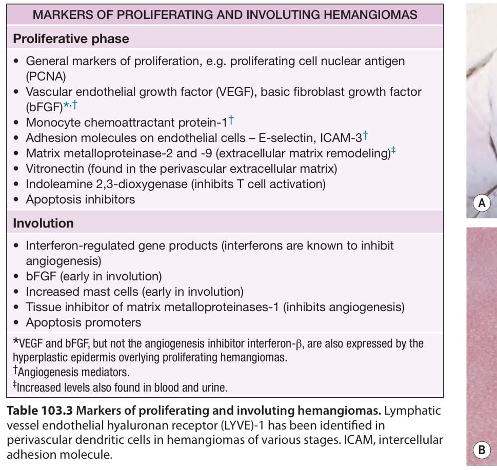
Table 103.3 Markers of proliferating and involuting hemangiomas. Lymphatic vessel endothelial hyaluronan receptor (LYVE)-1 has been identified in perivascular dendritic cells in hemangiomas of various stages. ICAM, intercellular adhesion molecule.