Proliferating Epidermoid Cyst
Synonyms: Proliferating epithelial cyst Proliferating infundibular cyst
Proliferating epidermoid cysts were first described in detail in 1995. Unlike proliferating tricholemmal cysts, proliferating epidermoid cysts are observed more commonly in men and only 20% occur on the scalp. The most common location is the anogenital region. They range in size from 0.5 to 15 cm. In a series of 30 patients for whom follow-up was available, 20% of the cysts recurred, some multiple times, and one patient died of intractable local disease. None of the patients developed metastatic disease.
Pathology
Histologically, almost half of cases show an epidermal connection, usually with a narrow opening or connection to a dilated follicle. Most tumors show areas of typical epidermoid cyst wall. In addition, areas of squamous proliferation with squamous eddy formation are seen, with formation of a granular layer and production of loose laminated keratin. The epithelium tends to proliferate peripherally into the surrounding
Well-circumscribed nodules of squamous epithelium in the dermis. Within their central portions, abrupt keratinization is seen. Courtesy Lorenzo Cerroni, MD.
dermis, rather than centrally as seen in proliferating tricholemmal cysts. Degrees of cellularity and atypia are variable, and frank carcinomatous change with an infiltrative growth pattern may be seen.
Treatment
Treatment is by complete surgical excision.
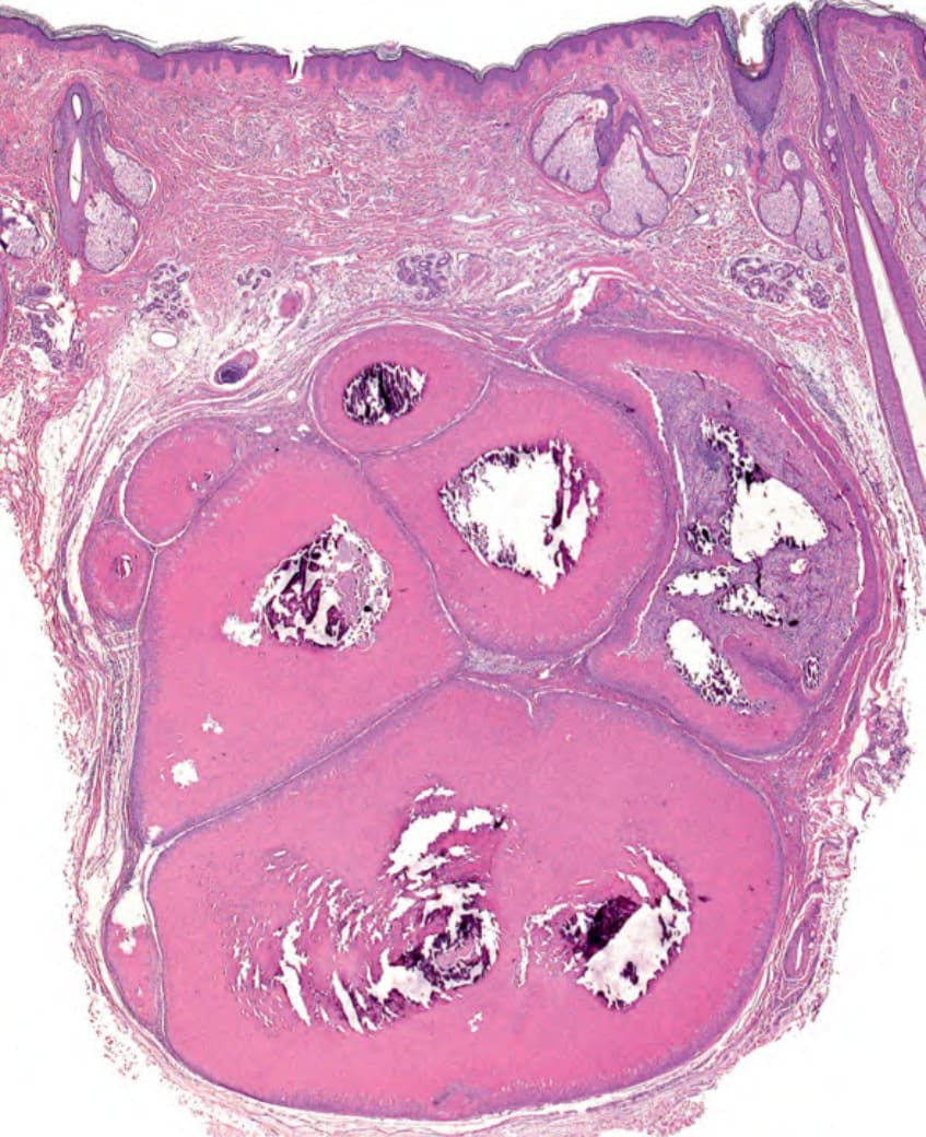
Fig. 110.10 Histopathology of a proliferating tricholemmal cyst.