DIAGNOSIS
Early detection is a key factor for improving survival in patients with melanoma. The clinical diagnosis of cutaneous melanoma continues to be based on visual inspection and dermoscopy. A history of change in the color, shape, or size of a pigmented skin lesion over the course of months or years is the most sensitive clinical sign for melanoma, and it may be detected by the patient or by serial screening examinations plus dermoscopy. In high-risk patients, the latter may be supplemented with comparison of stored digital and dermoscopic images and/or total body photography.
Public awareness campaigns have highlighted the ABCDEs of melanoma: Asymmetry, Border irregularity, Color variegation, Diameter >5 mm, and Evolution. It has been shown that patients more commonly recognize a melanoma by the development of a new pigmented lesion or by an increase in size or change in color of a pre-existing lesion, highlighting the importance of “E”. Additionally, the ABCD features broadly overlap with clinically atypical melanocytic nevi, including those that are banal and have low-grade histologic dysplasia, making ABCD less specific as warning signs for melanoma in certain patients.
Dermatologists often unconsciously rely on cognitive (overall pattern) and comparative (ugly duckling sign) processes rather than an algorithm of morphologic criteria (ABCD). A diagnostic clue is the so-called “ugly duckling” sign, which focuses on the morphologic differences between a single striking lesion and the surrounding benign “signature” nevi. There is a good interobserver reproducibility of this sign, supporting the significance of recognizing the “outlier” nevus.
Two other helpful clinical clues are the “Little Red Riding Hood” sign and the “EFG” rule. The former represents the finding of erythema or inflammation that at times will surround a cutaneous melanoma. “EFG” stands for an Elevated, Firm, or Growing lesion and is helpful to keep in mind for accurately diagnosing a clinically amelanotic nodular melanoma.
It is important to keep in mind that not all cutaneous melanomas present with typical clinical clues. Based on visual inspection alone, accuracy for the clinical diagnosis of melanoma does not exceed 75% and may be increased to up to 90% with the use of dermoscopy by experts. However, 10%–25% of melanomas may still be missed despite these diagnostic efforts. The term “featureless melanoma” has been coined for early and difficult-to-diagnose melanomas and may partially explain this statistic. Sometimes, patients are the first to recognize these featureless cutaneous melanomas, and there should be a low threshold for biopsying any persistent lesion that arouses patient concern for “being different” from the rest.
Differential Diagnosis
A variety of conditions may simulate melanoma, either clinically or histopathologically, or both. Awareness of these simulators is of great practical importance in order to avoid misdiagnosis or overdiagnosis of melanoma. Tables 113.6 and 113.7 list several melanocytic and non-melanocytic lesions that can mimic melanomas.
Dermoscopy
Dermoscopy, also known as skin surface microscopy or epiluminescence microscopy (ELM), is an essential, non-invasive tool for diagnosing pigmented skin lesions and for recognizing cutaneous melanoma (see Figs. 113.8, 113.10, 113.12, 113.14). Clinical diagnostic sensitivity
is significantly enhanced by the use of dermoscopy, and the correct diagnosis of melanoma is improved by nearly 50% in the hands of experienced/trained practitioners who utilize this diagnostic technique. It therefore can prevent unnecessary biopsies.
In one form of dermoscopy, a magnifying lens system is combined with a fluid interface in order to eliminate skin surface reflection. Hand-held lenses or cameras equipped with lenses and coupled with digital imaging systems are used. A glass plate makes direct contact with the skin, and ultrasound contact gel or a disinfectant spray can be used for the fluid interface. Morphologic structures within the epidermis, the dermal– epidermal junction, and the superficial dermis can be visualized with this technique. An alternative and more commonly employed form of dermoscopy uses polarized light to eliminate the surface reflection so that neither a liquid interface nor direct skin contact is required. The magnifications of these instruments range from 6- to 100-fold, but the most widely used dermoscope provides a 10-fold magnification, which is sufficient for routine assessment of most pigmented skin lesions.
The dermoscopic evaluation of cutaneous pigmented lesions involves a two-step algorithm. First, the observer determines whether the lesion under investigation is of melanocytic origin or not. For a lesion to be considered melanocytic, it needs to have at least one of the following dermoscopic structures/features: pigment network, streaks, aggregated globules, homogeneous blue pigment, or parallel pattern (acral lesions). If the lesion does not possess one of the aforementioned melanocytic features, then it needs to be evaluated further to determine if it has any dermoscopic characteristics consistent with other entities such as a seborrheic keratosis, dermatofibroma, or pigmented basal cell carcinoma (see Tables 0.14 & 0.15). However, if the lesion does not have any features of a melanocytic lesion and it does not have any features of a non-melanocytic tumor, then, by default, the lesion is considered to be of melanocytic origin.
Once a lesion is deemed to be melanocytic in origin, the second step of the algorithm is to differentiate between a benign melanocytic nevus and melanoma. To this end, multiple algorithms have been created, including pattern analysis, the ABCD rule, Menzies method, and the 7-point checklist, among others, which assist the clinician in deciding which lesions require a biopsy (see Tables 0.17–0.20). Several typical dermoscopic patterns have been described for benign nevi, including those with low-grade clinical atypia/histologic dysplasia, recognition of which can prevent unnecessary biopsy (see above).
The major dermoscopic criteria for melanoma can be broken down into global features, patterns, and local features. Global features of melanoma include dermoscopic asymmetry and the presence of multiple colors. Patterns seen by dermoscopy include reticular, globular, reticular–globular, homogeneous, reticular–homogeneous, and starburst. In melanoma, the most common patterns are the multi-component pattern (three or more dermoscopic structures distributed asymmetrically), asymmetric starburst pattern, and nonspecific pattern
(does not fit one of the known benign patterns). Lastly, the presence of any of the following local features should raise concern for melanoma: atypical network, negative pigment network, streaks, atypical dots or globules, irregular blood vessels, regression structures, crystalline structures, and blue–white veil (Table 113.8).
Over the past several years, dermoscopic criteria have been further refined in order to recognize difficult-to-diagnose melanomas such as ALMs and subungual melanomas. Additionally, diagnostic clues for early nodular melanomas and for amelanotic melanomas have been described, as these melanomas do not display the classic features (see above). As with clinical examination, not all melanomas can be clearly recognized by dermoscopy and “featureless” melanomas exist.
Photography
In high-risk patients, especially those with complex mole patterns, total body digital photography can be a helpful adjunct in the identification of new and changing lesions. The availability of baseline images for comparison, including dermoscopic images, permits the detection of melanomas that are growing or changing without manifesting obvious clinical characteristics, while simultaneously avoiding the removal of stable clinically atypical nevi. Future adoption of new and developing technologies, including automated two- and three-dimensional total body photography, sequential digital dermoscopic imaging, and computer-assisted diagnostic algorithms that incorporate artificial intelligence, may further enhance longitudinal evaluation of melanocytic lesions.
Non-Invasive Diagnostic Imaging
Non-invasive diagnostic imaging modalities are increasingly being utilized for melanoma detection. These technologies have the potential to augment clinical diagnosis and triage, decreasing unnecessary biopsies and excisions. However, their performance in “real-world” clinical settings remains to be seen, and they do not replace the gold standard of histopathologic diagnosis; widespread availability of novel imaging techniques is another limitation. While many are still in development, two modalities have been FDA-approved for clinical use: reflectance confocal microscopy and electrical impedance spectroscopy.
Reflectance confocal microscopy (RCM) uses a near-infrared, low-intensity diode laser designed for non-invasive in vivo imaging of the skin. Structures within the epidermis and papillary dermis are visualized with high resolution, producing images akin to traditional histology (see Ch. 0). This technique has been used to analyze various cutaneous tumors, including melanoma (Fig. 113.20), with a pooled sensitivity of 92% and pooled specificity of 70% for melanoma detection in a recent meta-analysis. RCM improves diagnostic accuracy for cutaneous melanoma compared to clinical and dermoscopic examination, preventing over half of benign lesions from unnecessary removal. RCM may also enable more accurate intraoperative assessment of subclinical extension of LMM, with excellent agreement (>80%) between RCM mapping and histopathologic margin assessment during staged excision. Additional applications of RCM include evaluation of dermoscopically equivocal lesions in difficult-to-biopsy situations (e.g. cosmetically sensitive areas, acral sites, young children) as well as short-term clinical monitoring of suspected lentigo maligna or amelanotic lesions whose initial RCM imaging appears negative for melanoma.
Electrical impedance spectroscopy (EIS) relies on the different electrical properties of normal and abnormal tissue to distinguish
By dermoscopy, the nevus has a regular cobblestone pattern (right) whereas the melanoma component has a disorganized pattern with an atypical pigment network as well as atypical dots and globules (left). B RCM of the nevus portion demonstrates regularly organized collections of reflective melanocyte cells within nests. C In the melanoma portion, there is architectural disorder of the epidermis with large round solitary atypical refractive (pagetoid) cells as well as bright atypical dendritic cells. Courtesy Anthony Rossi, MD and Ashfaq Marghoob, MD.
between benign nevi and melanoma. A hand-held probe with “microinvasive” electrode pins that penetrate the stratum corneum delivers an imperceptible low-level electrical current through the skin to measure the bioimpedance of the area of interest. EIS demonstrates high sensitivity: >96% melanoma detection for lesions deemed suspicious for melanoma on clinical and dermoscopic examination. However, specificity is much lower (~35% in one prospective trial). Notably, seborrheic keratoses may display a high false-positive rate with EIS, underscoring the importance of clinical judgment when adopting these imaging technologies.
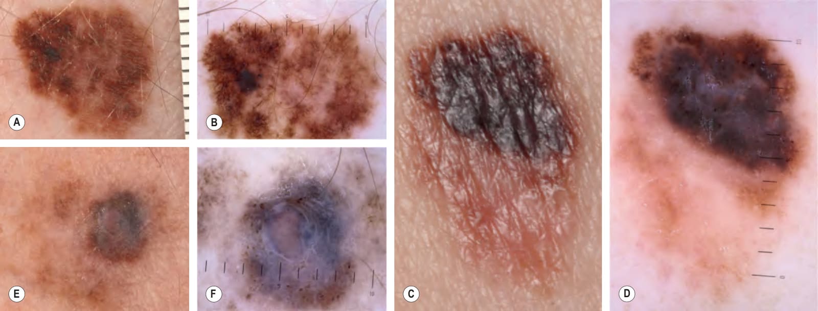
Fig. 113.8 Superficial spreading melanomas. Clinically, all of these lesions demonstrate asymmetry due to variation in color and irregularity in outline. Breslow depths for A, C, E were <0.5 mm, 0.58 mm, and 1.60 mm, respectively. B, D, F By dermoscopy, there is asymmetry, atypical pigment networks, irregular blotches, and multiple colors; in D and F a blue–white veil is present. Courtesy Claus Garbe, MD and Jürgen Bauer, MD.
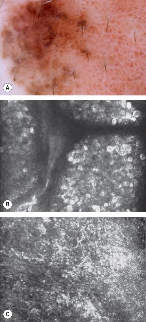
Fig. 113.20 Cutaneous melanoma arising within a compound melanocytic nevus – dermoscopy and reflectance confocal microscopy (RCM).A
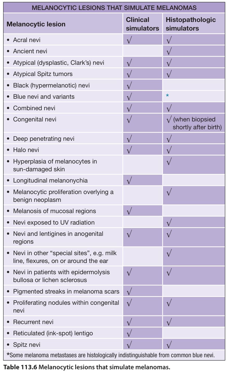
Table 113.6 Melanocytic lesions that simulate melanomas.
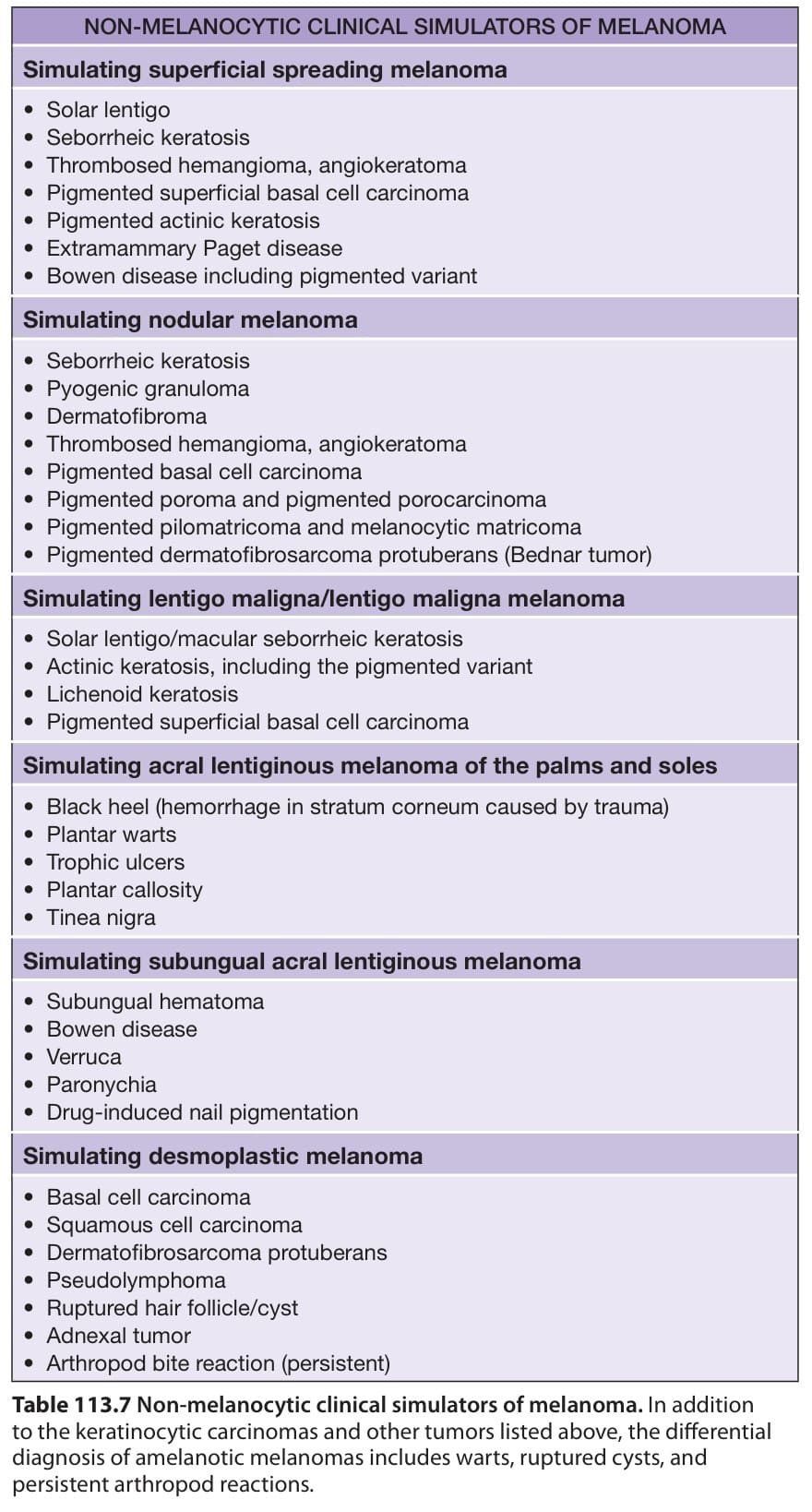
Table 113.7 Non-melanocytic clinical simulators of melanoma. In addition to the keratinocytic carcinomas and other tumors listed above, the differential diagnosis of amelanotic melanomas includes warts, ruptured cysts, and persistent arthropod reactions.
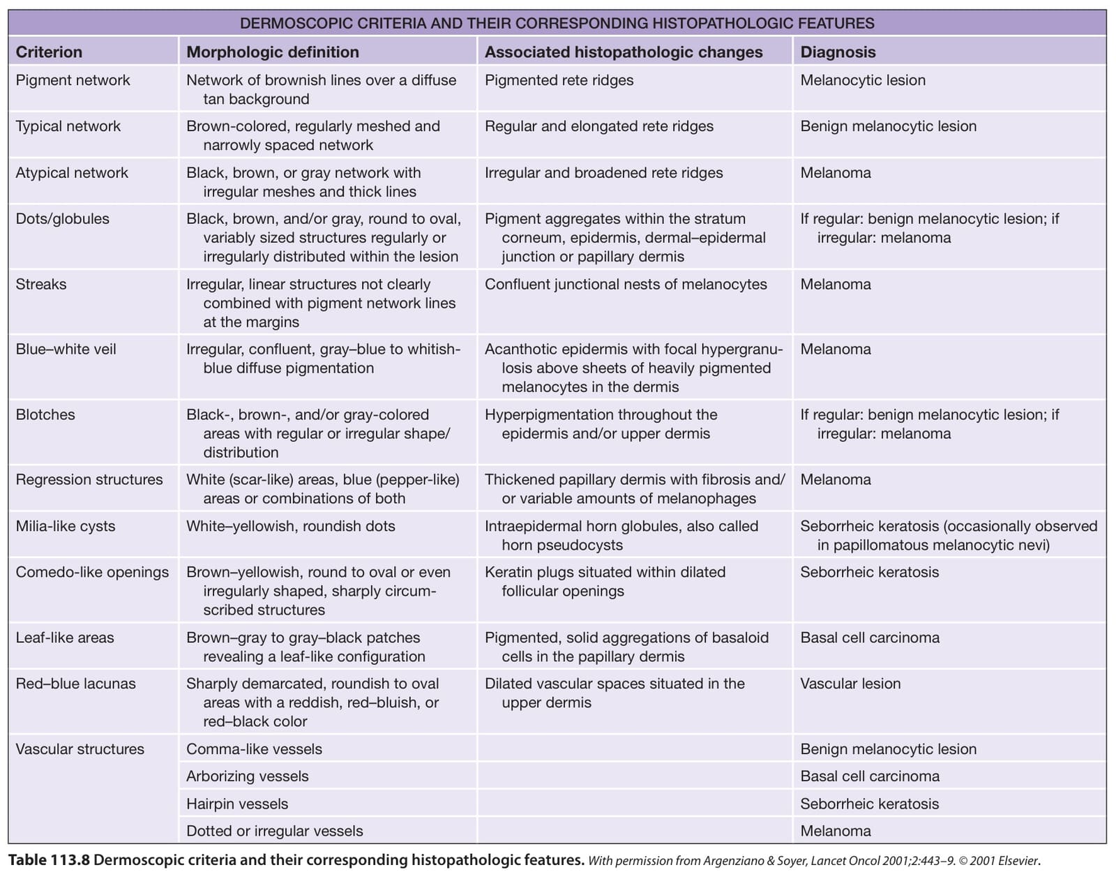
Table 113.8 Dermoscopic criteria and their corresponding histopathologic features.With permission from Argenziano & Soyer, Lancet Oncol 2001;2:443–9. © 2001 Elsevier.