PATHOLOGY
Histopathology remains the gold standard for melanoma diagnosis, although studies have demonstrated significant interobserver variability in the application of diagnostic criteria for melanocytic neoplasms ranging from moderately dysplastic nevi to in situ and thin (T1) melanomas. While most melanomas are readily distinguished from melanocytic nevi based on standard criteria (Table 113.9), some melanocytic lesions remain diagnostically challenging despite incorporation of immunohistochemistry and various molecular techniques.
It has been proposed that melanomas progress through two phases. The first is the radial (horizontal) growth phase (RGP), characterized by centrifugal spread of neoplastic melanocytes within the epidermis and infiltration of the papillary dermis by single cells or small nests. The second, vertical growth phase (VGP) is characterized by the presence of dermal nests/nodules of atypical melanocytes that are larger than and/ or cytologically distinct from their intraepidermal counterparts. It has been further postulated that the RGP lacks metastatic potential even in the presence of dermal invasion, whereas the VGP correlates with the capacity for metastasis. The major histologic types of melanoma described above differ with regard to the presence and morphology of the RGP. However, because the value of RGP versus VGP in assessing prognosis has not been confirmed by larger studies, the “growth phase” is not part of current classification schemes.
Typically, cutaneous melanoma is asymmetric, poorly circumscribed, and characterized by nests of melanocytes within the epidermis that are not equidistant from one another, vary in size and shape, and focally become confluent (Fig. 113.21). Within the epidermis, solitary melanocytes often predominate over nests. Some solitary melanocytes and nests of melanocytes are found well above the dermal–epidermal junction, at times extending into the upper epidermis, even the cornified layer; this is referred to as “pagetoid” scatter. These findings define melanoma in situ (Fig. 113.22). The presence of such features along with atypical melanocytes within the dermis constitute invasive melanoma. One element of histologic asymmetry in melanoma is the extension of the intraepidermal tumor cells far from the invasive intradermal component. Similar findings are present in the adnexal
(low-CSD) cutaneous melanoma.A Asymmetric melanocytic tumor with asymmetry in both distribution of melanocytic complexes and pigment. B Pagetoid melanocytes organized as solitary units and nests varying in size and shape are present throughout the entire epidermis. Neoplastic melanocytes extend into the dermis. There is absence of maturation at deeper levels of the dermis. Courtesy Lorenzo Cerroni, MD.
epithelium of folliculosebaceous units and eccrine ducts. Within the dermis, the melanocyte nuclei do not become smaller within the deeper portions of the tumor (absence of maturation). In parallel, the nests of melanocytes also do not become smaller with progressive descent.
In SSM, the radial growth phase predominates histologically with broad, horizontal extension of atypical melanocytic nests and single cells within the epidermis and superficial dermis. As these tumors frequently represent low-CSD melanomas, solar elastosis within the dermis is usually minimal. In nodular melanoma, the tumor extends vertically in the dermis with a comparatively limited involvement of the overlying epidermis (Fig. 113.23). Overall, the tumor may exhibit a nodular, polypoid, or pedunculated architecture.
LMM (high-CSD melanoma) differs from the prototypic melanoma by the presence of marked solar elastosis (a sign of chronic sun damage) as well as the tendency to have little pagetoid spread within the epidermis. Tumor cells show lentiginous spread, i.e. solitary tumor cells in the epidermal basal layer predominate over tumor cell nests within the epidermis. The atypical melanocytes are commonly present within the epithelium of adnexal structures, especially along the outer root sheath of hair follicles (Fig. 113.24). The invasive component is more often composed of spindle cells, although any morphology is possible. Reflecting the role of high CSD in the pathogenesis of this subtype, epidermal atrophy, in addition to solar elastosis in the upper dermis, is commonly observed.
ALM often has a proliferation of atypical melanocytes within the basal layer of a hyperplastic epidermis, i.e. a lentiginous spread of single melanoma cells (Fig. 113.25). These findings may be subtle in early lesions or at the tumor’s periphery. In volar and subungual sites, this lentiginous component may contain strikingly dendritic melanocytes with hyperchromatic, vertically elongated nuclei and irregular dendritic processes that may reach the mid spinous layers. Throughout the lesion, atypical melanocytes are arranged singly and in irregularly shaped nests at all levels of the epidermis (“pagetoid scatter”), with a predominance of single cells. In the cornified layer, numerous melanocytes and melanin granules are diffusely scattered. These findings are in contrast to the mild cytomorphologic atypia that may characterize benign acral nevi and nevi in other “special sites” (e.g. genitalia, milk line), a finding commonly referred to as “special site atypia”. For example, while some pagetoid scatter may be present in acral nevi, this is typically confined to the skin furrows (“columnar scatter”), whereas in melanoma it is present throughout the lesion, involving both ridges and furrows (“diffuse scatter”).
Irregular, sometimes confluent, nests of atypical melanocytes within a hyperplastic epidermis. Some of the melanocytes are hyperchromatic and are also present within the cornified layer. Note the thick stratum corneum typical of acral skin. Courtesy Lorenzo Cerroni, MD.
Microstaging
Of the histologic features in a primary cutaneous melanoma, Breslow depth (i.e. tumor thickness) is the strongest predictor of survival. Melanoma thickness is measured in millimeters from the top of the granular cell layer of the epidermis (or the base of an ulcer) to the deepest point of tumor penetration, using an ocular micrometer (Fig. 113.26). In addition to tumor thickness, a number of other histologic features should be noted such as ulceration and involvement of margins. Tumor thickness and ulceration are major determinants of T-stage in the current AJCC-8/UICC staging system, with ulceration defined histologically as the absence of an intact epidermis overlying a major portion of the primary tumor plus an associated host response (e.g. fibrinous exudate, acute inflammatory infiltrate).
Although dermal mitotic count (measured according to the “hot spot” method and reported as number/mm) does not currently upstage primary melanoma, it was viewed as a prognostic factor in the previous AJCC staging system. Guidelines for the histopathologic examination and reporting of melanoma features have been published by
Breslow method: measure from the granular layer of the epidermis to the deepest part of the tumor.
the International Collaboration on Cancer Reporting and the College of American Pathologists. However, a number of experienced dermatopathologists only report criteria essential for the current staging classification – tumor thickness, ulceration (if present), satellites (if present), and assessment of margins.
Immunohistopathology
Immunohistochemical studies can be helpful in the assessment of diagnostically difficult cases of primary melanoma as well as in the assessment of metastatic tumors of unknown origin (see Tables 0.13 & 122.4). However, the diagnosis of cutaneous melanoma is based upon routine H&E staining, as immunostaining patterns alone cannot clearly differentiate between benign and malignant lesions. A wide range of melanoma-associated antigens have been identified for which immunohistochemical stains are available. Melanocyte differentiation antigens, e.g. PMEL17/gp100 (HMB45), tyrosinase, MART-1/Melan-A, MITF, are useful for: (1) distinguishing cells of melanocytic lineage from other tumor types; (2) visualizing the full extent of tumor cells of a primary melanoma; and (3) identifying minute foci of melanoma in sentinel lymph node biopsies. HMB45, which recognizes the melanosome-specific glycoprotein PMEL17/gp100, has high specificity for melanocytes and nevus cells, but its utility is limited by its heterogeneous staining pattern and limited sensitivity. Transfer of melanosomes to keratinocytes can also lead to positive staining for tyrosinase and PMEL17/gp100 (HMB45) in keratinocytes. Because MITF is a transcription factor in melanocytes, it does not stain melanosome-containing keratinocytes. Of note, HMB45, tyrosinase, MART-1/Melan-A, and MITF may all be negative in desmoplastic melanoma, representing an important potential pitfall.
Staining for S100, a calcium-binding protein, has high sensitivity for melanocytes and melanoma, including desmoplastic or spindled cell melanoma. However, this protein is also expressed by Langerhans cells, other dendritic cells, eccrine glands, Schwann cells, chondrocytes, and adipose tissue. SOX10, a transcription factor, has emerged as a popular alternative to S100 and other melanocytic markers due to easier interpretation of its nuclear staining pattern. Although SOX10 is not entirely melanocyte-specific (expressed in myoepithelial cells and other cells of Schwannian lineage), it will identify the spindled component of most desmoplastic melanomas. There is also less background staining of dermal fibrohistiocytic cells by SOX10 compared to S100, making SOX10 useful in distinguishing residual desmoplastic melanoma from scar. In sum, the use of panels of these antibodies improves sensitivity and specificity, but remains diagnostically imperfect.
More recently, immunohistochemical staining for PRAME (PReferentially expressed Antigen in MElanoma) was introduced. Initially identified in tumor-reactive T cells from patients with cutaneous melanoma, PRAME was one of the few GEP genes that discriminated between melanoma and benign melanocytic neoplasms. Immunohistochemical staining for PRAME is generally negative in benign nevi, solar lentigines, melanocytic hyperplasia in sun-damaged skin, and other melanocytic lesions with inter-mediate histopathologic features. In contrast, the majority of primary cutaneous melanomas and their metastases demonstrate diffuse nuclear PRAME immunostaining, which appears to be strongly concordant with abnormal cytogenetic test results. PRAME may be a useful marker in the assessment of melanoma surgical margins (including LM), in distinguishing melanoma micrometastases from nodal nevi in sentinel lymph nodes, and in the evaluation of ambiguous melanocytic lesions. Potential exceptions include desmoplastic melanomas, of which only a third may be PRAME-positive, and spitzoid neoplasms, in which PRAME positivity may correlate less strongly with histopathologic features. Although it currently appears to be a promising immunohistochemical marker for melanoma, because a minority of melanocytic nevi are PRAME-positive, correlation with conventional histopathologic features is paramount for proper classification of both PRAME-positive and PRAME-negative lesions.
Molecular Analysis
Some cutaneous melanomas prove challenging to diagnose, and differentiation between atypical Spitz tumor (AST)/Spitz melanocytoma and spitzoid melanoma or between deep penetrating nevus or atypical blue nevus and melanoma can be difficult based upon histopathology. In such cases, CGH or FISH in addition to newer molecular techniques (NGS, GEP) may be helpful (Table 113.10).
CGH is a method for the genome-wide assessment of DNA copy number changes in tumor cells. Fluorescence-labeled tumor DNA and reference DNA from a healthy donor are hybridized onto metaphase chromosomes or on arrays of oligonucleotides (array CGH). From the relative fluorescence copy number, gains or losses within the tumor can be calculated. The oligonucleotide spots on the array can be mapped to the respective position within the human genome. CGH analysis of benign melanocytic nevi typically shows no clonally expanded chromosomal aberrations, while in the vast majority of melanomas, gains and losses of particular segments of chromosomes or entire chromosomes are found, including copy number gains of 1q, 6p, 7p, 7q, 8q,
17q, and 20q and losses of chromosomes 6q, 8p, 9p, and 10q. As an exception, ~20% of Spitz nevi show an increase in copy number of chromosome 11p (corresponding to HRAS activation) or an isolated loss of chromosome 3 (corresponding to loss of BAP1), aberrations not typically found in cutaneous melanoma.
FISH can also assist in differentiating between benign and malignant melanocytic tumors (see Fig. 3.8). These assays often focus on chromosomal loci that are frequently affected by copy number changes in melanoma. Studies have shown that FISH assays that target various combinations of 6p25 (RREB1), 6q23 (MYB), 8q24 (cMYC), 11q13 (CCND1), CEP6 (centromere of chromosome 6), and 9p21/CEP9 (CDKN2A) discriminate between benign nevi and melanoma with high sensitivity and specificity. The disadvantage of FISH is that only gains and losses involving a limited number of chromosomes are detected. However, compared to CGH, FISH is typically faster, less expensive, and requires less tumor tissue to perform.
NGS has also been used to further characterize the genomic landscape of multiple tumors, including melanoma and various spitzoid tumors. This method allows for the analysis of hundreds of genes in a single study (see Fig. 3.5), and it can potentially point to new prognostic markers or therapeutic targets. NGS has facilitated the discovery of various fusion kinases that underly the pathogenesis of some melanocytic neoplasms, including spitzoid neoplasms. Due to the immense data infrastructure and bioinformatics expertise required for NGS, its availability is currently limited to a few commercial laboratories and academic research centers.
A frequent application of the above molecular techniques is in the evaluation of AST versus spitzoid melanoma. One or multiple methods can be utilized given that complementary information is provided by each assay. For FISH, inclusion of the 9p21/CEP9 probe significantly increases sensitivity for spitzoid melanoma, as homozygous loss of this locus has been associated with advanced locoregional disease or death. Gains of 8q24 and 11q13 also correlate with an aggressive clinical course, but the risk is lower than with homozygous 9p21 deletions. In cases with ambiguous or negative FISH results, CGH may provide additional information regarding other copy number alterations supportive of melanoma. Identification of TERT promoter mutations via NGS is also suggestive of more aggressive clinical behavior, with one study demonstrating a significant association with extranodal metastasis and death. In the assessment of ASTs, identification of an underlying BRAF or NRAS mutation by NGS may favor a conventional melanoma with spitzoid features over AST. Regardless of which techniques are used, definitive classification of ASTs remains a significant challenge.
Gene Expression Profiling (GEP)
GEP has gained attention as a potentially useful molecular technique in the field of melanoma, and GEP assays for diagnosis as well as prognostication have been developed. Whereas diagnostic GEP aims to classify an undiagnosed melanocytic lesion as benign, intermediate, or malignant, prognostic GEP aims to provide further clinical risk stratification for melanomas beyond AJCC stage and other clinicopathologic factors. GEP assays typically use quantitative reverse transcriptase PCR (qRT-PCR) to measure the mRNA expression levels of a limited panel of genes; the latter have been correlated with pre-specified outcomes, such as benignity, tendency to metastasize, or SLNB risk prediction, and the goal is to accurately predict this outcome in the queried lesion. Ideally, during development of prognostic GEP assays, fully annotated cohorts of archival melanoma tissue samples would be analyzed, with subsequent prospective validation among separate melanoma cohorts as well as comparison against all known clinicopathologic factors and AJCC stage to determine prognostic accuracy and clinical utility.
Gene expression profiling for diagnosis
Development of diagnostic GEP assays was based on several DNA microarray studies that demonstrated disparate gene expression patterns when benign melanocytic lesions were compared to melanoma. There are currently two commercially available diagnostic GEP tests, a 3-gene assay (DermTech Smart Stickers™) that was initially a 2-gene assay, and a 23-gene expression signature (23-GES) assay (myPath® Melanoma). Both the initial 2-gene assay and the current 3-gene assay use adhesive tape stripping pre-biopsy to determine the likelihood of malignancy (and need for biopsy) of a melanocytic neoplasm based on melanoma-associated genes – LINC, PRAME, and more recently, TERT. The original 2-gene assay demonstrated a sensitivity of 91% and specificity of 69% in the classification of suspicious melanocytic lesions, with subsequent real-world studies demonstrating even higher specificity (91%) and high negative predictive value (99%) without decreased sensitivity for melanoma. The addition of TERT to the 3-gene assay was reported to increase diagnostic sensitivity to 97% in a study of 103 pigmented skin lesions clinically suspicious for melanoma.
The 23-GES assay uses formalin-fixed paraffin-embedded tissue samples from diagnostic biopsies, when histopathologic analysis is indeterminate for melanoma. Some studies of the 23-GES assay have demonstrated high sensitivity (93%) and specificity (96%) in distinguishing melanoma from benign nevi, with clinically proven outcomes. However, other studies have noted its limitations in more ambiguous melanocytic lesions, including spitzoid tumors. Given the novelty of diagnostic GEP assays and limited experience in clinical settings, current appropriate use criteria published by US dermatology and dermatopathology societies maintain an uncertain role for qRT-PCR-based assays in melanoma diagnosis.
Gene expression profiling for prognosis
Current AJCC-8 melanoma staging and international guidelines use primary tumor histologic factors, e.g. Breslow thickness, ulceration, and pathologic status of the regional lymph nodes (via SLNB) to estimate patient prognosis. Prognostic GEP aims to improve on AJCC melanoma staging by classifying metastatic risk and predicting disease recurrence, potentially identifying patients who would benefit from surveillance imaging and/or adjuvant therapy.
Currently, there are three commercially available prognostic GEP tests including the 31-gene DecisionDx®-Melanoma (US) and 11-gene MelaGenix (Europe). These two assays use a dichotomous class assignment of risk for metastasis (class 1 vs class 2 or low risk vs high risk) rather than calculating an estimated MSS as is done with AJCC staging and other risk-based predictive nomograms. The third Merlin Test combines an 8-gene GEP test with Breslow thickness and patient age to predict SLNB positivity or negativity, with the potential goal of reducing unnecessary SLNBs in patients at low risk of a positive result; prospective validation studies are ongoing. The 11-GEP MelaGenix assay was studied in Europe and reported as an independent prognostic factor for MSS, along with tumor thickness and age, in 291 stage I–III melanoma tumors, suggesting a potential role for this assay in clinical decisions regarding adjuvant therapy.
Currently, in the US, the majority of prognostic GEP testing is performed with the 31-gene panel. Most clinicians who order this test report an increased frequency of clinical or imaging surveillance in patients designated as having class 2 disease, regardless of AJCC stage. However, the clinical utility of prognostic GEP testing and its impact on patient outcomes remains uncertain, and routine prognostic GEP testing is not recommended in current US national guidelines (NCCN and AAD).
A major criticism of current GEP tests is their relatively low positive predictive value in stage I melanoma, particularly thin (T1) tumors. A meta-analysis of melanomas tested with 31-GEP demonstrated a true positive rate of 1% (6/623) and false positive rate of 10% (61/623) for stage I melanomas; this means that 100 stage I melanomas would need to be tested to identify one that will metastasize while 10 individuals would be falsely identified as having high-risk melanoma. Stage II melanomas had a better prognostic performance with a true positive rate of 28% (59/212) and false positive rate of 37% (78/212), suggesting that GEP testing may ultimately prove more useful in guiding adjuvant therapy use and/or surveillance imaging in this subset of patients. However, stage II melanoma patients are typically followed more intensely than those with stage I due to their increased chance of disease recurrence.
In thin melanomas, the lower positive predictive values of available GEP tests may be related to low numbers of metastases in the cohorts of stage I melanomas used to train and validate the models (e.g. 9% and 3%, respectively). Alternatively, predicting metastatic potential based upon mRNA expression may represent a biological obstacle for earlier stage melanomas, i.e. a small percentage of cells conferring metastatic potential is not detectable with existing technology platforms. An additional criticism is that designation of a dichotomous value (e.g. class 1 vs class 2) to a continuous variable (e.g. disease-free survival) leads to assignment of equal risk to heterogeneous populations. The major argument for testing thin melanomas with 31-GEP is the high negative predictive value (87%), which could provide additional reassurance to patients. However, the contribution of GEP testing beyond AJCC-8 staging and other known clinicopathologic factors (e.g. primary tumor mitotic rate, lymphovascular invasion, patient age, biologic sex) is unclear. In addition, the implications of a false-positive result should be discussed with the patient, preferably in a multidisciplinary care setting.
There is great interest in risk prediction for SLN-positivity in order to reduce the number of unnecessary SLNBs performed. Two commercially available GEP tests have been studied in this regard: (1) the 8-gene Merlin Test (combined with Breslow thickness and patient age); and (2) a modified i-31-GEP DecisionDx® that uses artificial intelligence to integrate a continuous GEP score with clinicopathologic factors (age, biologic sex, anatomic location, Breslow thickness, mitotic rate, ulceration, subtype, tumor infiltrating lymphocytes, deep margin transection). Three US and European validation cohort studies with 208, 210, and 421 patients have been performed, with the largest study reporting a 35% SLNB reduction rate (via a low risk score) in patients with T1–T2 melanomas (95% CI, 29.4–41.8), with a negative predictive value of 96.5% (95% CI: 90.0–99.3). In a validation cohort of 1674 T1–T4 melanomas from 25 surgical and 5 dermatology centers, the i-31-GEP model identified an additional 19% of tumors (compared with T-stage alone) with <5% SLNB risk, for which the procedure would not be recommended. A recent decision-analytic study suggested that the i-31-GEP model could help to risk stratify patients with T1b melanoma, identifying 23% who could safely forego SLNB (using a 5% risk threshold), though its role in patients with T1a and T2 melanoma requires further study. Various SLNB risk calculators have been developed worldwide that incorporate relevant clinicopathologic factors (e.g. Breslow thickness, ulceration, age, melanoma subtype, lymphovascular invasion) and may provide additional means to more easily predict SLN-positivity.
While current data are insufficient to support the use of available prognostic/predictive GEP tests to determine the need for SLNB, surveillance imaging, and/or adjuvant treatment, the field is rapidly evolving. There is consensus that further investigation of existing and novel GEP tests should meet certain criteria before being routinely incorporated into clinical practice. The latest NCCN guidelines note the need for ongoing study of large data sets of unselected patients to determine whether a combination of GEP testing and clinicopathologic variables provides clinically actionable information, given the previously cited limitations in stage I melanoma. As GEP testing is further refined and newer technologies (e.g. circulating tumor DNA) evolve, there is great promise that clinical decision-making will be guided by molecular tumor classifications.
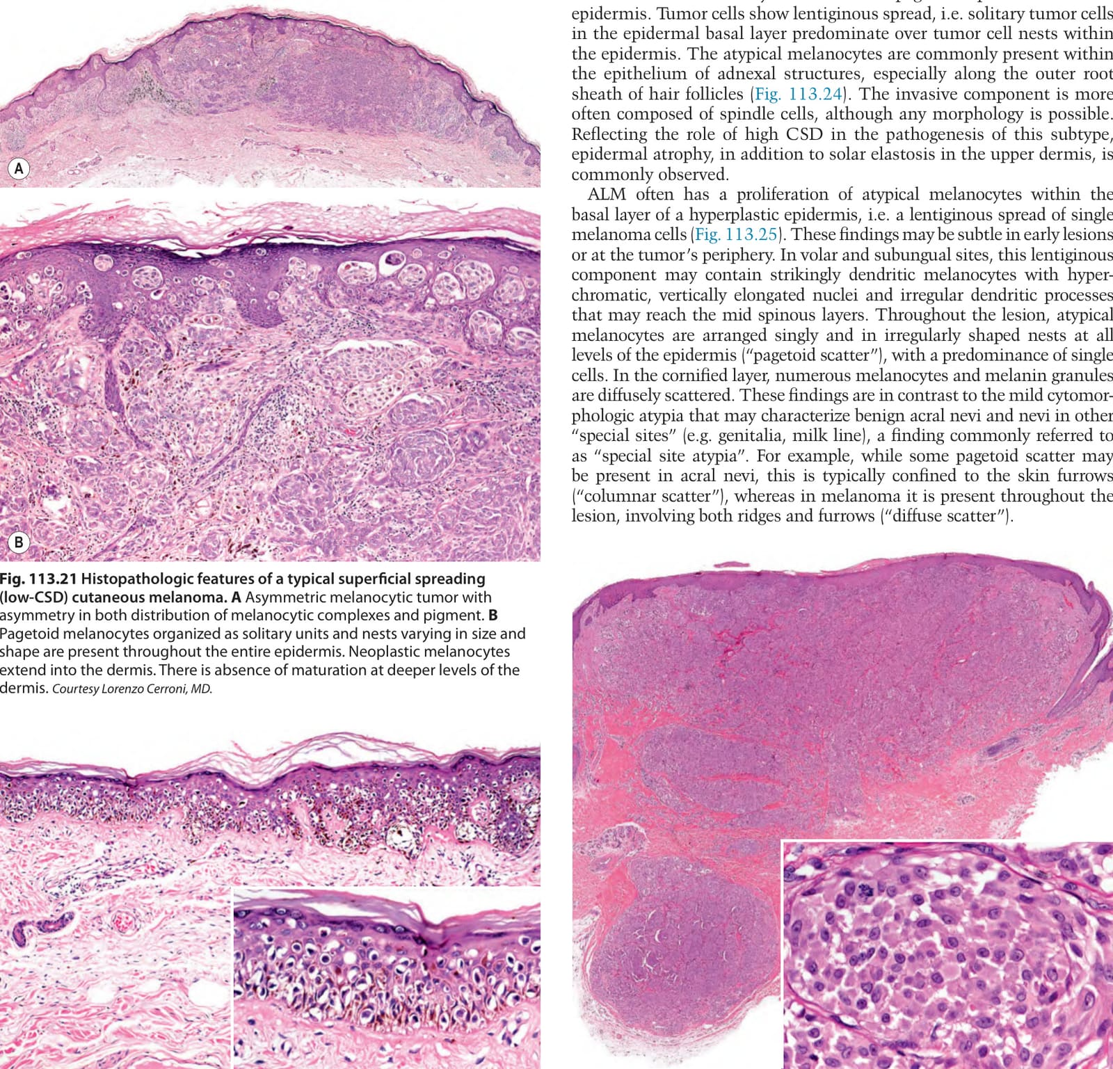
Fig. 113.21 Histopathologic features of a typical superficial spreading
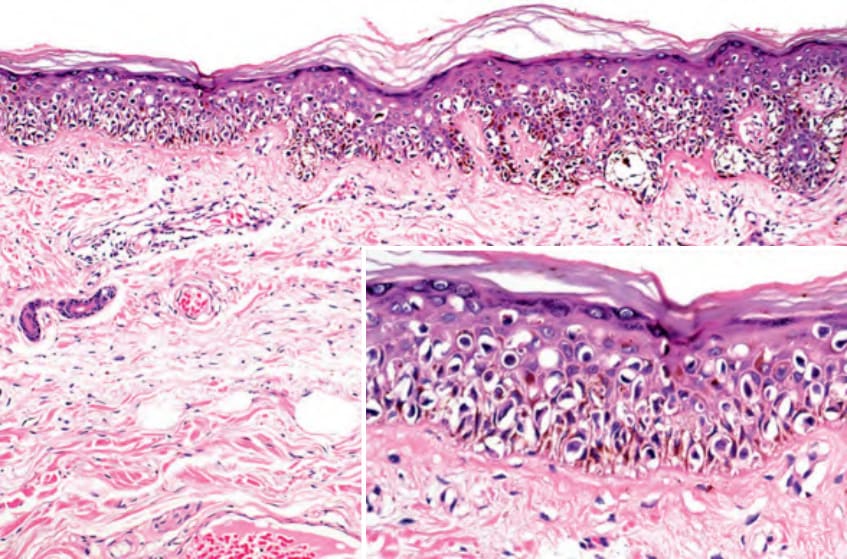
Fig. 113.22 Histopathologic features of a melanoma in situ. Increased number of melanocytes with atypical nuclei not only in the basal layer, but also at all levels of the epidermis. Note the predominance of single cells over nests. Courtesy Lorenzo Cerroni, MD.
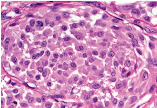
Fig. 113.23 Histopathologic features of a nodular melanoma. Relatively well-circumscribed tumor primarily within the dermis. Confluent complexes of atypical melanocytes extend throughout the entire dermis and into the super-ficial portion of the subcutaneous fat. Note the atypical epithelioid melanocytes and one mitotic figure (inset). Courtesy Lorenzo Cerroni, MD.
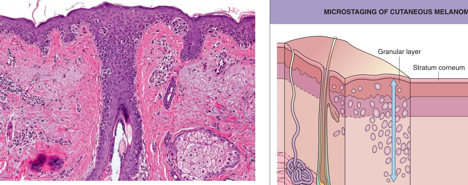
Fig. 113.24 Histopathologic features of a lentigo maligna (melanoma in situ of sun-damaged skin). Proliferation of atypical melanocytes within the epidermis, with single cells predominating over nests. Note the extension of the atypical melanocytes into the hair follicle epithelium and the solar elastosis in the dermis. Courtesy Lorenzo Cerroni, MD.
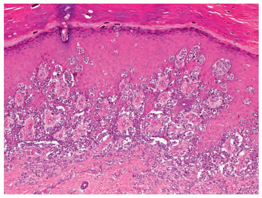
Fig. 113.25 Histopathologic features of an acral lentiginous melanoma.
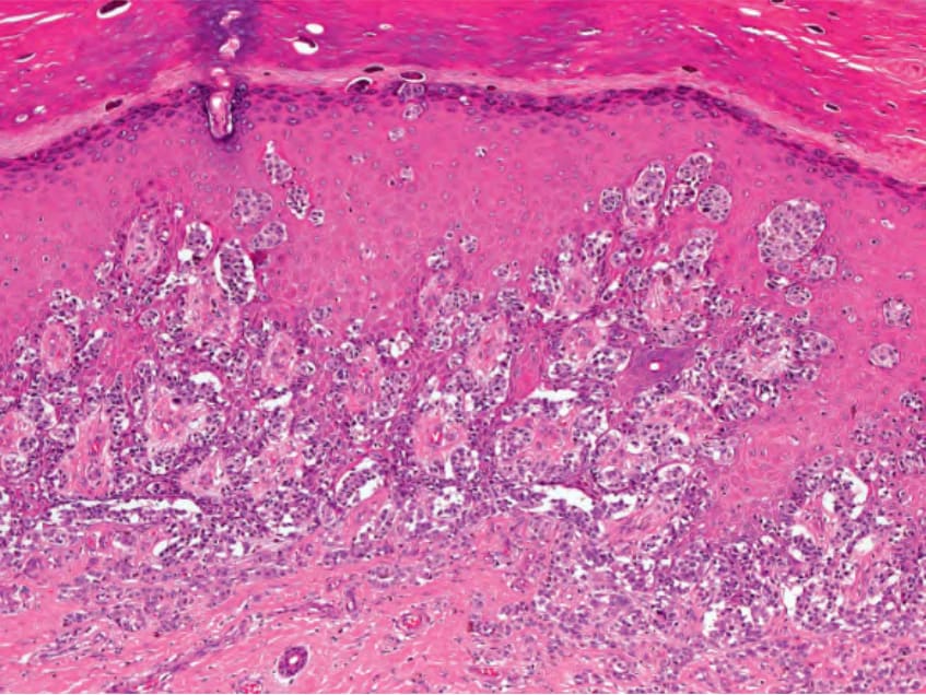
Fig. 113.26 Microstaging of cutaneous melanoma: Breslow tumor thickness.
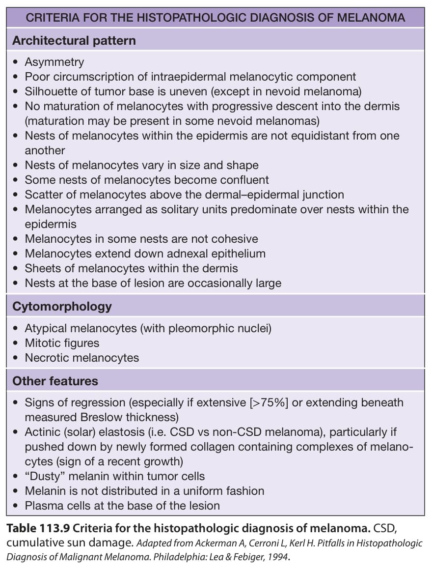
Table 113.9 Criteria for the histopathologic diagnosis of melanoma. CSD, cumulative sun damage. Adapted from Ackerman A, Cerroni L, Kerl H. Pitfalls in Histopathologic Diagnosis of Malignant Melanoma. Philadelphia: Lea & Febiger, 1994.
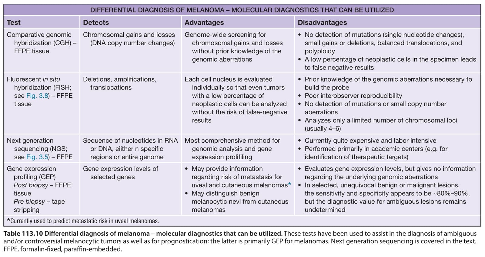
Table 113.10 Differential diagnosis of melanoma – molecular diagnostics that can be utilized. These tests have been used to assist in the diagnosis of ambiguous and/or controversial melanocytic tumors as well as for prognostication; the latter is primarily GEP for melanomas. Next generation sequencing is covered in the text. FFPE, formalin-fixed, paraffin-embedded.