PATHOGENESIS
In 1889, Dr Stephen Paget espoused the “seed and soil” hypothesis as the paradigm of metastatic disease – a malignancy must develop the capability to dissociate from its native environment, survive during transit, and then propagate in a new milieu. This process includes detachment, intravasation, circulation, stasis, extravasation, and finally proliferation. It is estimated that ≤0.01% of tumor cells shed into the vasculature form metastatic foci at distant sites.
The ability of a malignancy to acquire metastatic potential is thought to occur via one of two mechanisms. In the clonal expansion or linear progression model, selective growth advantages are acquired during the course of tumor development. Over time, subpopulations of cells acquire genetic characteristics that enable them to dissociate and then propagate in distant tissues. In this model, metastatic foci have similar phenotypes and genotypes to the original tumor. Alternatively, in the
variant or parallel progression model, rare, highly aggressive stem cell-like clones with high metastatic potential exist within the tumor and can cause dissemination at any point during tumorigenesis.
It is generally accepted that certain cancers tend to metastasize to distinct tissues. The mechanism by which they choose their final destination is related to hemostatic forces and surface protein expression patterns. When tumor emboli lodge in the first vascular bed they encounter, adhesion molecule interactions (e.g. integrins) facilitate extravasation and account for the commonly noted involvement of regional nodal basins. Yet, certain cancers have an affinity for specific tissues that are not anatomically convenient, such as prostate cancer spreading to bone and ocular melanoma metastasizing to the liver. Adhesion molecule expression seems likely to influence this tissuespecific homing of metastases. However, these mechanisms may be attenuated in highly aggressive, undifferentiated tumors that display a more random distribution of metastases.
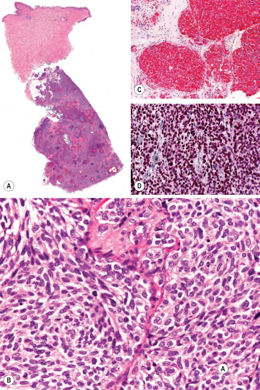
Fig. 122.2 Subcutaneous metastasis of small cell lung carcinoma.A Note sparing of the dermis. B Higher magnification demonstrating clusters of basaloid cells. C The tumor cells stain positively for CK7. D The tumor cells also stain positively for TTF-1 (thyroid transcription factor-1). Courtesy Lorenzo Cerroni, MD.