PHOTOTHERAPY WITH UVB
Historical Aspects
To date, the only dermatologist to be awarded a Nobel Prize in medicine was Dr Niels Finsen in 1903. The award recognized his demonstration of the beneficial effect of UV radiation on lupus vulgaris, and he eventually became known as the “father of photomedicine”. The combination of topical crude coal tar followed by UV irradiation for the treatment of psoriasis was introduced by Goeckerman in 1925 and became a standard therapy for psoriasis for half a century, particularly in the US. In the 1970s, it was observed that broadband (BB) UVB radiation alone, if given in doses that produce a slight erythema, could clear the milder forms of psoriasis, particularly the guttate type. In the 1980s, the discovery that the combination of psoralens with UVA (PUVA) exerts potent anti-inflammatory effects opened a new era of phototherapy. A further major advance was the development of fluorescent bulbs that emitted narrowband (NB) UVB radiation at 311–313 nm in the mid-1980s. This narrow spectrum is superior in clearing psoriasis and thus represents the most effective and most frequently used UVB phototherapy for psoriasis; it is also beneficial for a variety of other dermatoses that were previously treated with PUVA (see below).
Principles and Mechanisms
UVB phototherapy refers to the use of artificial UVB radiation without the addition of exogenous photosensitizers. The radiation is absorbed by endogenous chromophores, and photochemical reactions involving these UV-absorbing biomolecules mediate a variety of biologic effects, ultimately leading to the therapeutic effects. The most important chromophore for UVB is nuclear DNA. Absorption of UV by nucleotides causes the formation of DNA photoproducts, primarily pyrimidine dimers (see Ch. 86).
UVB exposure reduces DNA synthesis and thus is used to suppress the accelerated DNA synthesis found in psoriatic epidermal cells. UVB also induces the expression of the tumor suppressor gene TP53, and this can lead to either cell cycle arrest (allowing time for DNA repair) or apoptosis of keratinocytes (“sunburn cells”) if the DNA damage is too severe to be repaired. Through these mechanisms, p53 prevents photocarcinogenesis.
In addition to its effect on the cell cycle, UV induces the release of prostaglandins and cytokines. Interleukin (IL)-6 and IL-1, for example, seem to play important roles in producing the systemic symptoms of UV phototoxicity (sunburn) and immune suppression, respectively. There is also increasing evidence that UV radiation can affect, in addition to DNA, extranuclear molecular targets located in the cytoplasm and cell membrane. These targets include cell surface receptors, kinases, phosphatases, and transcription factors. It was shown that nuclear and cytoplasmic/membrane effects are not mutually exclusive – rather they independently contribute to the multiple biologic effects of UVB.
Many of the therapeutic effects of UVB may be due to its immunosuppressive properties which have been linked to the formation of pyrimidine dimers. UV radiation suppresses contact allergy, delayedtype hypersensitivity, and immune surveillance against UV-induced keratinocyte carcinomas in mice. Of note, Langerhans cells are very sensitive to UVB, which alters their antigen-presenting function. Keratinocytes also secrete soluble mediators such as IL-1 and IL-6, prostaglandin E, and TNF in response to UVB, which by themselves can alter the immune response. Therapeutic UVB suppresses the type 1 (proinflammatory) axis as defined by IL-12, interferon (IFN)-γ, and IL-8, and can selectively reduce proinflammatory cytokine production by individual T cells.
The interplay of the various photobiological pathways is far from being completely understood. In psoriasis, both epidermal keratinocytes and cutaneous lymphocytes may be targeted by UVB. Immune suppression, alteration of cytokine expression, and cell cycle arrest may all contribute to the suppression of disease activity in psoriatic plaques. In addition, a subset of T cells, Th17 cells, which seem to play a central role in the immunopathogenesis of psoriasis, are also downregulated by UVB. More recently, evidence was presented that keratinocyte apoptosis may be a key mechanism in the clearance of psoriatic plaques.
Action Spectrum and Radiation Sources
Parrish and Jaenicke demonstrated that for antipsoriatic activity, 304 and 313 nm were the optimally effective wavelengths (Fig. 134.2), even at suberythemogenic doses. Erythemogenic UVA doses are also therapeutically effective, but 1000 times higher fluences are needed
(as compared with UVB) which practically speaking is not feasible. The addition of UVA also does not enhance the therapeutic efficacy of UVB in psoriasis (unlike atopic dermatitis). The Philips TL01 fluorescent lamp that emits NB-UVB (311–313 nm) was introduced to optimally meet the requirements for antipsoriatic activity (Fig. 134.3).
Devices that contain UV radiation-emitting bulbs are available in a wide range of sizes and designs including enclosed whole-body cabinets, portable flat panels, and hand-held units. When reliable patients live far distances from physician-supervised phototherapy centers or for other reasons are unable to schedule multiple treatments per week, UVB units designed for home use can be utilized. Practical guidelines for home phototherapy have been published.
Exposure to natural sunlight is called heliotherapy. Natural sunlight contains sufficient UVB radiation such that it is sometimes used to treat photoresponsive dermatoses. However, due to variables that affect UVB irradiance, including time of day, cloud cover, season, altitude and latitude, appropriate exposure times cannot be standardized. As a result, there is a greater probability of sunburn and an inadequate therapeutic response.
Treatment Protocols
Over the past several years, a number of dermatologic societies have published guidelines for utilizing phototherapy. They all provide more or less similar recommendations with some slight variations which are due to national differences in the health care system, reimbursement,
legal requirements, availability/approval of alternative therapies, and of course traditions regarding prescribing practices. Table 134.1 lists some of the available guidelines. Most of these guidelines are updated regularly and are made available through online sources.
Before starting phototherapy, determination of the patient’s UV sensitivity via phototesting is frequently recommended, since skin typing by history alone does not always reflect the actual sensitivity of a particular individual. However, phototesting is not mandatory for the experienced therapist and, for practical reasons, is often not done. Testing is performed by exposing four to six small template areas (e.g. 1 cm diameter circles or squares) of usually non-sun-exposed skin (lower back, buttocks) to an incremental series of UVB doses. In successive test sites, the incremental increases can be fixed (e.g. 10 or 20 mJ/cm) or proportional (e.g. 40% higher). An example is given in Table 134.2. Note that the actual doses delivered as specified in fluence units (mJ/cm) strongly depend on the type of UVB unit; for example, NB-UVB is much less erythemogenic than BB-UVB and consequently higher numerical doses are required.
The minimal erythema dose (MED) is defined as the lowest dose that causes a minimally perceptible erythema reaction at 24 hours after irradiation. Sunbathing or exposure to tanning lamps should be avoided before phototesting. It is crucial to document the type of lamp used for the MED determination, since the values obtained with broadband and narrowband sources differ by up to ten-fold (see Table 134.2).
Despite continued discussion as to whether visual assessment of the MED is the optimal method for establishing the initial UVB dose, it
1 watt (W) = 1 joule (J) of energy per second.
is an easily performed procedure that does not require any specialized equipment. An initial therapeutic UVB dose equal to 70% of the MED is typically recommended. Subsequent treatments are given two to five times weekly. Since UVB erythema peaks within 24 hours after exposure, doses may be increased with each successive treatment. However, if treatments are given five times weekly, the doses should be increased every other treatment. The rate of increase depends on treatment frequency and the response to the preceding UVB exposure. The objective of the dose increments is to achieve a minimally perceptible erythema as a clinical indicator of optimal dosimetry. For example, with thrice-weekly exposures, doses are increased by a maximum of 40% if no erythema appears, and by a maximum of 20% upon development of slight erythema. If mild erythema persists, the dose should be maintained. With daily exposures, these rates are no more than 30%, 15% and 0%, respectively. If more intensive or painful erythema develops, irradiations are stopped until the symptoms disappear. Treatment is then continued until complete remission is achieved or no further improvement can be obtained with continued phototherapy (Fig. 134.4).
Whether maintenance treatment leads to a more prolonged remission time is still a matter of debate, since minimal precise data are available. In cutaneous T cell lymphoma (CTCL), most therapists perform maintenance treatment for several months to a year. For psoriasis, some centers use a 2-month maintenance phase with twice-weekly exposures for 1 month and once-weekly exposures for another month. The last effective UVB dose is given throughout the maintenance phase. If relapses occur during the maintenance phase, treatment frequency and UVB dose are again increased until clearing. Of note, there was some evidence in a randomized study that maintenance NB-UVB may lead to longer remission times in plaque-type psoriasis.
Phototherapy for Psoriasis
Guttate psoriasis and minimally elevated lesions of psoriasis respond most favorably and rapidly to BB-UVB, while chronic, plaque-type psoriasis is more resistant. In terms of clearing efficiency and duration of remission, BB-UVB is inferior to both NB-UVB and PUVA. As a result, in many countries, NB-UVB has largely replaced conventional BB-UVB phototherapy. Based on several publications, NB-UVB phototherapy appears to be somewhat less effective than PUVA.
In addition to the treatment protocols mentioned above, other regimens may be used. Some schedules utilize skin type-dependent starting doses and fixed increments regardless of skin reaction. Sometimes, the patient’s extremities (in particular the lower extremities which tend to respond more slowly) can be exposed to higher doses than the trunk.
Adjunctive topical agents and combination therapies are often used to improve efficacy and reduce the cumulative UVB dose, with the ultimate aim of minimizing the risk of long-term side effects. Narrowband UVB has been successfully combined with therapies such as anthralin and vitamin D analogues. The use of bland emollients before UVB treatment is laborious and time-consuming, and in our experience does not dramatically increase treatment efficacy. The concurrent use of topical corticosteroids may reduce remission times and thus is discouraged by some dermatologists.
Systemic drugs such as retinoids increase efficacy, particularly in patients with chronic plaque-type psoriasis. In addition, retinoids may reduce the carcinogenic potential of UVB phototherapy. There have been open-label studies and small comparison studies regarding the efficacy of combining UVB phototherapy with systemic immunomodulators (“biologics”). To date, there have been no large-scale studies, and long-term data, including remission times, are unavailable.
Phototherapy for Cutaneous T Cell Lymphoma, in Particular Mycosis Fungoides
For early stages (IA, IB, and IIA) of mycosis fungoides (MF), conventional treatment strategies include topical corticosteroids, UV radiation, topical retinoids (e.g. bexarotene), and topical cytotoxic agents (e.g. mechlorethamine [nitrogen mustard]). More advanced stages are treated with various combinations that include total body electron beam radiation therapy (see Ch. 139), local radiotherapy, systemic retinoids, PUVA, systemic chemotherapy (e.g. gemcitabine), and immunomodulators (e.g. brentuximab vedotin, mogamulizumab, IFN) (see Table 120.5), but none of these regimens have been demonstrated to induce permanent remission.
Lesions of MF frequently occur in non-sun-exposed areas of the body, and patients with early stage MF often benefit from exposure to natural sunlight. Response to phototherapy is related to the type (i.e. patch stage MF does better than the plaque type) but not the extent of skin involvement. In a more recent study, prolonged maintenance therapy (up to 30 months) resulted in a relapse-free period of 26 ± 10 months.
The proposed mechanisms for UVB phototherapy of MF include impairment of epidermal Langerhans cell function and alterations in cytokine production and adhesion molecule expression by keratinocytes. Moreover, NB-UVB can induce apoptosis of T lymphocytes, which may more specifically contribute to the beneficial effect of this light source.
UVB phototherapy is clearly less efficient than PUVA, and the duration of treatment in the clearing and maintenance phases is longer and thus requires higher patient compliance. Whether patients would benefit from adding UVA, as is sometimes recommended, remains questionable. Likewise, NB-UVB and BB-UVB therapy have not been compared in MF. It is possible that combining a systemic retinoid (e.g. bexarotene) with NB-UVB may increase efficacy. Lastly, a pilot study documented the efficacy of medium- to high-dose UVA1 therapy (see below) in patients with stage IA and IB MF. However, there is no evidence that UVA1 is superior to PUVA and no long-term follow-up is available.
Phototherapy for Vitiligo
Many patients observe follicular repigmentation in areas of vitiligo following sun exposure. Because of seasonal and weather-dependent variations in sunlight intensity in moderate climates, exposure to natural sunlight is often not a practical option for inducing repigmentation. In the past, PUVA was most frequently used for the treatment of vitiligo, but exposure to artificial UVB irradiation, in particular NB-UVB, can also provide an acceptable therapeutic effect, if given for a sufficiently long period. Although the action spectrum for phototherapy of vitiligo is not known, NB-UVB has clearly become quite popular in recent years. In comparison studies of NB-UVB with PUVA, NB-UVB was as effective as PUVA but had fewer side effects (Fig. 134.5). Also, in a randomized double-blind trial of non-segmental vitiligo, NB-UVB therapy was superior to oral PUVA therapy and the color match of repigmented skin was considered to be better in the patients treated with NB-UVB. UVA alone is of limited benefit.
Since intense erythema may induce the Koebner phenomenon and worsening of the disease, it is necessary to stay within a dose range that induces minimally perceptible erythema. Therefore, initial assessment of the MED in a vitiliginous area that is normally not sun-exposed (e.g. buttocks, lower back, abdomen) is recommended. The UVB doses have to be increased more carefully than in other disorders, because of the increased photosensitivity of depigmented skin.
The initial exposure is 70% of the MED in lesional skin, and subsequent doses are chosen according to the response in vitiligo areas, i.e. the goal
is to induce a barely perceptible erythema that has a light pink color. The presence or absence of faint erythema is the only useful parameter for determining dosage increments. The dose should not be increased more than once weekly by 5%–20% of the preceding dose. Despite the lack of pigment, vitiliginous skin does develop photoadaptation, probably as a result of epidermal hyperplasia and thickening of the stratum corneum. In contrast to previous assumptions, the risk of developing keratinocyte carcinoma is not enhanced in vitiliginous areas. Usually, a maximum dose will be reached during the first couple of months which is then used throughout the entire treatment period. Most commonly, two to three treatments per week are given. Although few controlled studies are available for any particular phototherapy regimen to be preferred over another, consensus recommendations are available.
More recently, the combination of an afamelanotide implant plus NB-UVB phototherapy resulted in clinically apparent, statistically significant superior and faster repigmentation compared with NB-UVB monotherapy. Afamelanotide is an analogue of α-melanocyte stimulating hormone that can induce diffuse hyperpigmentation of the skin, especially in sun-exposed sites. Of note, the response was more noticeable in patients with skin phototypes IV to VI. Enhanced repigmentation with a combination of NB-UVB plus topical 1.5% ruxolitinib cream twice daily has also been reported17a.
Phototherapy for Atopic Dermatitis
Broadband UVB (BB-UVB) phototherapy
Based on the empirical experience that sun exposure is beneficial for patients with atopic dermatitis, BB-UVB has been used to treat atopic dermatitis since the late 1970s.
UVA/UVB phototherapy
Subsequent studies suggested that a combination of UVA plus UVB irradiation (UVA/UVB therapy) is superior to conventional BB-UVB, conventional UVA, or low-dose UVA1 therapy in the management of chronic, moderate atopic dermatitis. A paired-comparison study demonstrated statistically significant differences in favor of UVA/UVB therapy, compared with BB-UVB therapy.
Narrowband UVB (NB-UVB) phototherapy
Earlier studies showed that NB-UVB irradiation not only improved the total clinical score, but also substantially reduced the need for topical corticosteroids. These beneficial effects were still present in the majority of patients 6 months after cessation of phototherapy. Narrowband UVB therapy has also been used successfully in conjunction with UVA1 therapy. However, due to the lack of baseline data on the duration and severity of atopic dermatitis, a final conclusion on preference of phototherapy in the acute or chronic phases cannot be drawn from available studies.
Phototherapy for Photodermatoses
In the majority of patients, polymorphous light eruption (PMLE) gradually abates with continual outdoor light exposure. For patients prone to PMLE, NB-UVB phototherapy can be used prophylactically to induce “hardening” of the skin before a vacation to a sunny climate or during the springtime (see Ch. 87). Two to three treatments per week for a total of 12–15 sessions are typically administered prior to an anticipated increase in exposure to sunlight. Despite using reduced UVB doses (as compared to those for psoriasis), this phototherapy regimen can induce a transient photoeruption; the latter usually responds to topical corticosteroids and smaller incremental increases in subsequent UVB doses. The precise mechanisms by which phototherapy induces adaptation to sunlight are not known, but may include thickening of the stratum corneum and modulation of cutaneous immune functions. This phenomenon is not due to hyperpigmentation, as was previously thought – patients with skin phototype I respond equally well to NB-UVB as other skin phototypes.
Narrowband UVB phototherapy has also been shown to be capable of inducing “hardening” in patients with erythropoietic protoporphyria, actinic prurigo, and solar urticaria. For solar urticaria, however, the UVA rush hardening regimen appears to be preferable, since effective protection can be achieved within a few days with this protocol.
Phototherapy for Pityriasis Lichenoides
Both pityriasis lichenoides acuta and chronica can have a prolonged course and be rather resistant to therapy. As sunlight may lead to some improvement, UVB phototherapy has also been associated with some success. However, PUVA seems to be more effective (see below), especially in the acute form of the disease (perhaps because of the deeper extension of the dermal inflammatory infiltrate). Although some authors have recommended PUVA as the treatment of choice for the acute form, PUVA may be reserved for cases of pityriasis lichenoides chronica that are UVB-resistant.
Phototherapy for Seborrheic Dermatitis
Facial and truncal seborrheic dermatitis generally improves during the summer and “sunny” vacations. Accordingly, UV radiation also seems to have a beneficial effect on this condition. However, flares of dermatitis may occasionally occur. In an open prospective study, NB-UVB was found to be an effective treatment in severe cases. Nonetheless, because topical therapies lead to improvement of seborrheic dermatitis in the majority of patients, phototherapy does not represent a first-line treatment.
Phototherapy for Pruritus
Both NB- and BB-UVB therapy can be of benefit in various forms of pruritus, particularly those associated with diabetes and renal or hepatic disorders, as well as in idiopathic pruritus. However, in patients with cholestatic pruritus, remissions are relatively short-lived. In a case series, NB-UVB led to improvement of uremic pruritus.
Side Effects of Phototherapy With UVB
Short-term side effects include erythema (Fig. 134.6), xerosis accompanied by pruritus, occasional blistering, and an increased frequency of recurrent herpes simplex viral infections. Painful erythema resulting from overexposure is treated with topical corticosteroids. Systemic nonsteroidal anti-inflammatory drugs and corticosteroids can prove useful in severe cases if administered early.
Long-term side effects include photoaging and carcinogenesis. Although UVB is a known carcinogen, its carcinogenic potential seems to be much lower than that of PUVA. In a British study involving 3867 patients who had been treated with NB-UVB, no significant association between NB-UVB treatment and basal cell carcinoma (BCC), squamous cell carcinoma (SCC), or melanoma was found. There was a small increase in BCCs amongst those patients who had also been previously treated with PUVA. To determine the true carcinogenic risk of NB-UVB phototherapy, additional longitudinal studies are essential.
UVB. Annular erythema is separated from the psoriatic plaques by Woronoff’s ring.
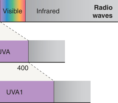
Fig. 134.1 Electromagnetic spectrum with expanded UV region.
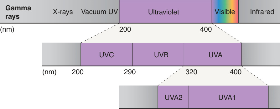
Fig. 134.2 Phototherapy action spectrum of psoriasis. The phototherapy action spectrum of psoriasis is plotted as the reciprocal of the lowest effective daily dose to clearing versus wavelength. Circles represent the reciprocal of the lowest effective daily dose for wavelengths that cleared psoriasis (295, 300, 304, 313, 365 nm); the arrows point to the wavelengths that were optimally effective (304, 313 nm). Presently, narrowband UVB sources that emit wavelengths between 311 and 313 nm are available. Adapted from Parrish JA, Jaenicke KF. Action spectrum for phototherapy of psoriasis. J Invest Dermatol 1981;76:359–62.
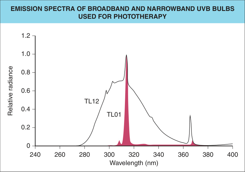
Fig. 134.3 Emission spectra of broadband and narrowband UVB bulbs used for phototherapy. Philips TL12, broadband UVB bulb; Philips TL01, narrowband UVB bulb.
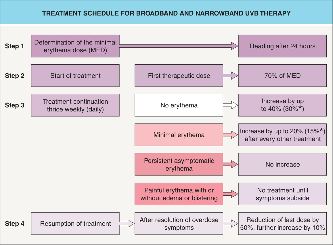
*Fig. 134.4 Treatment schedule for broadband and narrowband UVB therapy. If treatments are given five times per week or daily.
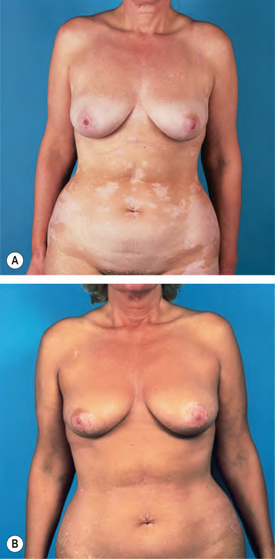
Fig. 134.5 Narrowband UVB phototherapy for vitiligo.A Before treatment. B After 10 months of treatment (twice weekly). Note the persistence of depigmentation of the areolae, an area which is particularly resistant to treatment.
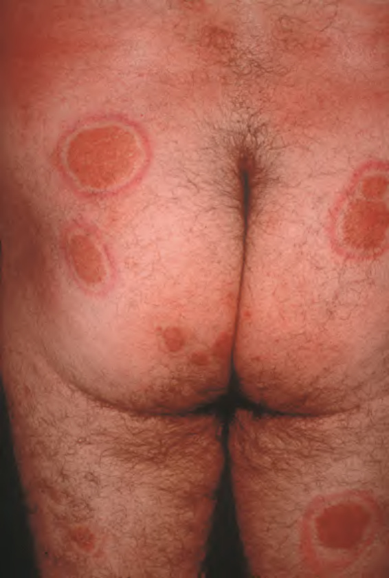
Fig. 134.6 Target-like appearance of psoriatic lesions secondary to phototoxicity due to
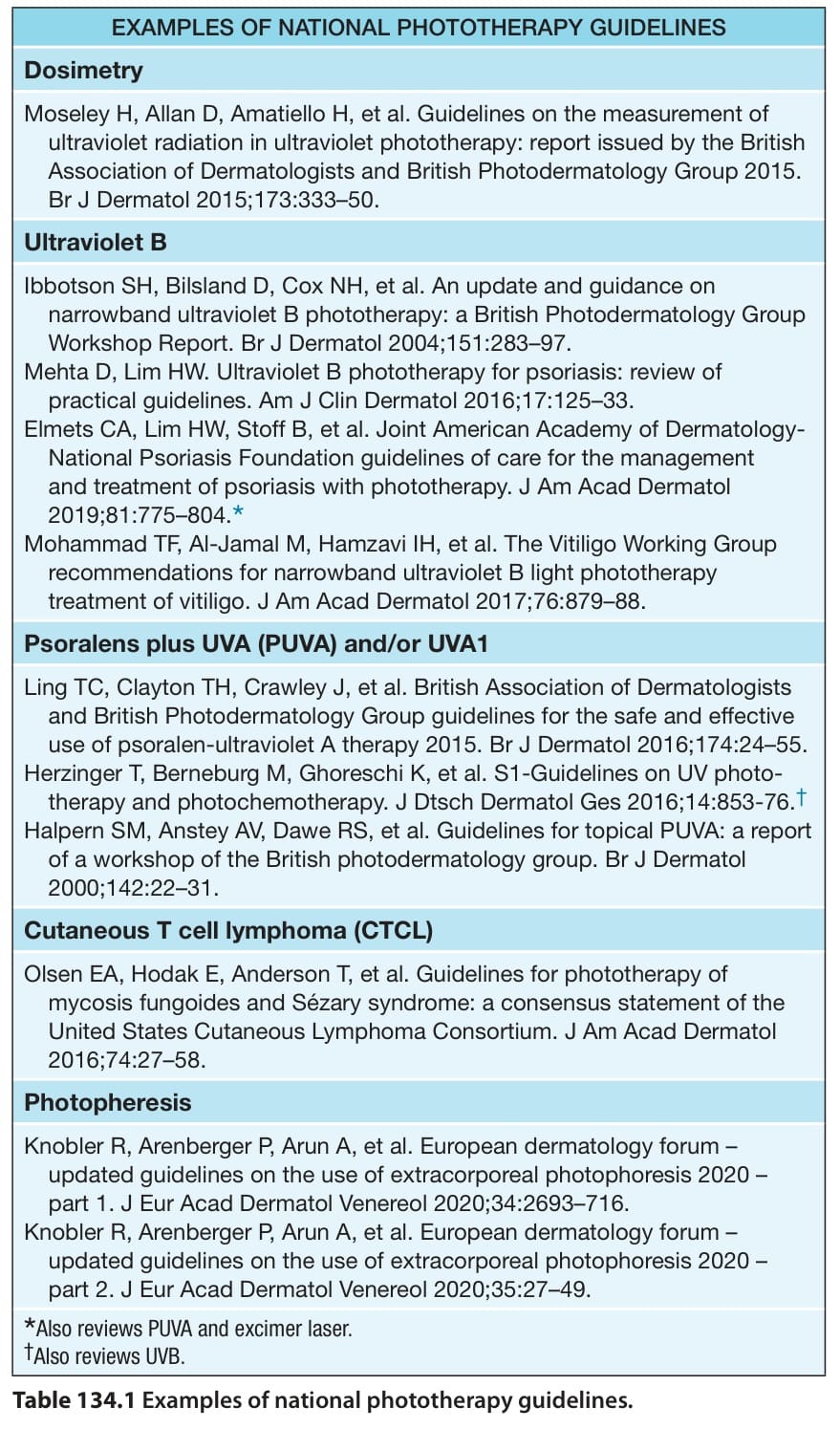
Table 134.1 Examples of national phototherapy guidelines.
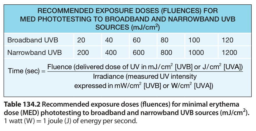
Table 134.2 Recommended exposure doses (fluences) for minimal erythema dose (MED) phototesting to broadband and narrowband UVB sources (mJ/cm).