HISTORY AND DEVELOPMENT
The photodynamic effect was originally studied in the early 1900s by a medical student who noted that cultures of Paramecium would die only if exposed to both an organic dye (acridine) and visible light. His professor discovered the oxygen-dependence of this effect and coined the term “photodynamic action” to describe the phenomenon. In humans, hematoporphyrin was subsequently shown by Meyer Betz to induce profound and persistent generalized photosensitivity, including when he self-injected this agent. In addition to its fluorescent properties when illuminated with a Wood’s lamp, hematoporphyrin was shown to localize to neoplastic tissue and then induce tumor involution when exposed to visible light.
The modern era of clinical PDT began in the 1970s when Dougherty and colleagues combined systemically administered hematoporphyrin derivative with visible light to treat cutaneous and subcutaneous malignancies in humans; they also confirmed the role of singlet oxygen. With the advent of lasers and fiberoptic endoscopes, the use of PDT was extended to carcinomas of the lung, bladder, and esophagus. Hematoporphyrin derivative and its subsequent porphyrin-based congeners require intravenous administration, and they induce marked generalized cutaneous photosensitivity that can last for days to weeks, necessitating strict avoidance of outdoor and bright indoor light.
Because they are relatively large molecules, porphyrins are poorly absorbed through the skin. The key breakthrough in developing PDT as a practical dermatologic therapeutic modality can be attributed to the Canadian team of Kennedy and Pottier. In the 1980s, they noted that the topical application of a naturally occurring porphyrin precursor, 5-aminolevulinic acid (ALA), as a prodrug could photosensitize the skin by inducing local intracutaneous production of protoporphyrin IX (PpIX; see Fig. 135.1). Their successful treatment of keratinocyte cancers using topical ALA plus broadband red light abrogated the two major disadvantages associated with the use of systemic porphyrin photosensitizers (see above).
The elegance of using topical ALA for PDT lies in how it takes advantage of the endogenous porphyrin biosynthetic pathway (which is present in all nucleated cells) to generate PpIX from ALA. The simplicity of this approach greatly facilitated clinical PDT research, culminating in the FDA approval of topical ALA plus blue fluorescent light for the treatment of actinic keratoses (AKs) in 1999. Five years later, topical methyl aminolevulinate (mALA), an esterified derivative of ALA, in combination with red light-emitting diode (LED) light was approved in several countries for dermatologic PDT. It was subsequently shown that natural daylight could safely and effectively activate ALA for skinbased PDT.
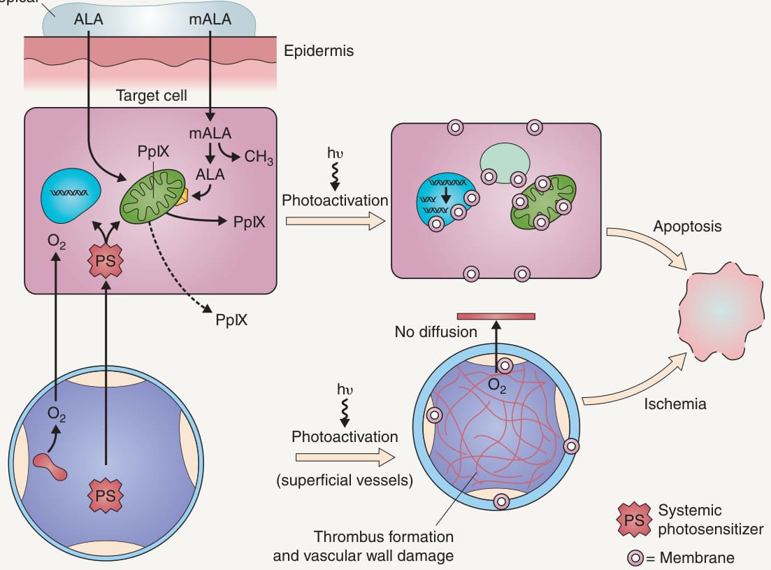
Fig. 135.1 Photosensitizer delivery and photodynamic effects on target cells and tissue vasculature.