RADIOTHERAPY OF IN SITU CUTANEOUS MALIGNANCIES
Squamous Cell Carcinoma In Situ (Bowen Disease)
Radiotherapy can be used, albeit infrequently, to treat SCC in situ. The malignancy is limited to the epidermis and its appendages, arises more commonly in sun-exposed sites, and usually presents as a slowly growing, scaly, erythematous plaque. A biopsy is important to exclude the possibility of eczema or psoriasis. Only a small minority (<5%) become invasive SCCs, although this may be higher (10%–30%) in genital lesions such as erythroplasia of Queyrat. Patients are treated with 40–50 Gy in 10–20 fractions using superficial low-energy photons (110–150 kVp), with reported local five-year cure rates of 98%–100%.
Patients with widespread actinic keratoses have been treated with radiotherapy under special circumstances. Given that other very effective therapies are readily available (see Ch. 108), radiotherapy should not be considered a first-line treatment modality.
Lentigo Maligna
Lentigo maligna (melanoma in situ arising within photodamaged skin) often presents as a longstanding and large-sized hyperpigmented patch on the face of elderly patients (see Ch. 113). The size and location often preclude simple excision, and radiotherapy remains a well-established, evidenced-based option with excellent five-year cure rates of 90%–95% (Fig. 139.3). Reported doses range from 35 to 100 Gy administered in 5–10 fractions, i.e. large doses per fraction. However, dose fractionation schedules of 40–50 Gy in 10–20 fractions would also be efficacious. Field margins of 1–1.5 cm are recommended, demarcated by Wood’s lamp examination or confocal microscopy (if available), and treating to a depth of at least 5 mm.
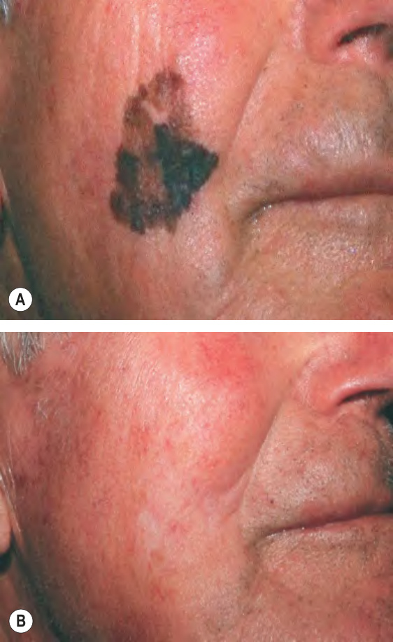
Fig. 139.3 Lentigo maligna (melanoma in situ).A An 82-year-old man with a lentigo maligna on his right cheek. B Three months after superficial radiotherapy (45 Gy in 15 daily fractions, 120 kVp, HVL 4.2 mm Al).