ADVANCED WOUND THERAPIES
Scientific and clinical advances have increased the therapeutic options for chronic wounds. For example, topical growth factors and tissue-engineered skin equivalents can be combined with standard treatments.
Topical Growth Factors
Growth factors are critical to the wound healing process, as they stimulate fibroblasts, promote angiogenesis, and encourage migration of keratinocytes. Fluid from acute wounds contains multiple growth factors including platelet-derived growth factor (PDGF) and epidermal growth factor (EGF). Maintaining contact of this fluid with the under-lying wound bed via the use of occlusive dressings has been shown to augment wound healing. A topical formulation of recombinant human PDGF, becaplermin (Regranex®) gel, has been shown to increase the complete closure rate of diabetic foot ulcers by 43%. Its current FDA indication is for non-healing diabetic neuropathic foot ulcers. Unfortunately, in vivo results with other growth factors failed to translate into improved wound healing in human clinical trials, highlighting the complex microenvironment of a chronic wound.
Tissue-Engineered Skin Equivalents or Skin Substitutes
Advances during the 1970s in tissue culture techniques for keratinocytes and fibroblasts, including the ability to generate large quantities of these cells, resulted in the production of a new group of wound dressings. A variety of tissue-engineered skin equivalents, both cellular and acellular, are commercially available and Table 145.5 outlines some of these products. A true skin substitute provides both the physiologic and mechanical functions akin to an autologous skin graft. Advantages of tissue-engineered dressings include no painful donor sites and the potential for providing coverage of large areas.
Chronic wounds treated with engineered dressings often undergo an “edge effect”, characterized by promotion of epithelialization from the edge of the ulcer toward its center, likely due to a release of cytokines by the donor cells. There are currently three types of skin substitutes: epidermal grafts, dermal replacements (acellular or cellular), and composite grafts (with both an epidermal and dermal component). Tissue-engineered dressings can also be classified as autologous, allogeneic (human donor), or xenogeneic (usually bovine or porcine). Currently, the primary allogeneic sources are cadaveric dermis and neonatal foreskin. Cellular and tissue-based products, which includes skin equivalents and substitutes, is a term used by the FDA to signify those products regulated by the Center for Biologics Evaluation and Research.
Epidermal Grafts
The first cultured epidermal autografts were successfully cultivated in 1975 by Rheinwald and Green. They have been used successfully in a variety of clinical situations including burns, chronic ulcers, vitiligo, and postsurgical wounds.
Epicel® represents an epidermal autograft in which a skin biopsy is obtained from the patient and then the autologous keratinocytes are co-cultured with irradiated murine fibroblasts that serve as a feeder layer. A sheet of keratinocytes two to eight cells thick forms and is attached to petrolatum gauze. The graft is then sutured in place and the gauze backing is removed at 1 week when the keratinocytes have attached to the wound. Skin biopsies obtained 6–12 months post-grafting show anchoring fibrils and neovascularization. This epidermal autograft is approved for use in partial- and full-thickness burns. Disadvantages include the several-week period required to culture the keratinocytes, graft fragility, a short shelf life, and the cost of processing.
For decades, the roofs of suction blisters created in uninvolved skin have been used to repigment vitiligo. More recently, an automated system for epidermal autologous grafting was introduced that uses heat and suction to produce small epidermal microdomes, with consistent cleavage at the lamina lucida. Advantages include no requirement for anesthesia or a highly skilled surgeon, performance in an outpatient setting, minimal donor site pain, and lack of scarring. The grafts have been applied to a variety of chronic and acute wounds and pathergy has not been observed in patients with pyoderma gangrenosum.
Dermal Replacements
There are two types of dermal replacements: xenogeneic and allogeneic (Table 145.6). Neither of these is permanent: DNA analysis of wounds after the application of non-autologous skin substitutes shows almost complete disappearance of the grafted cells after two months. The goal of these dermal grafts is to provide a temporary biologic scaffold in order to stimulate the healing process. They are placed over the wound, extending slightly onto normal skin, and then bolstered into place. Secondary dressings must be applied. It is advisable to keep the primary dressing in place for up to a week following its placement and to follow the individualized product instructions. The major component of dermal replacements is collagen; other elements of the extracellular matrix, e.g. glycosaminoglycans (GAGs), may be included and in the case of cellular products, fibroblasts.
Xenogeneic
Although other sources are available, xenogeneic grafts are usually composed of porcine or bovine collagen (see Table 145.6). The advantages of these products are: (1) their ability to effect hemostasis and to provide immediate closure as well as cosmetically acceptable scars with second intention healing; (2) safety from potential human pathogen transmission due to their animal origin; and (3) an adequate shelf life, facilitating off-the-shelf access.
Oasis® Wound Matrix and Biobrane® are examples of acellular matrices derived from porcine collagen (Fig. 145.11). They have significant elasticity, enabling a full range of motion of the covered body part, and there is no requirement for dressing changes once it has adhered to the wound bed. However, Biobrane® may adhere to the wound so tightly that it can damage newly developed epithelium.
INTEGRA® Dermal Regeneration Template is composed of two layers: an outer silicone layer overlies a layer composed of bovine tendon
collagen and shark cartilage GAGs. The latter is thought to simulate dermal matrix. Because INTEGRA® has a complicated application procedure, the FDA requires that all physicians complete a companysponsored training program.
Allogeneic
Allogeneic dermal grafts are composed of cadaveric dermis or neonatal foreskin fibroblasts (see Table 145.6). The advantages of these products include their ability to provide immediate closure and cosmetically acceptable scars with second intention healing, in addition to an adequate shelf life, facilitating off-the-shelf access. However, there is a risk of graft rejection due to T cell recognition of donor T cells. Examples include AlloDerm® Regenerative Tissue Matrix which consists of decellularized cadaveric dermis and GRAFTJACKET NOW™ Acellular Dermal Matrix which is derived from processed acellular cadaveric skin (see Table 145.5). The FDA currently categorizes both AlloDerm® and GRAFTJACKET NOW™ as banked human tissue for transplantation. Dermagraft® is a biodegradable scaffold seeded with neonatal foreskin fibroblasts and is FDA-approved for the treatment of diabetic foot ulcers.
Composite Grafts
Engineered composite grafts are bilayered skin equivalents. Current products consist of an epidermal layer composed of human keratinocytes and a dermal layer composed of bovine collagen matrix seeded with human fibroblasts. In Apligraf®, both cell types are derived from human neonatal foreskins (Fig. 145.12). This composite graft produces its own matrix proteins and growth factors and, if wounded, it can repair itself. Although FDA-approved for venous leg ulcers and diabetic foot ulcers, Apligraf® has been used to treat acute and chronic wounds that failed to heal by standard means, including those due to epidermolysis bullosa, pyoderma gangrenosum, burns, and surgical excisions. Whereas several applications of the material may be required to completely heal chronic wounds, a single application may be sufficient to initiate a satisfactory healing response. Chronic venous ulcers that were randomized to receive compression plus Apligraf® healed in a shorter time period compared to compression alone, especially in the case of larger defects.
More recent evidence suggests that Apligraf® modulates an array of cellular pathways in the epidermis at the wound’s edge, ultimately transforming a chronic wound into an acute one. In venous leg ulcers, Apligraf® also stimulates antifibrotic changes within the wound bed. This includes decreased expression of gene targets of profibrotic TGF-β1, increased levels of the TGF-β inhibitor decorin, and upregulation of fibroblast-derived matrix metallopeptidase 8.
A point-of-care delivery system named RECELL® was recently developed that allows delivery of autologous skin to the wound bed via a cell spray containing keratinocytes, melanocytes, Langerhan cells, and papillary dermal fibroblasts. This onsite cell harvesting device can create 1 ml of cell suspension from a 1 cm2 piece of split-thickness donor tissue; the former can then be sprayed onto an area of up to 80 cm2. The maximum per device is 24 ml of cell suspension. In a randomized controlled study in burn patients that compared standard split-thickness skin grafts (STSGs) versus the application of RECELL® Spray-On Skin™ cells to widely meshed STSGs, the cell spray had similar efficacy and safety but decreased the donor site area by ~30%.
Apligraf® and similar products are costly, but they have an excellent safety profile as well as the advantage of accelerating healing and reducing long-term costs.
Negative Pressure Wound Therapy (NPWT)
NPWT, developed in the 1990s, is based on the delivery of sub-atmospheric pressure to the wound bed. In this vacuum-assisted closure (VAC) system, a polyurethane or polyvinyl alcohol foam is cut and placed on the wound surface. The foam is then sealed over by a transparent drape in order to provide a closed airtight system (Fig. 145.13). A vacuum pump, connected to this space via a plastic tube, provides a negative pressure environment. NPWT removes interstitial fluid, stimulates angiogenesis, and enhances circulation as well as lymphatic drainage. There is evidence to support the use of NPWT for acute postsurgical wounds. Although there is some evidence that NPWT improves healing of diabetic foot ulcers, more information regarding appropriate patient selection, duration of treatment, and use of combined treatments is required.
Nowadays there are portable, often single-use, systems that can deliver effective negative pressure to a variety of low-to-moderately exudative wounds. Some of these systems use superabsorbent dressings, rather than a canister, to retain wound exudate. The dressing is placed under a sealed film covering and is linked to a battery-operated, portable pump that can be harnessed around the thigh, allowing for patient mobility. Single-use NPWT systems appear to lead to similar, if not improved, wound healing and they reduce the cost of care given the outpatient setting. Another modification is lavage of the wound bed by a wound cleanser or antiseptic while the NPWT is functioning.
Oxygen Therapy
Because multiple studies have pointed to hypoxia as one of the major factors leading to a delay in wound healing, both hyperbaric oxygen therapy (HBOT) and topical oxygen therapy have been utilized.
In HBOT, oxygen is administered at pressures greater than one atmosphere. Patients are placed in a pressure chamber where they breathe oxygen at this increased pressure. Systemic HBOT sessions last 45–120 minutes and are conducted once or twice daily for 20–30 sessions. This oxygenation of hypoxic tissue induces vasoconstriction and reduces edema and congestion. At a cellular level, there is increased fibroblast replication, collagen synthesis, and neovascularization as well as upregulation of growth factors.
To screen for a potential response to HBOT, the patient has transcutaneous oxygen levels measured in the peri-wound region both at baseline and while breathing 100% oxygen; at least a doubling of the levels is preferred. While a systematic review concluded that there was short-term but not long-term improvement of diabetic ulcers with HBOT, randomized controlled trials with small sample sizes reported the effectiveness of HBOT. A data analysis of 6259 patients did not support the use of HBOT for diabetic foot ulcers, highlighting the need for large randomized controlled trials. Additional methods for oxygenation of wounds include topical pressurized oxygen, topical continuous diffusion of oxygen, and dressings that release oxygen, but proper selective application of these interventions is required.
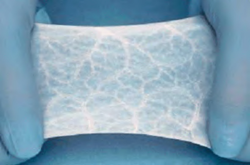
Fig. 145.11 An acellular xenogeneic dressing. This particular wound matrix is derived from porcine small intestine submucosa (Oasis®) and serves as a dermal graft.
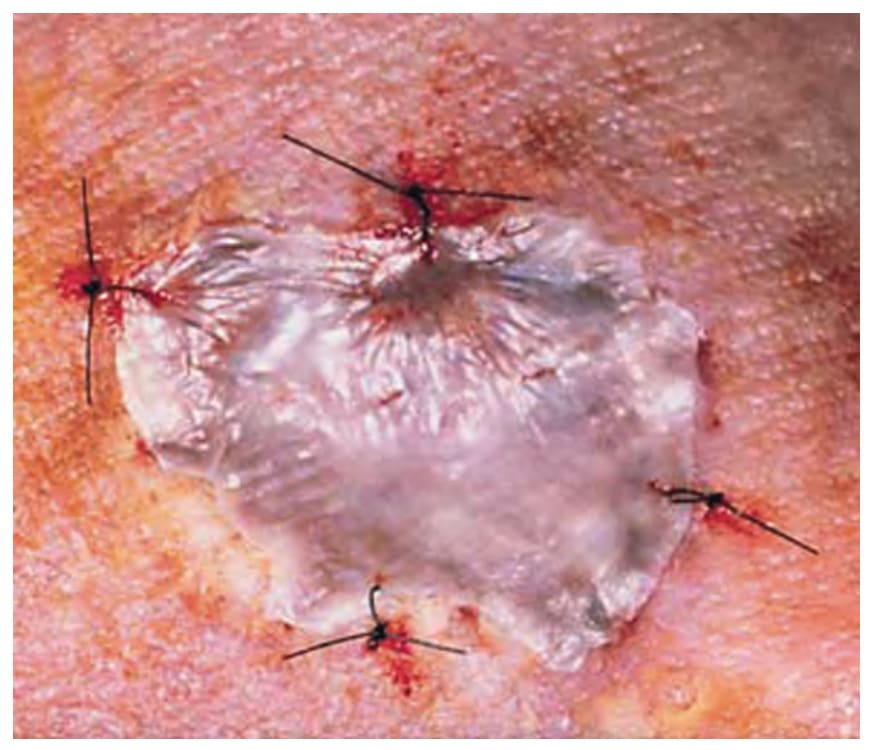
Fig. 145.12 Example of an engineered composite (xenogeneic and allogeneic) graft sutured into place (Apligraf®).Courtesy Gregg M. Menaker, MD.
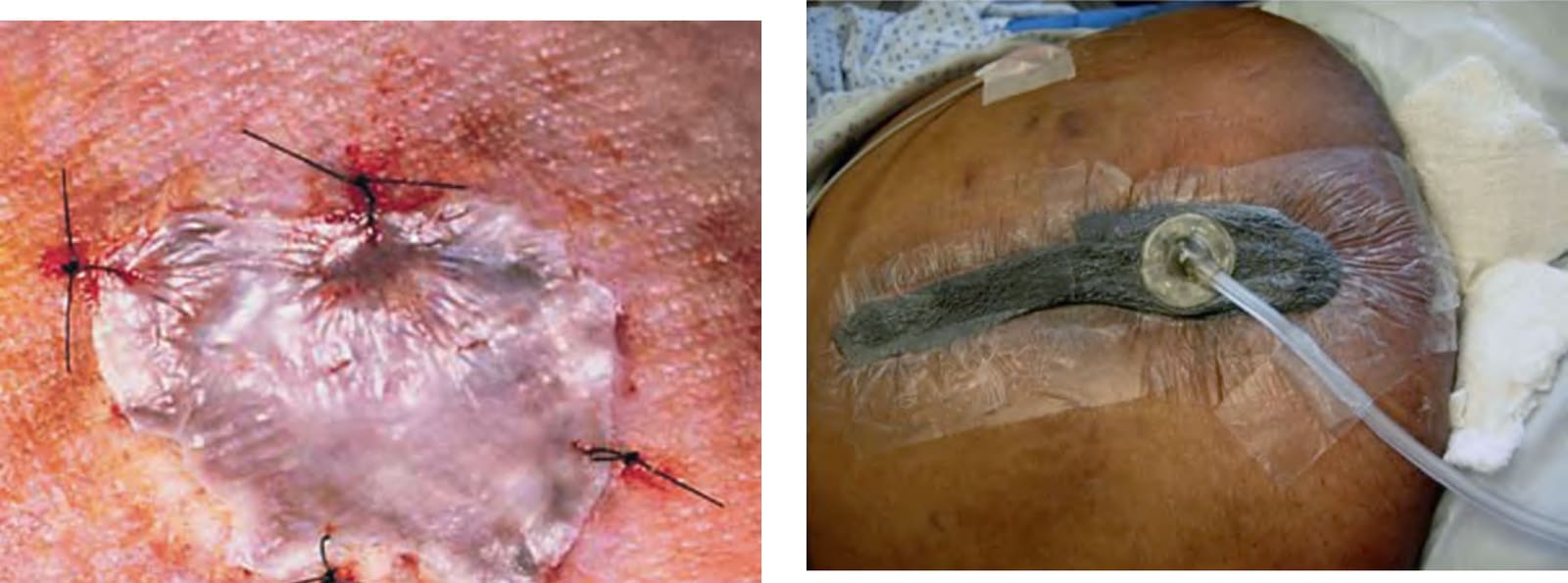
Fig. 145.13 Negative pressure wound therapy. In negative pressure wound therapy, a polyurethane (this patient) or polyvinyl alcohol foam is cut and placed on the wound surface. The foam is then sealed over by a transparent drape. A vacuum pump, connected via a plastic tube, provides a negative pressure environment. The sterile polyurethane foam has large pores and stimulates granulation tissue.
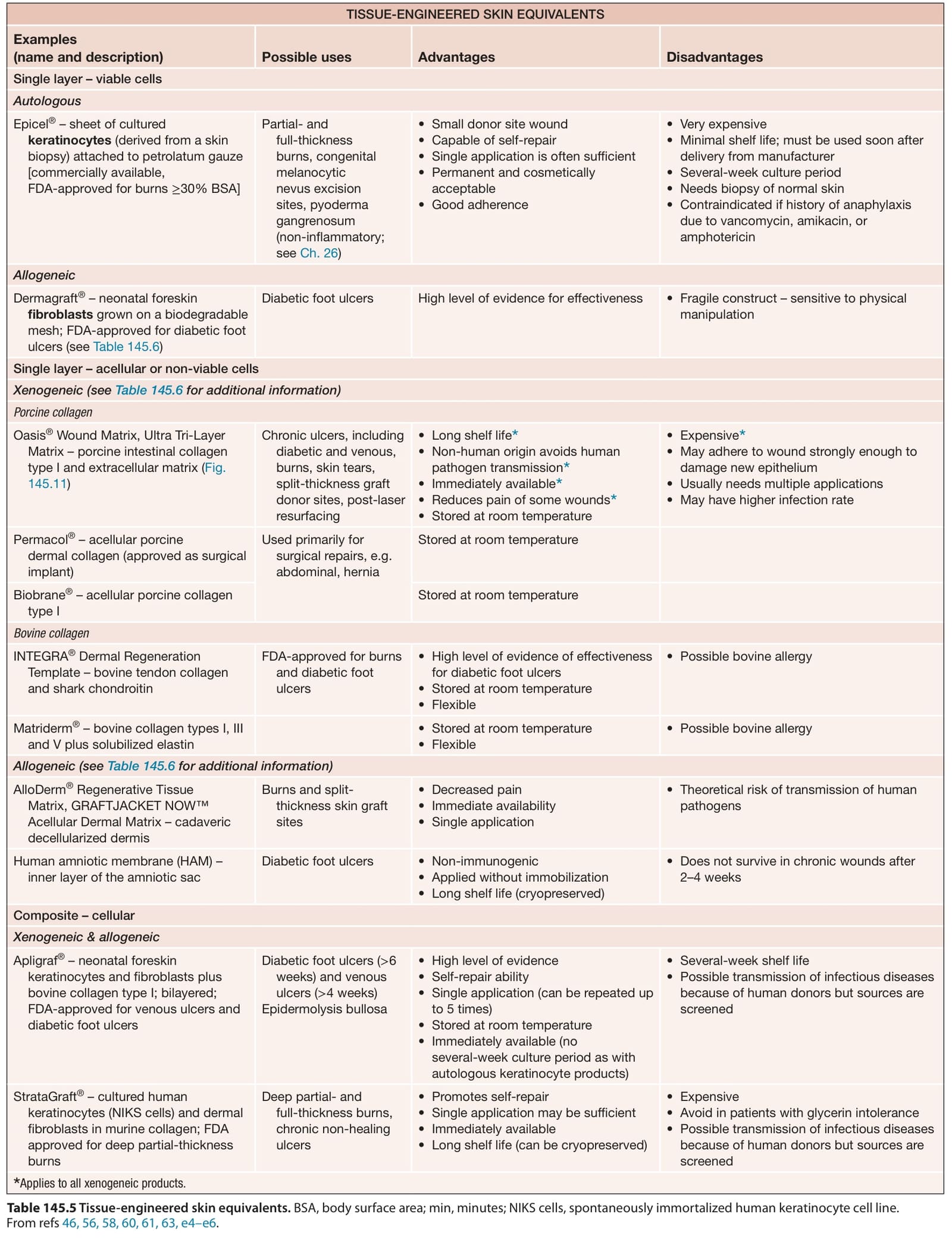
Table 145.5 Tissue-engineered skin equivalents. BSA, body surface area; min, minutes; NIKS cells, spontaneously immortalized human keratinocyte cell line. From refs 46, 56, 58, 60, 61, 63, e4–e6.
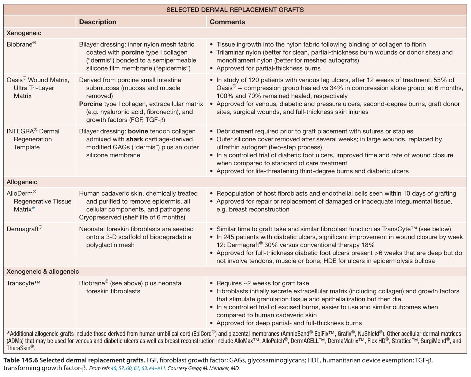
Table 145.6 Selected dermal replacement grafts. FGF, fibroblast growth factor; GAGs, glycosaminoglycans; HDE, humanitarian device exemption; TGF-β, transforming growth factor-β.From refs 46, 57, 60, 61, 63, e4–e11. Courtesy Gregg M. Menaker, MD.