FUTURE TRENDS
For the foreseeable future, histologic examination of tissue specimens will remain the mainstay of dermatologic diagnostic testing. Excisions likewise will be frequently utilized, both to remove a specimen for diagnosis as well as to clear pathologic tissue completely. Hopefully, in the future, there will be improved means of anesthesia and improved wound care with the development of affordable agents that speed healing and minimize scarring. Advances in dermoscopy, confocal laser microscopy, and ultrasound allow visualization of microscopic tissue architecture and cellular detail in vitro, but for diagnostic purposes, this technology has not replaced standard biopsy techniques. However, they may direct site selection for biopsies, improving diagnostic accuracy.
Additional figure available in our eBook (see inside front cover for access code).
- McMurray CL, Hardy KJ, Verlander NQ, Hawkey PM.
biopsies in the management of cutaneous melanoma. Australas J Dermatol. 2019;60:209–213.2. Messingham MJ, Arpey CJ. Update on the use of antibiotics
- McGuinness JL, Goldstein G. The value of preoperative
Antibiotic surgical prophylaxis increases nasal carriage of antibiotic resistant staphylococci. J Med Microbiol. 2015;64:1489–1495.6. Classen DC, Evans RS, Pestotnik SL, et al. The timing in cutaneous surgery. Dermatol Surg. 2005;31:1068–1078.3. Wilson W, Taubert KA, Gewitz M, et al. Prevention of biopsy site photography for identifying cutaneous lesions. Dermatol Surg. 2010;36:194–197.9. Alam M, Lee A, Ibrahimi OA, et al. A multistep approach to improving biopsy site identification in dermatology: physician, staff and patient roles based on Delphi consensus. JAMA Dermatol. 2014;150: 550–558.10. Darouiche RA, Wall MJ, Itani KMF, et al. Chlorhexidine- of prophylactic administration of antibiotics and the risk of surgical wound infection. N Engl J Med. 1992;326:281–286.7. Bode LGM, Kluytmans AJW, Wertheim HFL, et al.
infective endocarditis: guidelines from the American Heart Association. Circulation. 2007;116:1736–1754.4. Meyer GW, Artis AL. Antibiotic prophylaxis for ortho- pedic prostheses and Gl procedures: report of a survey. Am J Gastroenterol. 1997;92:989–991.
Preventing surgical-site infections in nasal carriers of Staphylococcus aureus. N Engl J Med. 2010;362:9–17.
alcohol versus povidone-iodine for surgical site antisepsis. N Engl J Med. 2010;362:18–26.
of tension at the line of closure, and the use of multiple subcutaneous sutures can reduce the likelihood of this complication.
Scars may pursue a prolonged course of healing with erythematous thickening at the line of closure. These hypertrophic scars do resolve with time. To speed their resolution, practitioners often advise massage or inject triamcinolone acetonide (10–40 mg/ml) into the scars. Silicone dressings are also recommended, but they are probably no more effective than non-silicone gel dressings. In addition, scars may have a fragile surface (due to reduced dermal–epidermal attachments), pruritus, or paresthesias, most of which resolve with time.
A true keloidal scar is one that grows beyond the boundaries of the original surgical injury and appears as a thickened erythematous or hyperpigmented nodule or plaque (see Ch. 98). It occurs in predisposed individuals who may have a history of previous keloid formation. Keloidal scarring is most commonly observed when procedures are performed on the upper trunk and shoulders and may be very difficult to treat.
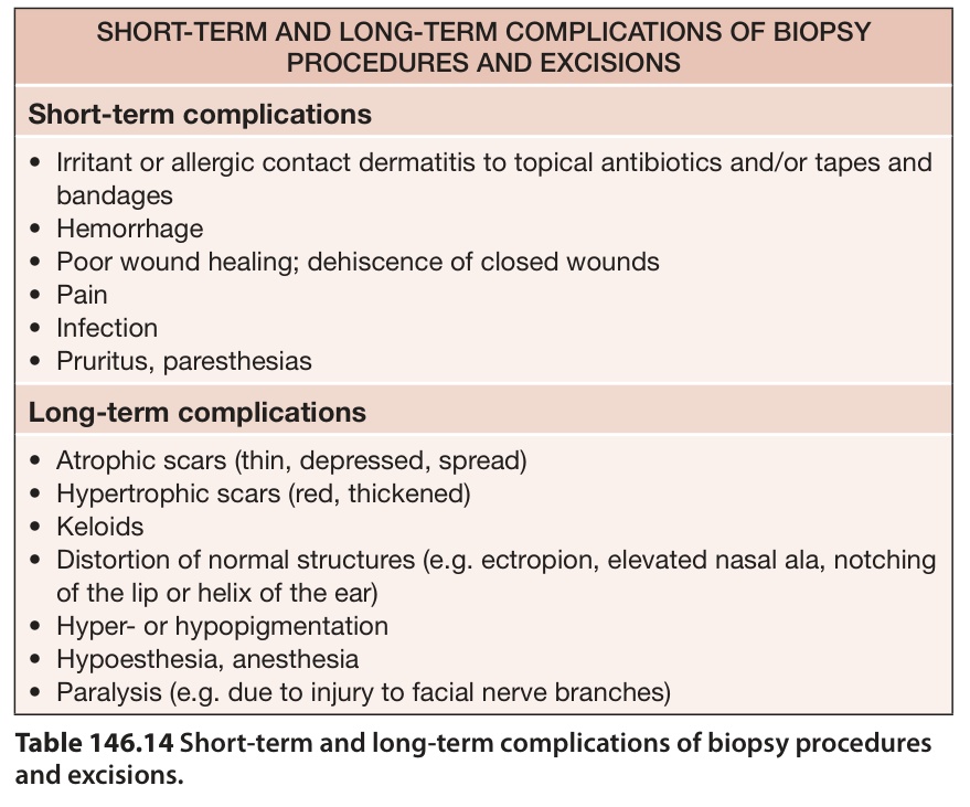
Table 146.14 Short-term and long-term complications of biopsy procedures and excisions.