TECHNIQUE
Table 150.5 provides a flow sheet for performing MMS and Fig. 150.7 depicts the procedure. (Also see Video 150.1)
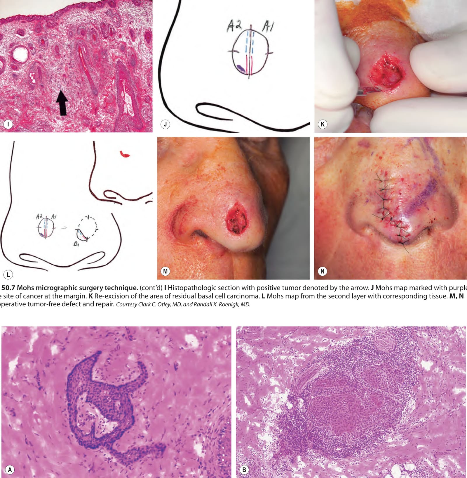
Fig. 150.7 Mohs micrographic surgery technique.A Preoperative appearance of a basal cell carcinoma on the nose. B Debulking of the tumor via curettage. C Beveled excision (with scalpel at a 45° angle) of all clinically evident tumor plus a small margin of normal-appearing skin. D Placement of skin notches at 3, 6, 9 and 12 o’clock positions in the specimen and adjacent surrounding tissue. E Orientation of the excised specimen on an anatomic card. F Specimen sectioned into two pieces and flattened. G Marking of the cut margins of the specimen with red and blue inks. H Matching Mohs map and tissue specimen ready for histologic preparation. A red mark on the gauze denotes the location of the first specimen with the second specimen always placed below. Different systems are used but precision is common to all.
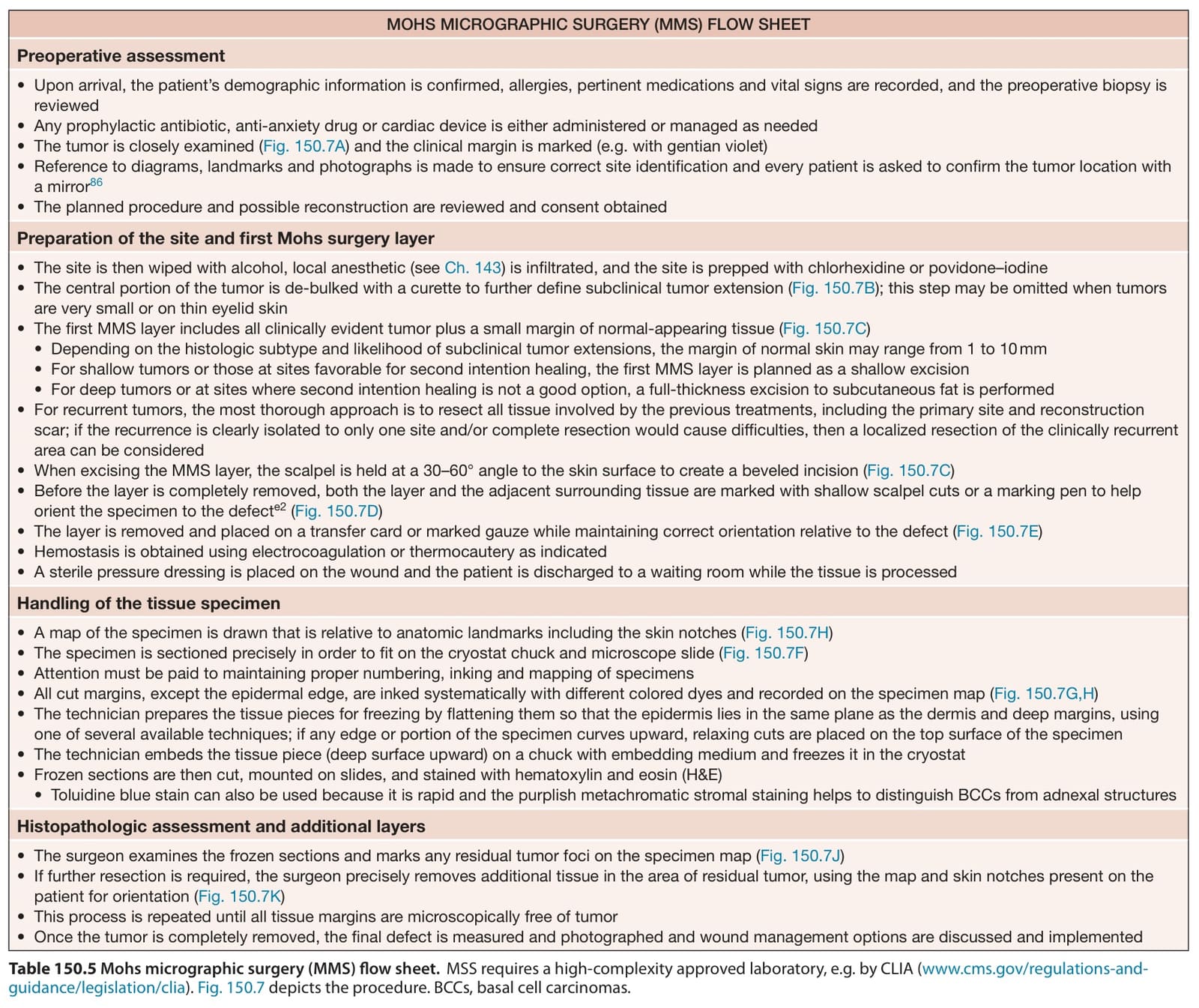
Table 150.5 Mohs micrographic surgery (MMS) flow sheet.MSS requires a high-complexity approved laboratory, e.g. by CLIA (www.cms.gov/regulations-andguidance/legislation/clia). Fig. 150.7 depicts the procedure. BCCs, basal cell carcinomas.