VARIATIONS/REFINEMENTS
There are several modifications/variations of MMS that are used to improve: the speed of tissue processing; the efficiency of tumor removal; and the recognition of individual tumor cells for better margin control. For descriptions of staged-excision (“slow Mohs”) techniques, see the Cutaneous Melanoma section.
The single section method can be used for smaller tumors. It consists of a highly beveled specimen with relaxing incisions on the superior, tumor-bearing portion to allow all peripheral edges to fold outward into a single plane. This method is particularly useful for tumors in which a shallow Mohs layer can be taken and then the surgical defect allowed to heal by second intention. The unaltered examination of the complete deep margin also reduces the possibility of missed deep margins due to tissue subdivision.
A non-beveled 90° incision can be used for MMS on the extremities. The resulting vertical edges are ideal for reconstruction. Although a traditional beveled edge can be trimmed prior to reconstruction, a true, sharp, straight 90° angle is more difficult to obtain. Disadvantages include excision of more tissue and the necessity to manipulate the specimen to make the tissue flatten into a single plane.
In wide excision MMS (“wide Mohs”), a larger, clinically tumor-free margin (5–20 mm) is taken. It can be used for tumors that have a high propensity for recurrence or metastases (including satellite metastases), in particular melanoma, Merkel cell carcinoma, and cutaneous sarcomas. In these scenarios, margin control is prioritized over tissue sparing.
During MMS, rapid immunohistochemical (IHC) staining can be utilized to assist in histologic evaluation (see Table 150.4). Some surgeons find it especially helpful for challenging and high-risk tumors. However, there has been rather limited adoption of IHC staining by Mohs surgeons due to the additional cost and time required as well as concerns regarding reliability. In a 2013 survey of Mohs surgeons, only 22% of the respondents (n = 378) incorporated IHC staining into their practice following fellowship training.
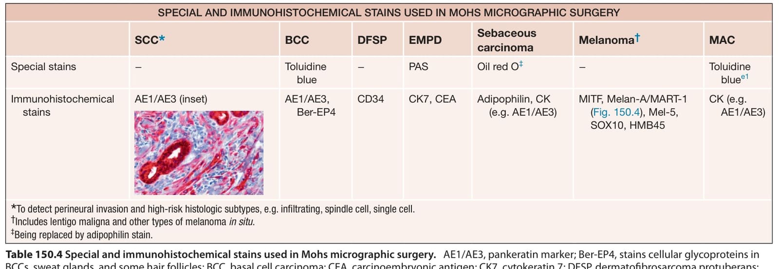
Table 150.4 Special and immunohistochemical stains used in Mohs micrographic surgery.