EOSINOPHIL BIOLOGY
The eosinophil is a leukocyte ~10–16 microns in diameter that possesses a segmented (usually bi-lobed) nucleus. It is produced in the bone marrow and circulates transiently (8–18 hours) in the peripheral blood. Under normal conditions, circulating eosinophil counts range from 0 to 500 cells/mcl of human blood. Except for the bone marrow, gastrointestinal tract (distal to the esophagus), and lymphoid tissues (spleen, thymus, and lymph nodes), eosinophils are limited to diseased tissues.
In 1879, Paul Ehrlich named the “eosinophil” because of the intense staining of its distinctive cytoplasmic granules with the acidic dye, eosin. Ultrastructurally, eosinophils are characterized by membranebound, cytoplasmic granules that have an electron-dense core and less dense matrix (Fig. 26.1). The eosinophil produces multiple factors that make it a highly proinflammatory and pro-fibrotic leukocyte. It also expresses a number of receptors on its cell surface that are important for activation, prolonged survival, and targeted migration into tissue compartments (see Fig. 26.1).
Eosinophils are attracted into tissues by the coordinated actions of: (1) eosinophil activating cytokines (commonly referred to as “priming” cytokines); (2) tissue-released chemokines; and (3) endothelial adhesion molecules. Eosinophils express receptors for granulocyte–macrophage colony-stimulating factor (GM-CSF), interleukin (IL)-3, and IL-5 which are important for bone marrow production, maturation, activation, and prolonged survival. Several lines of investigation indicate that eosinophils are recruited to and activated within tissues by cytokines that largely come from a type 2 immune response (IL-33) or from Th2 cells (IL-4, IL-5, and IL-13). Natural killer cells also produce IL-5, and mast cell-derived cytokines contribute indirectly to eosinophil activation via induction of IL-5 and GM-CSF.
Eosinophils themselves elaborate important inflammatory and regulatory cytokines, including IL-1α, TGF-α and TGF-β, GM-CSF, IL-3, IL-5, IL-6, IL-8, TNF, and macrophage inflammatory protein 1α (CCL3; see Fig. 26.1). As a result, eosinophil activation also occurs in an autocrine manner. In cytotoxicity assays, eosinophils are maximally activated by GM-CSF, followed by IL-3, IL-5, TNF and IL-4, in order of potency. Eosinophil numbers in the circulation and tissues is a function of balance of mediators that prolong survival versus those that induce apoptosis, like glucocorticoids. Glucocorticoids can counteract the prolonged survival induced by IL-3, IL-5 and GM-CSF, which may in part explain their therapeutic efficacy in most eosinophil-driven conditions. More recently, the surface receptors sialic acid-binding immunoglobulin-like lectins (Siglec) -8 and -10, which are expressed on eosinophils and mast cells, have been shown to mediate apoptosis.
Several members of the C-C chemokine gene superfamily are chemotactic for eosinophils, including the eotaxins 1, 2, and 3 (CCL11, CCL24, and CCL26). These chemokines signal primarily through C-C chemokine receptor (CCR)-3, which is highly expressed on eosinophils. In addition to their chemotactic properties, these chemokines also induce the release of reactive oxygen species. Eotaxins are produced by tissue resident cells positioning them as upstream mediators of cutaneous inflammation.
Eosinophils enter tissues by traversing blood vessels. Similar to other leukocytes, this transmigration involves adhering to endothelial adhesion molecules within the selectin and immunoglobulin supergene families. In particular, eosinophils constitutively express very late antigen (VLA)-4, the ligand for vascular cell adhesion molecule (VCAM)-1; the latter is a cytokine-inducible endothelial adhesion molecule (see Fig. 102.11). After entering tissues, eosinophil activity is influenced by interactions between integrins on the eosinophil cell surface and both extracellular proteins (fibronectin, laminin, or collagen) and glycosaminoglycans (hyaluronic acid or chondroitin sulfate), which are induced under inflammatory conditions.
Granular Contents
Upon activation, eosinophils release their granular contents into the extracellular spaces by three mechanisms: cytolytic degranulation, piecemeal degranulation, and regulated secretion. Cytolytic degranulation is characterized by organelle rupture, chromatolysis of nuclei with loss of morphologic integrity, and extensive deposition of eosinophil granules and granule products within tissue; this process occurs in many inflammatory disorders, including atopic dermatitis, as well as in affected organs (including the skin) in patients with hypereosinophilic syndromes (HES).
Eosinophil major basic protein 1 (eMBP1), the constituent of the granules’ crystalline cores (see Fig. 26.1), directly damages helminths and mammalian cells, exemplified by its ability to cause exfoliation of bronchial epithelial cells. In addition, eMBP1, but none of the other eosinophil granule proteins, induces basophil histamine release. Furthermore, eMBP1 stimulates neutrophils, leading to the release of superoxides and lysozyme. Eosinophil cationic protein (ECP or RNase3) and eosinophilderived neurotoxin (EDN or RNase2) are members of the RNase family. ECP is a potent toxin for parasites through a different mechanism than eMBP1 and is more effective at killing certain helminths than eMBP1. EDN, as its name implies, is a neurotoxin and also has antiviral activity against RNA viruses.
Eosinophil peroxidase (EPO) kills numerous microorganisms in the presence of hydrogen peroxide (generated by eosinophils and other phagocytes) and halides. This combination of products also initiates mast cell secretion. Binding of EPO to microbes, including Staphylococcus aureus, greatly potentiates their killing by phagocytes. Similarly, EPO-coated tumor cells are spontaneously lysed by activated macrophages, and eMBP1 is also toxic to tumor cells. EPO and eMBP1 are potent platelet agonists that lead to release of 5-hydroxytryptamine (serotonin) and promote clotting.
In summary, the highly basic granule proteins released by activated eosinophils have evolved to not only protect us from parasitic helminths but also some bacteria and viruses, and they may in some settings possess anti-tumor properties.
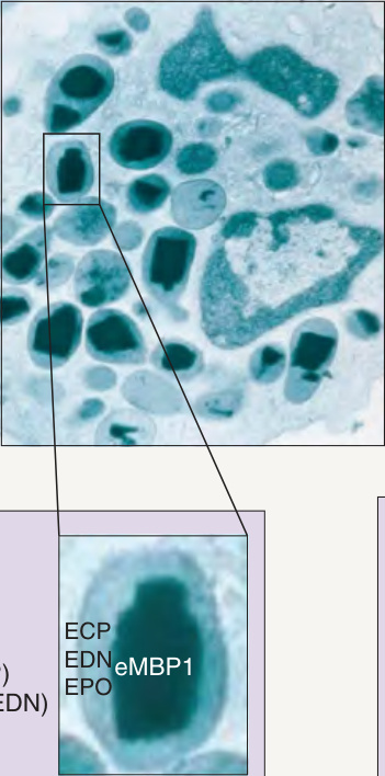
*Fig. 26.1 Products of eosinophils and localization of specific granule proteins. The eosinophil produces myriad products that determine its role in inflammation. Specific granules contain major basic protein 1 and 2 (in the electron-dense core) and other toxic proteins (in the matrix). Receptor for eotaxins and RANTES. §Charcot–Leyden crystal protein. §§Forms mitochondrial DNA traps with ECP and eMBP1. CCL, chemokine ligand; CCR, CC chemokine receptor; GM-CSF, granulocyte–macrophage colonystimulating factor; HETE, hydroxyeicosatetraenoic acid; IFN, interferon; IL, interleukin; MIP, macrophage inflammatory protein; MMP, matrix metalloproteinase; OSM, oncostatin M; P2X4, P2X purinoceptor 4; PAI-2, plasminogen-activator inhibitor; PDGF, platelet-derived growth factor; RANTES, regulated on activation, normal T cell expressed and secreted; Siglec, sialic acid-binding immunoglobulin-type lectins; TGF, transforming growth factor; TLR, toll-like receptor; TNF, tumor necrosis factor.
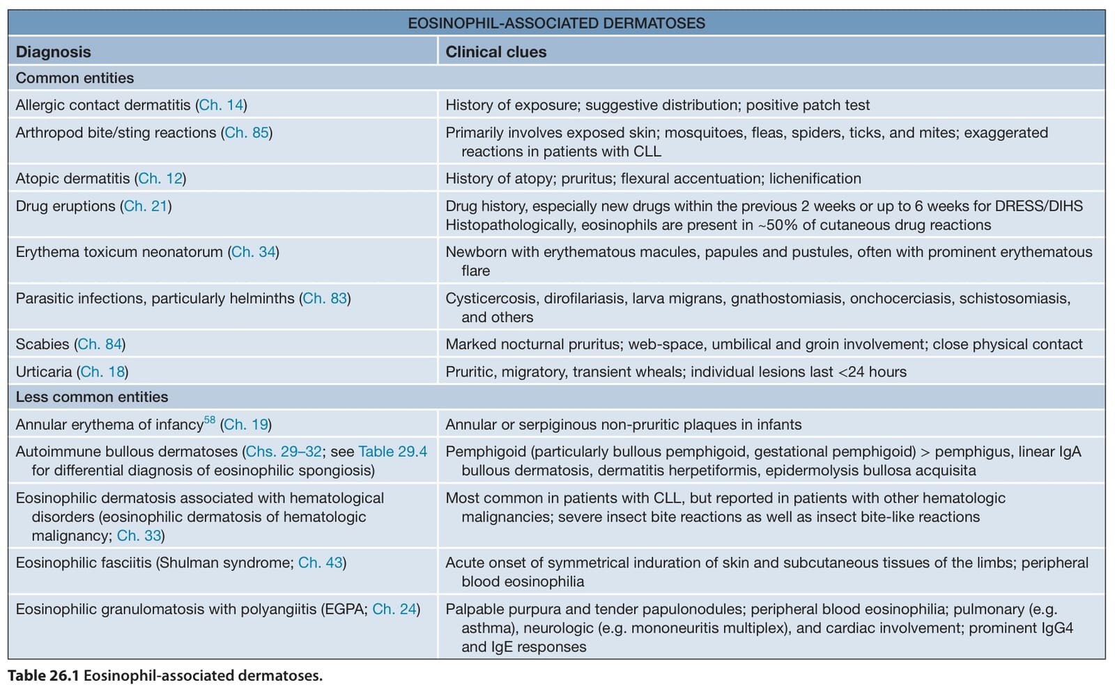
Table 26.1 Eosinophil-associated dermatoses.