INTRODUCTION
Basement membranes are specialized structures located between cells and their underlying stroma or between different cell types. Basement membranes of various tissues differ ultrastructurally, biochemically, and functionally. All basement membranes contain an electron-dense, somewhat granular matrix of variable thickness called the lamina densa. Major components of the lamina densa include type IV collagen, laminins, and heparan sulfate proteoglycans. Other components of the lamina densa vary depending upon the tissue type.
Basement membranes serve a variety of functions that are manifest in a tissue-specific manner. Functions common to virtually all basement membranes include (1) substrates for cell attachment; (2) templates for tissue repair; (3) matrices for cell migration; (4) substrata that influence differentiation, morphogenesis, and apoptosis of epithelial cell layers; and (5) permeability barriers for cells and macromolecules. In the skin, the two major basement membrane complexes are found at the epidermal–dermal interface and around the dermal microvasculature. This chapter will deal largely with the epidermal basement membrane.
The epidermal basement membrane is a highly specialized structure that contains numerous tissue-specific elements (Table 28.1). The first characterization of this ultrastructural region in the skin was made via electron microscopy, which identified four distinct subregions of the epidermal basement membrane (Fig. 28.1). From superior to inferior, they are:
●basal keratinocytes with their cytoskeletons, hemidesmosomal plaques, and plasma membranes;
●the lamina lucida, an electron-lucent region that contains delicate anchoring filaments; the latter connect hemidesmosomes (HDs) in basal keratinocytes to the underlying lamina densa;
●the lamina densa; and
●the sublamina densa region that contains anchoring fibrils, anchoring plaques, and filamentous proteins of the papillary dermis. These subregions of the epidermal basement membrane, readily identified in fixed skin samples by transmission electron microscopy, have served as a useful conceptual model of a highly complex adhesion unit that preserves the integrity of the skin (Fig. 28.2). Interestingly, specialized transmission electron microscopy studies of cryopreserved skin have suggested that the lamina lucida may actually represent an artifact of tissue dehydration. Nonetheless, the classic laminated model of the epidermal basement membrane serves as a useful framework for understanding its structure, function, and impairment in disease.
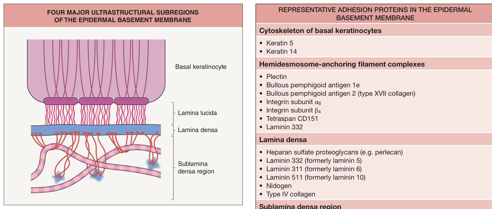
Fig. 28.1 Four major ultrastructural subregions of the epidermal basement membrane. The major ultrastructural subregions of the epidermal basement membrane consist of the following: (1) cytoskeleton, hemidesmosomal plaques, and plasma membranes of basal keratinocytes; (2) electron-lucent lamina lucida; (3) lamina densa; and (4) sublamina densa region of the papillary dermis.
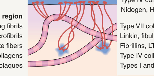
Fig. 28.2 “Laminated” model of the epidermal basement membrane. In the laminated model, keratin intermediate filaments attach to electrondense hemidesmosomes on the basal plasma membranes of keratinocytes. Hemidesmosomes connect to the underlying lamina densa by small threadlike strands termed anchoring filaments. The lamina densa and the overlying epidermis are tethered to the papillary dermis by anchoring fibrils, a series of looping elements along the underside of the lamina densa that serve as attachment sites for fibrillar proteins in the papillary dermis. HSPG, heparan sulfate proteoglycans; LTBP, latent TGF-β-binding proteins.
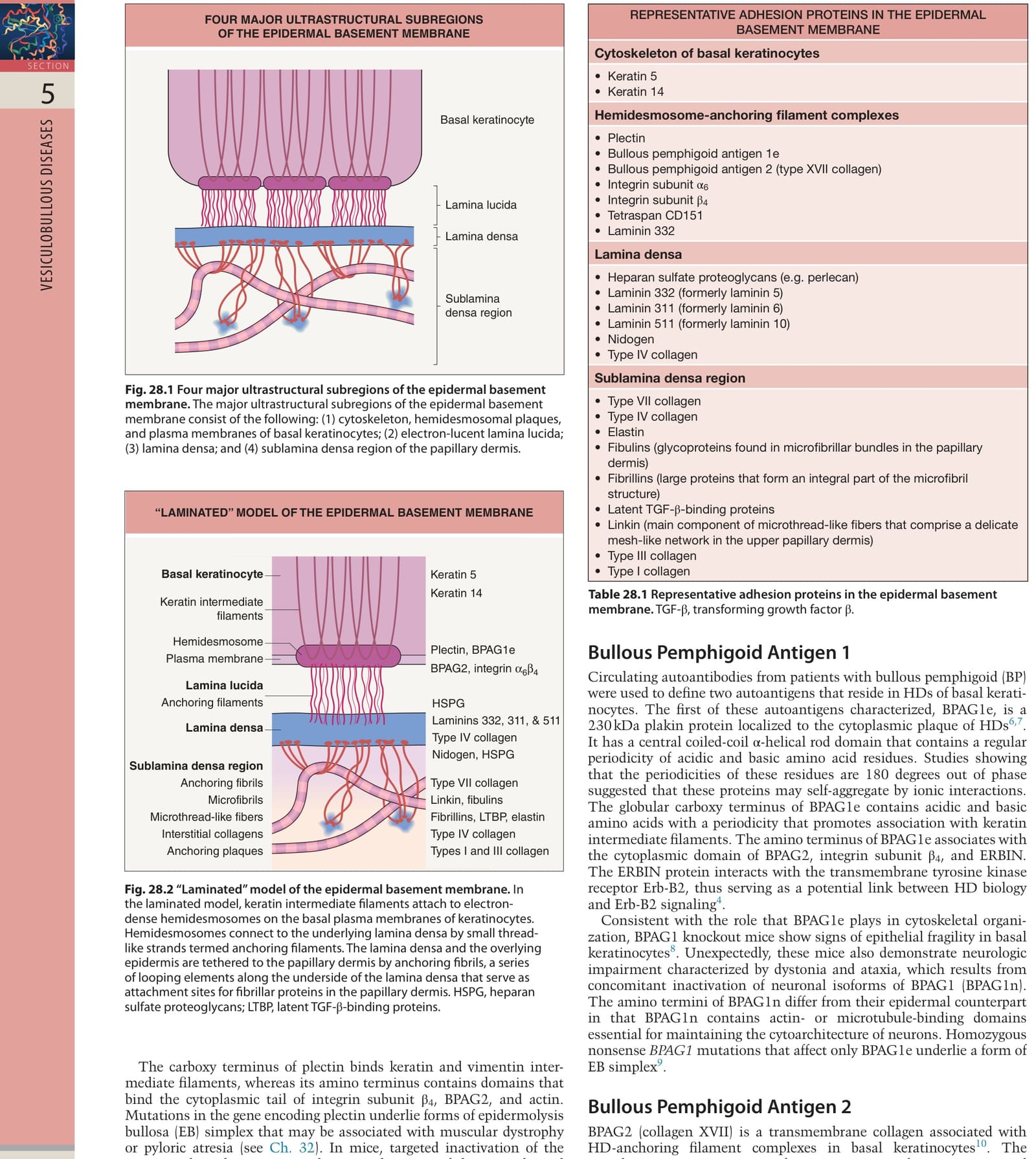
Table 28.1 Representative adhesion proteins in the epidermal basement membrane. TGF-β, transforming growth factor β.