SECONDARY CUTANEOUS MUCINOSES
Secondary cutaneous mucinoses include all those diseases in which mucin deposition represents only an additional histologic finding and not the main feature. As an epiphenomenon, a wide range of inflammatory disorders and neoplasms may have mucin deposition in the epidermis and/or dermis or within the follicular unit. Secondary mucinoses are outlined in Table 46.3.
Additional figures available in our eBook (see inside front cover for access code).
-
Rongioletti F. Primary paediatric cutaneous mucinoses.
eds. Clinical and Pathological Aspects of Skin Diseases in Endocrine, Metabolic, Nutritional and Deposition Disease. New York: Springer; 2010:146–147.2. Sorrell JM, Carrino DA, Baber MA, Asselineau D, Caplan
-
Kudlak N, Schuler A, Dong J, Crowe D. Pretibial
Br J Dermatol. 2020;182:29–38.17a. Stammler R, Bessis D, Meunier L, et al. Jadassohn- myxedema treated with intralesional hyaluronidase and triamcinolone. JAAD Case Rep. 2020;6:810–811.33a. Mervis JS, Maeng MM, Kirsner RS, Wester ST.
Dössekker’s atypical tuberous myxedema: report of three cases and review of literature. J Eur Acad Dermatol Venereol. 2023;37:e968–e970.18. Rongioletti F, Ghigliotti G, De Marchi R, et al.
AI. A monoclonal antibody which recognizes a glycosaminoglycan epitope in both dermatan sulfate and chondroitin sulfate proteoglycans of human skin. Histochem J. 1999;31:549–558.3. Neudecker BA, Stern R, Mark LA, Steinberg S.
Teprotumumab as a novel treatment for pretibial myxoedema. Br J Dermatol. 2022;187:795–797.34. Burman KD, McKinley-Grant L. Dermatologic aspects of
Cutaneous mucinoses and HIV infection. Br J Dermatol. 1998;139:1077–1080.19. Rongioletti F, Rebora A. Cutaneous toxic mucinoses. J
Scleromyxedema-like lesions of patients in renal failure contain hyaluronan: a possible pathophysiological mechanism. J Cutan Pathol. 2005;32:612–615.4. Rongioletti F, Rebora A. Updated classification of papular thyroid disease. Clin Dermatol. 2006;24:247–255.35. Rongioletti F, Merlo V, Riva S, et al. Reticular erythem- atous mucinosis: a review of patients’ characteristics, associated conditions, therapy and outcome in 25 cases. Br J Dermatol. 2013;169:1207–1211.36. Cinotti E, Merlo V, Kempf W, et al. Reticular erythem-
Am Acad Dermatol. 1992;26:789–790.20. Chen KY, Tzeng IS, Lee YY, et al. Scleromyxedema and lichen myxedematosus: is it associated with viral hepatitis? J Dermatol. 2019;46:879–885.21. Rongioletti F. New and emerging conditions of mucinosis, lichen myxedematosus, and scleromyxedema. J Am Acad Dermatol. 2001;44:273–281.5. Rongioletti F, Merlo G, Cinotti E, et al. Scleromyxedema:
atous mucinosis: histopathological and immunohistochemical features of 25 patients compared with 25 cases of lupus erythematosus tumidus. J Eur Acad Dermatol Venereol. 2015;29:689–697.37. Dallo C, Lee K, Cragun WC, Lee M. A case of papulo- acquired cutaneous mucinoses in adults. J Eur Acad Dermatol Venereol. 2022;36:1016–1024.22. Hermans C, Goldscheider I, Ruzicka T, Rongioletti F.
a multicenter study of characteristics, comorbidities, course, and therapy in 30 patients. J Am Acad Dermatol. 2013;69:66–72.6. Mecoli CA, Talbot Jr CC, Fava A, et al. Clinical and molecular
Atypical discrete papular lichen myxedematosus associated with monoclonal gammopathy: report of four cases and review of the literature. J Eur Acad Dermatol Venereol. 2016;30:2048–2051.23. Luchsinger I, Coulombe J, Rongioletti F, et al. Self-healing phenotyping in scleromyxedema pretreatment and posttreatment with intravenous immunoglobulin. Arthritis Care Res (Hoboken). 2020;72:761–767.7. Kalli F, Cioni M, Parodi A, et al. Increased frequency of nodular mucinosis in a patient with systemic lupus erythematosus. Am J Dermatopathol. 2020;42:280–282.38. Perel-Winkler AC, Derk CT. Diffuse cutaneous mucinosis in dermatomyositis: a case report and review of the literature. Case Rep Dermatol Med. 2014:938414.39. Van Zander J, Shaw JC. Papular and nodular mucinosis juvenile cutaneous mucinosis: clinical and histopathologic findings of 9 patients: the relevance of long-term follow-up. J Am Acad Dermatol. 2018;78:1164–1170.24. Deza G, García-Martínez P, Gallardo F, et al. Self-healing interleukin-4 and reduced frequency of interferon-γ and IL-17-producing CD4+ and CD8+ cells in scleromyxedema. J Eur Acad Dermatol Venereol. 2020;34:1092–1097.7a. Gallo R, Trave I, Cotzia E, et al. Dermato-neuro syndrome as a presenting sign of progressive systemic sclerosis. J Am Acad Dermatol. 2002;46:304–306.40. Cohen PR, Erickson CP, Calame A. Case report and cutaneous mucinosis in adulthood: the adult counterpart of the juvenile variant of the disease? Am J Dermatopathol. 2019;41:60–64.25. Rongioletti F, Kaiser F, Cinotti E, et al. Scleredema. A after COVID-19 vaccination in a patient with scleromyxoedema previously controlled with bortezomib and intravenous immunoglobulins. Acta Derm Venereol. 2022;102:adv00836.8. Rongioletti F, Rebora A. Cutaneous mucinoses: micro- review of solitary cutaneous focal mucinosis: a unique primary cutaneous mucinosis unrelated to mucinosisassociated systemic diseases. Dermatol Online J. 2020;26:13030/qt33m815p3.41. Kempf W, Von Stumberg B, Denisjuk N, et al. Trauma- multicentre study of characteristics, comorbidities, course and therapy in 44 patients. J Eur Acad Dermatol Venereol. 2015;29:2399–2404.26. Ferreli C, Gasparini G, Parodi A, et al. Cutaneous scopic criteria for diagnosis. Am J Dermatopathol. 2001;23:257–267.9. Rongioletti F, Cozzani E, Parodi A. Scleromyxedema with induced cutaneous focal mucinosis of the mammary areola: an unusual presentation. Dermatopathology (Basel). 2014;1:24–28.42. Rongioletti F, De Lucchi S, Meyes D, et al. Follicular manifestations of scleroderma and scleroderma-like disorders: a comprehensive review. Clin Rev Allergy Immunol. 2017;53:306–336.27. Venturi M, Damevska K, Ferreli C, et al. Scleredema of an interstitial granulomatous-like pattern: a rare histologic variant mimicking granuloma annulare. J Cutan Pathol. 2010;37:1084–1087.10. Dinneen AM, Dicken CH. Scleromyxedema. J Am Acad mucinosis: a clinicopathologic, histochemical, immunohistochemical and molecular study comparing the primary benign form and the mycosis fungoidesassociated follicular mucinosis. J Cutan Pathol. 2010;37:15–19.42a. Jin MF, Ali NS, Davis DMR, et al. An update on long-term
Buschke associated with lichen sclerosus: three cases. Indian J Dermatol Venereol Leprol. 2020;86:272–277.28. Knobler R, Moinzadeh P, Hunzelmann N, et al. European
Dermatol. 1995;33:37–43.11. Guarneri A, Cioni M, Rongioletti F. High-dose intra- dermatology forum S1-guideline on the diagnosis and treatment of sclerosing diseases of the skin, Part 2: scleromyxedema, scleredema and nephrogenic systemic fibrosis. J Eur Acad Dermatol Venereol. 2017;31:1581–1594.29. Hong KR, Hong JY, Chung EH, et al. Monoclonal venous immunoglobulin therapy for scleromyxoedema: a prospective open-label clinical trial using an objective score of clinical evaluation system. J Eur Acad Dermatol Venereol. 2017;31:1157–1160.12. Mahévas T, Arnulf B, Bouaziz JD, et al. Plasma cell-directed outcomes of pediatric follicular mucinosis over a 20-year period: a retrospective cohort study. Int J Dermatol. 2023;62:1056–1059.43. Khalil J, Kurban M, Abbas O. Follicular mucinosis: a review. Int J Dermatol. 2021;60:159–165.44. Cerroni L, Fink-Puches R, Bäck B, Kerl H. Follicular gammopathy-associated scleredema adultorum of Buschke in a patient with diabetes mellitus successfully treated with intravenous immunoglobulin and narrowband ultraviolet B phototherapy: a case report. Ann Dermatol. 2021;33:586–588.30. Mirza FN, Leventhal JS. The rash in life-threatening therapies in monoclonal gammopathy-associated scleromyxedema. Blood. 2020;135:1101–1110.13. Haber R, Bachour J, El Gemayel M. Scleromyxedema treatment: a systematic review and update. Int J Dermatol. 2020;59:1191–1201.14. Rongioletti F, Zaccaria E, Cozzani E, Parodi A. Treatment mucinosis: a critical reappraisal of clinicopathologic features and association with mycosis fungoides and Sézary syndrome. Arch Dermatol. 2002;138:182–189.45. Cinotti E, Basso D, Donati P, et al. Urticaria-like follicular mucinosis: four new cases of a controversial entity. J Eur Acad Dermatol Venereol. 2013;27:e435–e437.46. Lee MY, Byun JY, Choi HY, Choi YW. Mucinous nevus. Ann metabolic and endocrine disorders. Clin Dermatol. 2020;38:79–85.31. Ferreli C, Atzori L, Rongioletti F. Obesity-associated of localized lichen myxedematosus of discrete type with tacrolimus ointment. J Am Acad Dermatol. 2008;5:530–532.15. Rongioletti F, Rebora A, Crovato F. Acral persistent
Dermatol. 2018;30:465–467.47. Rongioletti F, Barnhill RL. Deposition disorders. In:
lymphedematous mucinosis and stasis mucinosis. Clin Dermatol. 2021;39:229–232.32. Kotwal A, Turcu AF, Sonawane V, et al. Clinical papular mucinosis: a new entity. Arch Dermatol. 1986;122:1237–1239.16. Rongioletti F, Ferreli C, Atzori L. Acral persistent papular
Barnhill RL, ed. Dermatopathology. 4th Edition, New York: McGraw-Hill; 2020:485–486.48. Sarfo A, Helm K, Flamm A. Cutaneous myxomas and a experience with rituximab and intravenous immunoglobulin for pretibial myxedema: a case series. Thyroid. 2019;29:692–699.
mucinosis. Clin Dermatol. 2021;39:211–214.
psammomatous melanotic schwannoma in a patient with Carney complex. J Cutan Pathol. 2019;46:93–96.
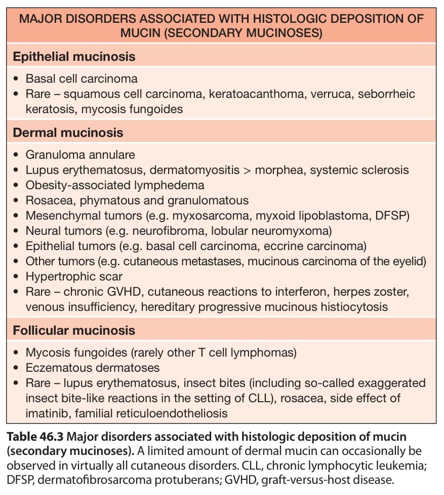
Table 46.3 Major disorders associated with histologic deposition of mucin
(angio)myxomas are true neoplasms, although benign, which can recur after incomplete excision. When an (angio)myxoma is located in the genital region, it needs to be distinguished from an aggressive angiomyxoma, a rare neoplasm with potential for local recurrence and very rarely for distant metastases. Aggressive angiomyxomas usually stain positively for estrogen and progesterone receptors and are characterized by a different vascular component with variable thickness of vessel walls.