EPIDERMAL DIFFERENTIATION
Epidermal differentiation is a tightly regulated process that involves the transformation of proliferating cells in the basal layer into the dead corneocytes of the stratum corneum. During this process, the keratins and junctional proteins (see below) that are expressed are highly specific for the state of differentiation (Fig. 56.7). The mitotically active keratinocytes in the basal layer primarily express the keratin pair KRT5 and KRT14, with less abundant expression of KRT15. KRT15 is also able to assemble with KRT5, thereby providing mechanical stability to the keratinocyte in the absence of K14.
As keratinocytes move suprabasally to the spinous layer, they withdraw from the cell cycle. This process is associated with a downregulation of KRT5 and KRT14 and an induction of the differentiationspecific keratins, KRT1 and KRT10. Further maturation of spinous keratinocytes into granular keratinocytes results in expression of KRT2, a reinforcement keratin. With further maturation, filaments containing the suprabasal keratins are bundled parallel to the surface and, eventually, keratinocytes lose their cytoplasmic organelles and differentiate into lifeless corneocytes that are shed into the environment.
Interestingly, a number of epidermal keratins have a more restricted anatomic distribution pattern. For example, KRT9 is specifically expressed in the suprabasal cells of palmoplantar skin. KRT6, KRT16, and KRT17 are expressed in the palmoplantar epidermis as well as in the keratinocytes of the nail bed, hair follicle, and sebaceous and sweat glands. In addition, this group of keratins is rapidly induced by injury and ultraviolet radiation and appears in hyperproliferative conditions.
Regulatory Pathways Involved in Epidermal Development and Differentiation
The regulatory pathways necessary for normal epidermal differentiation include those that: (1) establish and maintain basal keratinocytes; (2) initiate and execute keratinocyte terminal differentiation; and (3) form the embryonic epidermis. Examples of these pathways are discussed below.
Genes required for establishing and maintaining basal
The importance of TP63 in epidermal and adnexal development became apparent with the generation of knockout mouse models. The p63 protein encoded by this gene has at least six different isoforms
The p63 protein regulates the transcription of multiple genes in basal keratinocytes. For example, p63 induces expression of epidermal keratins KRT5 and KRT14 while simultaneously repressing expression of KRT18, a keratin expressed in single-layered epithelia. Another key function of p63 is to maintain the proliferative state of basal keratinocytes by repressing cell cycle inhibitors.
Genes required for differentiation in mature epidermis
After basal keratinocytes have undergone a few rounds of cell division, they irreversibly withdraw from the cell cycle, move suprabasally, and become spinous keratinocytes. This process is also regulated by p63, in particular its ΔNp63α isoform. In this context, p63 mediates cell cycle exit and induces expression of KRT1. The importance of p63 for normal epidermal development and differentiation is further underscored by the finding that p63 mutations underlie a subset of ectodermal dysplasias characterized by abnormalities in the skin and its adnexae (see Ch. 63).
Notch signaling is also required for the formation of the spinous layer. In mouse models, ablation of Notch signaling resulted in the development of an extremely thin spinous layer, whereas constitutively active Notch signaling resulted in an expanded spinous layer. Notch signaling is essential for controlling the balance between proliferation and differentiation in the developing and postnatal epidermis.
The extracellular concentration of Ca+ also plays an important role in keratinocyte differentiation. In a mature epidermis, there is a gradient of increasing extracellular Ca+ concentration from the basal layer to the cornified layer. Increasing the Ca+ concentration in the media of cultured keratinocytes can induce a differentiation program similar to that of keratinocytes in vivo, with successive expression of markers of keratinocyte terminal differentiation.
Several Ca+-responsive proteins in the epidermis have key roles in the formation of the granular cell layer. For example, the protein kinase C (PKC) family is activated by Ca+ signaling and functions specifically in the transition from spinous to granular cells. PKC proteins have a dual role, contributing to the downregulation of KRT1 and KRT10 expression while also inducing markers of granular keratinocytes such as loricrin, filaggrin, and transglutaminases.
In addition to the PKC family, other proteins that undergo conformational changes upon binding to Ca+ are expressed in mouse and human epidermis. Of these, the calcium-sensing receptor is specifically expressed in granular keratinocytes. Mice lacking a functional calciumsensing receptor fail to properly form a granular layer, while overexpression of the calcium-sensing receptor in basal keratinocytes leads to expansion of the spinous and granular cell layers.
Genes required for development of the embryonic
During epidermal morphogenesis, basal keratinocytes do not directly differentiate into spinous keratinocytes as they do in postnatal skin. Instead, basal keratinocytes initially differentiate into intermediate keratinocytes that, like spinous cells, express KRT1. However, unlike spinous keratinocytes, intermediate keratinocytes still undergo proliferation. The intermediate cell layer exists only transiently during epidermal morphogenesis, and intermediate keratinocytes ultimately differentiate into spinous and granular keratinocytes, which then undergo further terminal differentiation.
The importance of the intermediate cell layer for normal epidermal development has been demonstrated by mouse models in which inter-mediate cells fail to mature into spinous and granular cells. Such a block in differentiation occurs in mice lacking expression of inhibitor of κB kinase-α (IKKα), interferon regulatory factor 6 (IRF6), or ovo-like 1 (Ovol1), as well as in mice expressing a mutant form of the 14–3–3σ protein. In all instances, an expanded intermediate cell layer develops, terminal differentiation is disrupted, and the consequent failure to establish barrier function results in neonatal lethality.
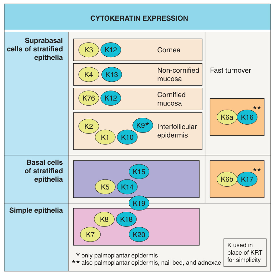
Fig. 56.7 Cytokeratin expression. Type II (basic) keratins are in yellow (KRT1–KRT8; KRT76) and type I (acidic) keratins are in blue (KRT9–KRT20). Keratin disorders are presented in Fig. 56.5 and Table 56.4 that activate or repress transcription, depending on the cellular context. p63 is essential for epidermal morphogenesis, as highlighted by the failure of Trp63-null mice to develop a stratified epidermis. After birth, exposure of these mice to the environment leads to rapid death due to dehydration. The failure of p63-deficient epithelial cells to adopt an epidermal fate underscores the critical role of p63 in specifying an epidermal lineage.