HUTCHINSON–GILFORD PROGERIA SYNDROME
Synonyms: Progeria Hutchinson–Gilford syndrome
Key features
Rare, occurring in 1 in 4 to 8 million births
Due to mutations in LMNA, which encodes nuclear lamins A and C
Normal motor and mental development
Characteristic facies with prominent scalp veins and a beaked nose
Wrinkled as well as scleroderma-like skin, alopecia, and poor growth
Generalized atherosclerosis with premature death from cardiovas- cular and cerebrovascular disease at a median age of 14 years
Lonafarnib, an oral farnesyltransferase inhibitor that reduces the accumulation of farnesylated mutant prelamin A (“progerin”), is an FDA-approved therapy
Introduction
Hutchinson–Gilford progeria syndrome (HGPS) is a genetic disease characterized by accelerated aging that begins during infancy. The term “progeria” originates from the Greek word for old age, “geras”, and was proposed by Gilford when he delineated the clinical features and course of the disorder in 1904.
History
The syndrome was first reported in 1886 by Jonathan Hutchinson, with Hastings Gilford subsequently documenting the postmortem features. In 2003, mutations in the gene that encodes lamin A were first described.
Epidemiology
HGPS is estimated to occur in 1 in 4 to 8 million births, with an overall prevalence of ~1 in 20 million people. The male : female ratio is 1.2 : 1, and it has been reported worldwide in patients with a wide variety of ethnic backgrounds.
In the 64 cases reviewed by DeBusk, median and mean paternal ages were advanced, consanguinity was uncommon, and the abortion
rate in affected families was normal. These observations favored a de novo dominant mutation, which was later confirmed by genetic studies (see below). Since HGPS patients usually do not become sexually mature and typically succumb to the disease early in the second decade of life, familial occurrence is rare; however, it can occur in the setting of gonadal mosaicism. Consanguineous families with autosomal recessive variants of HGPS have been described, but additional clinical features such as clavicular aplasia/hypoplasia in affected individuals suggest an overlap with mandibuloacral dysplasia, which is caused by biallelic mutations in LMNA (Table 63.10).
Pathogenesis
The vast majority of patients with both classic and atypical forms of HGPS have a de novo heterozygous point mutation that affects splicing of the lamin A gene (LMNA). This gene encodes two protein products, lamin A and lamin C, that represent major components of the nuclear membrane lamina and contribute to the structural scaffolding of the nucleus. Classic HGPS (80%–90% of affected individuals) is caused by a particular mutation, c.1824C>T, which activates a cryptic splice site within exon 11 and leads to an internal deletion of 50 amino acids. This deletion includes a cleavage site that is necessary for the conversion of prelamin A to mature lamin A (Fig. 63.13). As a result, the cells contain a dominant-mutant prelamin A (“progerin”) that disrupts the nuclear scaffolding. In the patients’ cells, including lymphocytes and fibroblasts, there is evidence of altered nuclear sizes and shapes, as well as lobulation of the nuclei, extrusion of chromatin, and nucleolar alterations. Such structural changes result in functional defects such as abnormal DNA damage repair, mitochondrial impairment and aberrant cell-cycle regulation, which lead to premature aging.
Other heterozygous progerin-producing LMNA mutations cause either atypical HGPS with clinical findings similar to classic HGPS or autosomal dominant progeroid laminopathies with distinct phenotypes that share some features with HGPS (see Table 63.10). The severity of these disorders correlates with the ratio of progerin to lamin A, and extremely high progerin levels have been associated with neonatalonset progeria and death in the first few years of life. In families with autosomal recessive progeroid laminopathies, including the progeria– mandibuloacral dysplasia overlap syndromes, biallelic mutations in LMNA may affect both lamin A and C, disrupting interactions with nuclear proteins.
Other cellular and molecular changes that contribute to the pathogenesis of HGPS include altered epigenetic regulation and gene expression, shortened telomeres, and increased cellular senescence. Of note, progressively decreasing telomere length is seen with normal aging and is associated with progerin production and low levels of telomerase activity. In a telomerase knockout mouse model (see Ch. 4), some clinical features of accelerated aging have also been observed. Additional abnormalities associated with the normal aging process, such as insulin resistance and decreased survival of fibroblast cultures, are seen in patients with HGPS.
Clinical Features
HGPS is a multisystem disease with prominent involvement of the skin, bones, skeletal muscle, adipose tissue, and cardiovascular structures. Although affected children usually appear normal at birth, growth failure and other clinical features typically appear rather abruptly during the first year of life. Weight gain is very slow, with marked loss during episodes of illness. Linear growth proceeds at half the normal pace and does not undergo the normal acceleration around puberty. Sexual maturation is absent in most patients, and affected individuals have both short stature and a low weight for height.
At birth or during early infancy, patients may have thick, inelastic, scleroderma-like skin, usually on the lower abdomen, flanks, thighs, and buttocks. Following the onset of growth failure, the skin becomes thin and dry, with less hair than normal. Some areas may appear taut and shiny, whereas others (especially the fingers and toes) lax and wrinkled. Infantile fat is rapidly lost with the onset of growth failure, resulting in prominent superficial veins and the appearance of perioral cyanosis. As the aged appearance progresses, irregular brown pigmentation becomes evident in sun-exposed areas.
At the onset of growth failure, patients begin to develop characteristic facial features, including a disproportionately large cranium, frontal bossing, a large open anterior fontanelle, pronounced scalp veins, prominent eyes due to relatively slow growth of the facial bones, a thin beaked nose with a sculpted appearance, and micrognathia. The facial stigmata tend to become marked by age 2 to 3 years, giving a “pluckedbird” appearance.
Alopecia develops during the first year of life and becomes diffuse and generalized, with later hair growth tending to be fine and lightly pigmented. Eyebrows and eyelashes are often sparse or absent. Although the nails may be normal, dystrophy in the form of small, short and thin nails is common. Marked delay in the eruption of primary and secondary dentition has also been noted in most patients. Teeth may be crowded, rotated, overlapped and maloccluded, and the voice tends to be high-pitched and piping.
Truncal features include a narrow pyriform thorax, shoulders with thin and short clavicles, and prominent thoracic kyphosis giving a stooped appearance. Prominence of the abdomen (relative to the chest) and hypoplastic nipples also contribute to the distinctive habitus. The limbs are usually proportionate and become progressively thinner with increasing prominence of the joints, especially the knees, elbows, and small joints of the hand. Coxa valga is usually present by age 2 to 3 years and, in combination with increasing joint stiffness, contributes to a wide-based gait.
Patients are of normal intelligence and may be self-conscious about their appearance. Early onset of progressive coronary and cerebral atherosclerosis results in a median lifespan of approximately 14 years. Other complications include osteopenia, low-frequency conductive hearing loss, corneal dryness, and hyperopia. Although mild insulin resistance occurs in up to half of patients, overt diabetes mellitus is rare.
Pathology and Laboratory Findings
Cutaneous histopathology varies with the site and the age of the patient, and it is usually not helpful in diagnosing the condition. The epidermis is fairly normal with minimal hyperkeratosis and a slight
increase in melanin in the basal cell layer. Although dermal elastic tissue is normal, dermal collagen tends to be thickened and hyalinized. Adnexal structures are normal or decreased in density and arrector pili muscles are usually prominent.
Radiographic findings include hypoplasia of the facial bones, thinned cranial bones, open fontanelles, and hypoplasia of the mandible with crowding of the teeth. Progressive resorption of bone from the distal phalanges of the fingers and toes is also characteristic, but not diagnostic, of HGPS.
Differential Diagnosis
The differential diagnosis is outlined in Table 63.9 and includes Werner syndrome, “atypical Werner syndrome”, Néstor–Guillermo progeria syndrome, metageria, and acrogeria. In contrast to HGPS, patients with Werner syndrome have premature canities, cataracts, and an increased incidence of malignancy. HGPS should also be distinguished from the following disorders: Cockayne syndrome (Ch. 87), Rothmund– Thomson syndrome (Ch. 87), ataxia–telangiectasia (Ch. 60), Kindler syndrome (Ch. 32), Wiedemann–Rautenstrauch syndrome (neonatal progeroid syndrome), and forms of Ehlers–Danlos syndrome and cutis laxa with progeroid features (Ch. 97). Lastly, clinical overlap may be seen with other disorders that are due to mutations in LMNA, such as early-onset myopathy with progeroid features, restrictive dermopathy (Ch. 34), and mandibuloacral dysplasia (see Table 63.10).
Treatment
Management is directed toward the prevention and treatment of complications, particularly cardiovascular, cerebrovascular, and musculoskeletal. Regular cardiovascular monitoring is recommended and low-dose aspirin may be of benefit. No dietary regimen has been shown to alter the disease course. Although growth impairment in HGPS is not typically related to growth hormone deficiency, administration of exogenous growth hormone may increase weight and, to a lesser degree, height. Physical and occupational therapy can help to maintain joint mobility, and patients and families may benefit from psychological and genetic counseling.
Because farnesylation of the dominant-mutant progerin form of prelamin A causes it to be anchored to the nuclear membrane, thereby disrupting nuclear structure (see Fig. 63.13), inhibitors of farnesylation were identified as a potential treatment for HGPS. Initial studies found that administration of farnesyltransferase inhibitors (FTIs) restored normal nuclear morphology to human HGPS fibroblasts in vitro, and these inhibitors improved body weight, bone density, strength, and survival in mouse models of progeria. Additional investigation in mouse models showed benefit from treatment with the combination of a bisphosphonate and statin, which inhibit farnesyl pyrophosphate synthase and HMG-CoA reductase, respectively. Each of these enzymes functions in the protein prenylation pathway essential to both farnesylation and the alternative pathogenic geranylgeranylation that occurs in the setting of FTI monotherapy.
Treatment of HGPS patients with the FTI lonafarnib, either alone or together with the bisphosphonate zoledronate and pravastatin, has been shown to result in small but significant increases in survival. Mechanistic target of rapamycin (mTOR) inhibitors (e.g. sirolimus, everolimus) increase autophagic degradation of progerin, and a clinical trial investigating the combination of everolimus and lonafarinib is underway. Antisense oligonucleotides that reduce progerin transcript and protein levels were recently shown to partially rescue the cardiovascular phenotype and increase lifespan in an HGPS transgenic mouse model. Other possible therapies being explored include inhibition of progerin–lamin A binding with the small molecule progerinin, inhibition of isoprenylcysteine carboxyl methyltransferase to mislocalize progerin away from the nuclear rim, vitamin D to potentially reduce progerin production, remodelin to inhibit N-acetyltransferase-10 and rescue nuclear shape, tocilizumab to reduce oxidative stress by neutralizing interleukin-6, and inhibition of the NLRP3 inflammasome. Lastly, gene correction via base editing or CRISPR/Cas9-based methods is being investigated in HGPS mouse models.
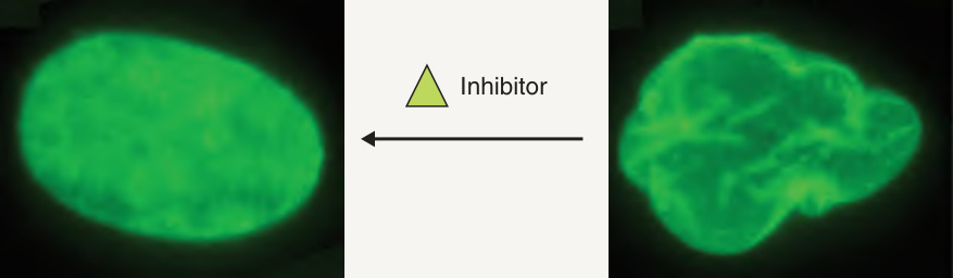
Fig. 63.13 Pathogenesis of Hutchinson–Gilford progeria syndrome (HGPS). Patients with classic HGPS have a C-to-T point mutation that activates a cryptic splice site and leads to an internal in-frame deletion of 50 amino acids. Because the deleted region includes a cleavage site for the ZMPSTE24 zinc metalloproteinase, removal of a farnesylated and methylated portion of prelamin A by this enzyme in order to produce mature lamin A does not occur in HGPS patients. Mutant “progerin” therefore remains farnesylated and, as a result, anchored to the nuclear membrane, where it disrupts nuclear scaffolding and causes blebbing. Administration of farnesyltransferase inhibitors can prevent this disruption of nuclear structure and function by progerin. Of note, ZMPSTE24 mutations underlie some human cases of restrictive dermopathy and mandibuloacral dysplasia (see Table 63.10).
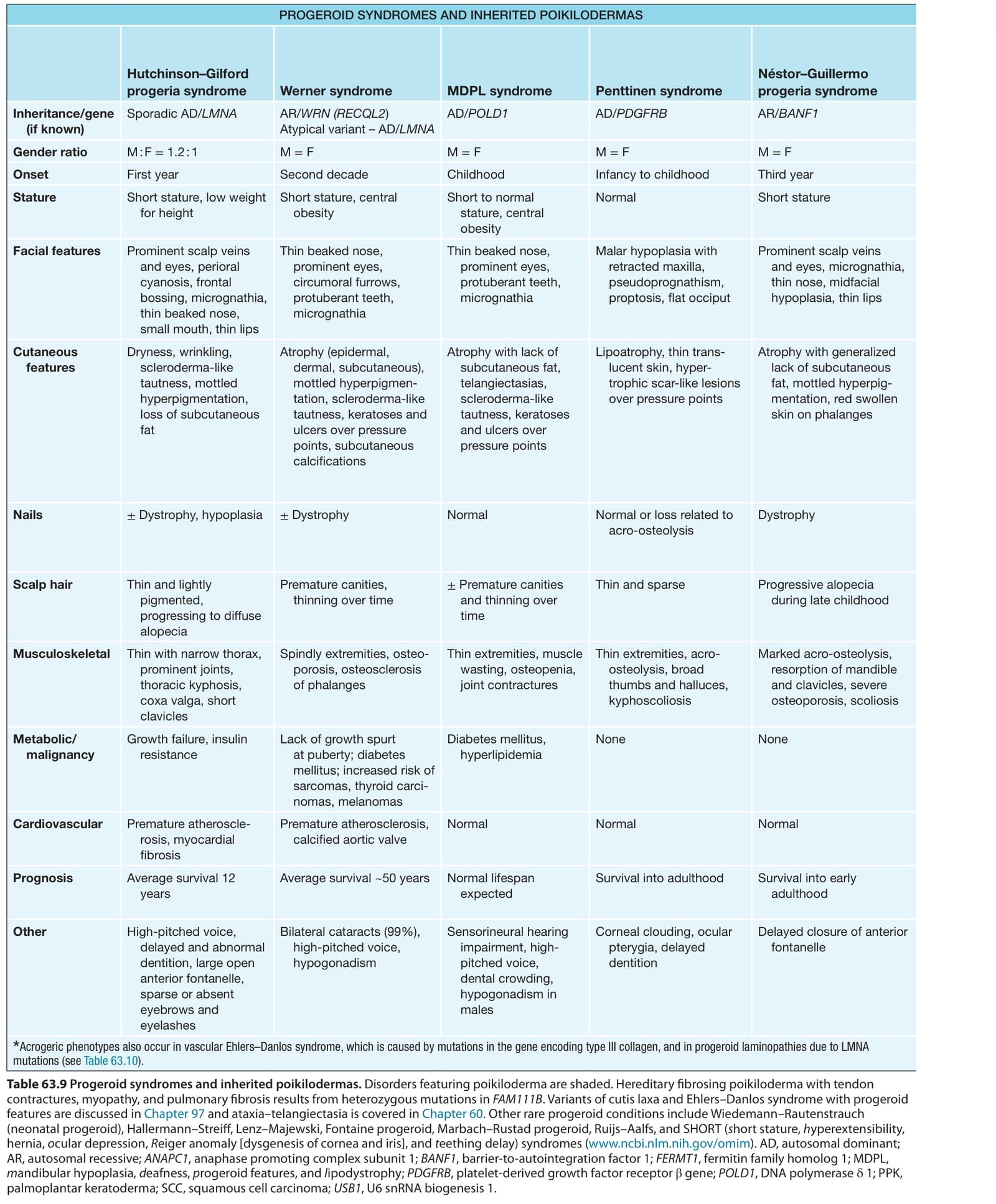
Table 63.9 Progeroid syndromes and inherited poikilodermas. Disorders featuring poikiloderma are shaded. Hereditary fibrosing poikiloderma with tendon contractures, myopathy, and pulmonary fibrosis results from heterozygous mutations in FAM111B. Variants of cutis laxa and Ehlers–Danlos syndrome with progeroid features are discussed in Chapter 97 and ataxia–telangiectasia is covered in Chapter 60. Other rare progeroid conditions include Wiedemann–Rautenstrauch (neonatal progeroid), Hallermann–Streiff, Lenz–Majewski, Fontaine progeroid, Marbach–Rustad progeroid, Ruijs–Aalfs, and SHORT (short stature, hyperextensibility, hernia, ocular depression, Reiger anomaly [dysgenesis of cornea and iris], and teething delay) syndromes (www.ncbi.nlm.nih.gov/omim). AD, autosomal dominant; AR, autosomal recessive; ANAPC1, anaphase promoting complex subunit 1; BANF1, barrier-to-autointegration factor 1; FERMT1, fermitin family homolog 1; MDPL, mandibular hypoplasia, deafness, progeroid features, and lipodystrophy; PDGFRB, platelet-derived growth factor receptor β gene; POLD1, DNA polymerase δ 1; PPK, palmoplantar keratoderma; SCC, squamous cell carcinoma; USB1, U6 snRNA biogenesis 1.
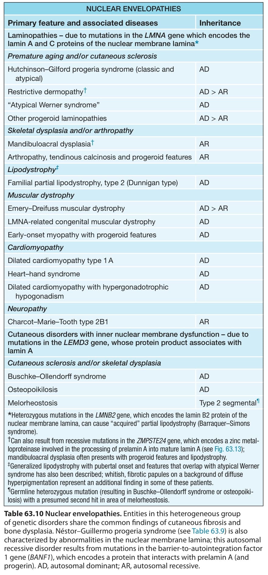
Table 63.10 Nuclear envelopathies. Entities in this heterogeneous group of genetic disorders share the common findings of cutaneous fibrosis and bone dysplasia. Néstor–Guillermo progeria syndrome (see Table 63.9) is also characterized by abnormalities in the nuclear membrane lamina; this autosomal recessive disorder results from mutations in the barrier-to-autointegration factor 1 gene (BANF1), which encodes a protein that interacts with prelamin A (and progerin). AD, autosomal dominant; AR, autosomal recessive.