THE MIDLINE LESION
A number of critical events occur during the third to fifth weeks of embryologic development (see Ch. 2). These include formation of the neural tube, separation of neuroectoderm from surface ectoderm, and interposition of mesodermal elements. Inherent in such a complicated process is the potential for error, which spans a spectrum from obvious, fulminant neural tube defects to subtle cutaneous anomalies. However, the latter may represent a sign of a significant underlying malformation.
Classically, closure of the human neural tube was thought to be a continuous process, beginning at a single focus in the cervical region and progressing bidirectionally in a “zipper-like” fashion to culminate in the closure of the anterior and subsequently posterior neuropores. However, evidence suggests that in humans as well as other mammals, the cranial segment of the neural tube closes in an intermittent or multisite pattern. Four distinct initiation sites of neural tube closure have been identified in the mouse, and a similar pattern of closure has been proposed for humans, with an additional caudal site (Fig. 64.1). In support of the multisite closure model, neural tube defects (e.g. cephaloceles, myelomeningoceles, heterotopic brain/meningeal tissue) cluster either entirely within one closure site or at the junction of two sites, such as the nasal root (2–3) or vertex (2–4; see Fig. 64.1). Neural tube
closure in the posterior cranial area is thought to occur via a membrane rather than a midline fusion of folds, and the appearance of this site during embryologic development in animals resembles that of human membranous aplasia cutis. Specific genes may control individual closure sites, explaining repetitive events within families.
Midline Lesions of the Head, Neck, and Anterior Trunk
The pathogenesis, differential diagnosis, and management of midline lesions of the nose (Fig. 64.2) and scalp are discussed below, followed by a review of the individual developmental anomalies that may present with a midline lesion on the head, neck, and anterior trunk. Midline cysts of this region (e.g. thyroglossal duct, bronchogenic) are discussed in Chapter 110.
Midline nasal lesions
It is important for physicians to recognize the potential intracranial connection of a midline nasal mass or pit. Dermoid cysts, dermal sinuses, cephaloceles, and nasal glial heterotopia (nasal “gliomas”) resulting from faulty embryologic development of the frontobasal skull and/or adjacent ectodermal/neuroectodermal tissues may present as congenital nasal lesions (Table 64.1). As a group, these midline nasal masses occur in ~2–5 per 100 000 live births. They are often misdiagnosed as more familiar entities such as epidermoid cysts and infantile hemangiomas.
The various congenital midline nasal masses have similar developmental origins. As the bones of the skull form during the second month of gestation, diverticula of dura mater project: (1) inferiorly into the prenasal space that extends from the base of the skull to the nasal tip,
deep to the nasal bone and skin but superficial to the nasal cartilage; and (2) anteriorly through the nasofrontal fontanelle into the glabellar and inner canthal areas. These diverticula may make connections with the overlying dermis, but usually they regress completely, with subsequent bony fusion to form the base of the skull and the cribriform plate. However, if the dura mater does not separate from the dermis, an ectodermal residue, with or without a patent intracranial connection, can persist; furthermore, if faulty neural tube closure results in a bony defect, there may be herniation of brain tissue. Such ectopic elements can occur either within the prenasal space (nasoethmoidal, “intranasal”) or superiorly at the glabella (nasofrontal, “extranasal”). Depending on the contents of the lesion (e.g. cutaneous versus neural tissue, cyst versus tract only) and the patency of the intracranial connection, a dermoid cyst, dermal sinus, cephalocele, or nasal glial heterotopia may result (see below).
Many of these midline nasal lesions have the potential for intra-cranial communication and therefore present a risk for the development of serious CNS infections such as meningoencephalitis or a brain abscess. In addition, recurrent inflammation and progressive expansion of the mass may result in bony atrophy and nasal distortion. Early and complete surgical excision with repair of associated bony and dural defects is imperative. In order to prevent ascending infections, a biopsy, needle aspiration, or other preoperative manipulations should be avoided.
A thorough radiologic evaluation prior to surgery is mandatory. Ultrasound may detect some bony defects but has limited sensitivity. While CT is the most sensitive method for defining bony skull defects, false-positive studies are common in young children undergoing bone maturation. MRI is superior in demonstrating intracranial masses and other soft tissue changes and may aid in planning surgical approaches. In addition, surgical exploration is often necessary to definitively exclude the presence of an intracranial connection, as small fibrous tracts may not be detectable with imaging studies.
Midline lesions of the scalp
Subcutaneous nodules of the scalp are common, and in adults they are usually acquired lesions, such as epidermoid and tricholemmal
cysts. However, midline scalp lesions that are noted at birth or in early childhood have a high likelihood of representing developmental defects, such as dermoid cysts, cephaloceles, heterotopic brain or meningeal tissue, sinus pericranii, and aplasia cutis congenita. In one series of 70 children presenting with a solitary, non-traumatic scalp nodule, 61% of the lesions were dermoid cysts and 4% cephaloceles. More importantly, 37% of the lesions were found to extend intracranially to the dura or brain.
In the evaluation of a patient with a subcutaneous nodule on the scalp, clinical clues that heighten suspicion of a developmental anomaly with potential intracranial extension include:
●a patient who is an infant or child
●a lesion first noted at birth or during early childhood
●a family history of neural tube defects
●a history of meningitis
●the presence of neurologic signs or symptoms
●location of the lesion along the midline or over the suture lines of the scalp
●lesions that pulsate, transilluminate, or fluctuate in size with crying or straining
●lesions with a central pore in young children, who are less likely to have epidermoid cysts
●an overlying capillary malformation
●a positive hair collar sign or hair tuft (see below). A congenital midline lesion, or a scalp nodule with other concerning features, requires imaging studies prior to surgical excision or biopsy. As noted previously, CT is superior in detecting bony defects and MRI is more sensitive in evaluating transcranial extension of soft tissue. Ultrasound is sometimes utilized as an initial study in infants, but it has a relatively high false-negative rate. A neurosurgical referral is indicated if these studies are positive and, as small communications may be missed, complete surgical removal may be prudent even if they are negative.
The hair collar sign consists of a ring of long, dark, coarse hair surrounding a congenital scalp lesion. The presence of a hair collar or a tuft of hair in the midline scalp is a relatively specific marker for ectopic neural tissue. Hair collars are also seen in membranous aplasia cutis (Figs. 64.3 & 64.4), a disorder thought to represent a forme fruste of a neural tube defect (i.e. without the presence of ectopic neural tissue). Drolet et al. hypothesized that cerebral herniation produces aberrant shearing forces during the formation of hair follicles early in development, causing them to point outward from the defect. The proximity of neuroectoderm expressing neural adhesion molecules may also alter normal epidermal–dermal interactions, thereby inducing the development of large, abnormal follicles. These speculations correlate with the histologic and dermoscopic finding of numerous horizontally oriented, hypertrophic follicles emerging from the edge of scalp lesions to form the hair collar. The differential diagnosis and evaluation of a positive hair collar sign is presented in Fig. 64.5.
Other developmental anomalies of the scalp
Sinus pericranii is a rare disorder characterized by a congenital, or occasionally acquired, epicranial venous malformation of the scalp that communicates with an intracranial dural sinus through dilated diploic cranial veins. The lesion is usually located near the midline, especially in the frontal region. Most are asymptomatic and appear as a compressible, fluctuant mass that varies in size with changes in intracranial pressure (Fig. 64.6). The differential diagnosis includes a simple venous or arteriovenous malformation, meningocele, and encephalocele; of note, sinus pericranii can also occur in association with heterotopic meningeal or brain tissue.
Hairless congenital scalp lesions such as a nevus sebaceus (see Chs. 62 & 111) and nevus psiloliparus (a hallmark of encephalocraniocutaneous lipomatosis; see Ch. 62) due to mosaic activating mutations may mimic developmental anomalies of the scalp.
Dermoid cysts
Dermoid cysts result from sequestration of ectodermal tissue along embryonic fusion planes during development. They are a separate entity from benign cystic teratomas, neoplasms that are also referred to as dermoids. Although dermoid cysts are congenital defects and may be recognized at birth, these lesions often escape notice until they become enlarged, inflamed, or infected. This typically occurs by early childhood, but diagnosis may be delayed until as late as the sixth decade of life. Dermoid cysts present as firm, non-compressible, non-pulsatile subcutaneous nodules that often reach a size of 1–4 cm in diameter.
The lesions usually do not transilluminate. They are most commonly located around the eyes, particularly the lateral eyebrow (Fig. 64.7A), but may also be found on the nose (Fig. 64.7B), scalp (often over the anterior fontanelle or the midline occiput), neck, sternum, sacrum, and scrotum.
Dermoid cysts on the nose or midline scalp have a much higher likelihood of intracranial extension than those in a periocular location. Nasal lesions, which account for <10% of cervicofacial dermoid cysts, occur anywhere from the glabella to the tip of the nose (see above) and can result in a widened nasal bridge. The presence of a sinus ostium, which may be heralded by protruding hairs or a sebaceous discharge, increases the risk of intracranial involvement of a nasal dermoid cyst to almost 50%, versus ~25% overall. A sinus ostium without an underlying subcutaneous mass may also connect with an intracranial dermoid cyst. These lesions favor the midline occipital area but can occur elsewhere on the face and scalp; recurrent drainage and inflammation may represent signs of deeper extension. In addition to infectious complications, a chemical meningitis or even hydrocephalus due to release of keratinous and sebaceous material into the cerebrospinal fluid (CSF) can occur with intracranial dermoid cysts. Lastly, dermoid cysts and dermal sinuses overlying the spine may be associated with occult dysraphism (see below) and if the tract connects with the subarachnoid space, an increased risk of meningitis.
Histologically, dermoid cysts in any location are lined by keratinizing stratified squamous epithelium and contain mature adnexal structures (see Ch. 110). Treatment is by excision, and preoperative imaging studies are required for lesions located on the nose, midline scalp, or posterior axis or those with other suspicious features (e.g. a draining sinus ostium) in order to exclude a connection to the CNS.
Cephaloceles
Cephalocele is a general term for congenital herniation of intra-cranial structures through a defect in the skull. Cranial meningoceles are herniations in which the sac contains meninges and CSF, while encephaloceles contain both meninges and brain (Fig. 64.8). Therefore, by definition, all cephaloceles have an intracranial connection. These lesions result from faulty closure of the cephalic portion of the neural tube during development, and they are much less common than their caudal counterparts (e.g. myelomeningoceles). Cephaloceles occur in ~1–3 per 10 000 live births, with a higher incidence in those with a family history of neural tube defects and an ~5% risk of neural tube defects in subsequent siblings. They are usually midline or paramedian, occurring most frequently at the occiput or vertex (75%) (see Fig. 64.1), followed by extranasal (e.g. glabella or medial orbit; 15%) and intranasal or pharyngeal (10%) locations. Encephaloceles are generally noted early in life due to the resultant deformity. Occasionally, small frontal lesions are misdiagnosed as deep infantile hemangiomas, vascular malformations, hypertelorism, or nasal polyps; of note, the latter are almost never seen in children younger than 3 years of age.
Cephaloceles typically appear as a soft, compressible, pulsatile mass with a bluish color. They may be covered with either normal skin or a thin, glistening epithelial membrane. The size of cephaloceles varies tremendously, from 1 cm nodules to lesions containing nearly half of the brain, and the size does not necessarily correlate with the extent of the underlying bony defect. Patients with intranasal cephaloceles often present with a broadened nasal bridge but no palpable mass, whereas
lesions on the scalp may have an overlying capillary malformation and/ or a surrounding hair collar (see above, Midline lesions of the scalp). Transillumination frequently indicates the presence of CSF; in addition, increased flow of CSF into lesions with crying, the Valsalva maneuver, or compression of the jugular veins (Furstenberg test) may result in transient expansion. Leaks of CSF can occur (e.g. CSF rhinorrhea), and patients may have a history of meningitis. Nasal cephaloceles may develop in conjunction with facial clefts and other midline defects, while encephaloceles are often associated with neurologic abnormalities.
On histologic examination, encephaloceles show mature neuroglial tissue embedded in varying amounts of fibrous stroma. Heterotopic brain tissue (see Fig. 64.8) demonstrates similar features, and these lesions cannot be reliably distinguished histologically. When a patient is suspected of having a cephalocele, imaging studies and subsequent neurosurgical repair should be performed.
Nasal glial heterotopia
Synonyms: Nasal glioma Nasal neuroglial heterotopia Nasal heterotopic brain tissue
Nasal glial heterotopia (NGH) refers to extracranial rests of brain tissue located extranasally (e.g. at the nasal root or glabella; 60%), intranasally (30%), or both (10%). The previous term for NGH, nasal glioma, is a misnomer since this lesion represents a developmental anomaly rather than a neoplasm; it is also entirely separate from the similarly named glioma, a malignant brain tumor. NGH results when herniated brain tissue is sequestered extracranially by the bony fusion that forms the base of the skull during development (see Fig. 64.8). Although a vestigial fibrous stalk connecting with the intracranial space persists in 15%–25% of cases, unlike encephaloceles, there is no communication with the intracranial leptomeninges
or the CSF. NGH is present at birth, accounting for ~5% of congenital nasal midline masses, and it typically enlarges in proportion to the growth of the child.
Extranasal NGH typically presents as a firm, non-compressible, non-pulsatile, non-transilluminating mass at or just lateral to the nasal root (Fig. 64.9; see Fig. 115.20). The overlying skin is often red with prominent telangiectasias, which may result in misdiagnosis as a hemangioma. Intranasal NGH manifests as a widened nasal bridge or a polypoid nodule within the nasal cavity, sometimes protruding from a nostril. It occasionally causes nasal obstruction resulting in respiratory distress, and lesions involving the nasopharynx may be associated with a cleft palate. Excessive lacrimation or strabismus may occur ipsilateral to the NGH.
Histologically, NGH demonstrates well-circumscribed nodules composed mostly of astrocytes, admixed with other glial cells and loosely textured intercellular glial substance interwoven with fibrous tissue (see Ch. 115). Neurons may or may not be present focally, and there are often prominent dilated blood vessels in the lesion and overlying skin. Immunoperoxidase staining for S100 protein and glial fibrillary acidic protein (GFAP) is positive. After imaging studies to determine whether an intracranial extension is present, NGH should be surgically excised.
Heterotopic brain tissue and rudimentary meningoceles
Synonyms: Heterotopic brain tissue – neuroglial heterotopia, neuroglial choristoma Rudimentary meningoceles – heterotopic meningeal tissue, sequestrated meningoceles, meningeal hamartomas, meningothelial hamartomas
Heterotopic brain tissue and rudimentary meningoceles are uncommon congenital anomalies thought to represent encephaloceles or meningoceles that have lost their intracranial attachment (see Fig. 64.8). The bone underlying these lesions is usually intact. However, in inter-mediate forms known as atretic encephaloceles and meningoceles, a fibrotic stalk that extends intracranially with some degree of luminal ablation remains. Heterotopic brain tissue may be located in the scalp, orbit, lip and oropharynx, as well as in the nasal area (nasal glial heterotopia; see above); rudimentary meningoceles are usually found on the scalp or overlying the spine.
Heterotopic brain tissue and rudimentary meningoceles typically present as a 1–4 cm solid or cystic subcutaneous nodule, often with a blue–red hue. Occasionally, rudimentary meningoceles have a bullous appearance and are clinically indistinguishable from membranous aplasia cutis. Lesions are often located in the midline occipital or parietal scalp, where they can manifest with alopecia of the overlying skin and/or a surrounding hair collar. An overlying capillary malformation also may be present.
Histologic features of heterotopic brain tissue are as described for nasal glial heterotopia. Although classic findings such as cystic cavities lined by meningothelial cells are present in some cases of rudimentary meningocele, the histologic features are often subtle, with meningeal elements simulating the appearance of vascular or loose connective tissue. While signs such as the tendency of meningocytes to encircle collagen bundles and the presence of psammoma bodies may be helpful,
immunohistochemical staining for epithelial membrane antigen (EMA) and vimentin are critical in establishing the diagnosis. Because the presence or absence of an intracranial connection cannot be reliably determined by clinical examination, imaging studies should be performed prior to surgical excision.
Midline anterior neck inclusion cyst
A midline anterior neck inclusion cyst (MANIC) presents as a congenital milia-like papule on the midline lower neck superior to the sternum. The small, superficial, yellow to white papule is not associated with underlying anomalies (Fig. 64.10). Histologically, a MANIC is lined by stratified squamous epithelium with intact keratohyalin granules, resembling an epidermoid cyst.
Midline cervical cleft
A midline cervical cleft is a rare congenital anomaly resulting from incomplete fusion of the branchial arches in the ventral midline of the neck. Approximately 200 cases have been reported worldwide, with a female-to-male ratio of 2 : 1. Midline cervical clefts may be isolated or found in association with other defects, including other midline clefts (e.g. lower lip, tongue, mandible) and thyroglossal, branchial cleft, or bronchogenic cysts.
Patients present at birth with a vertically oriented, linear cleft on the midline neck at any point between the inferior edge of the mandible and the sternum. The lesion is covered with atrophic skin, which is often weeping in the neonatal period. Additional features may include a blind mucosal sinus tract inferiorly and a skin protuberance superiorly. A subcutaneous fibrous cord deep to the latter may extend upward to the chin, resulting in progressive cervical contracture and/or webbing of the neck with extension (pterygium colli medianum).
Histologically, midline cervical clefts consist of parakeratotic stratified squamous epithelium without normal adnexal structures. The subcutis often contains dense connective tissue, and fibrous cords may include bundles of skeletal muscle. Following ultrasonography to evaluate for an associated thyroglossal duct cyst (see Ch. 110), complete surgical excision and closure via Z-plasty (to reduce the risk of cicatricial contracture) should be performed during the first two years of life.
Sternal clefts and supraumbilical raphae
Sternal clefts are rare congenital malformations that result from defective embryologic fusion of paired mesodermal bands in the ventral midline. These lesions most frequently involve the superior sternum, but inferior clefts and a complete bifid sternum can also occur. The skin overlying sternal clefts may be atrophic (Fig. 64.11), scarred, or ulcerated, and a dermal sinus tract is occasionally present. Approximately 40% of sternal clefts are associated with PHACE syndrome (posterior fossa malformations; hemangiomas; arterial, cardiac and eye anomalies; see Ch. 103). The sternal abnormality in PHACE syndrome may be limited to cutaneous atrophy or a pit in the presternal area, with intact underlying bone. For sternal clefts associated with a substantial bony defect, surgical repair can be performed to protect the mediastinal organs and improve respiration as well as cosmesis. A supraumbilical raphe is a rare midline abdominal defect that presents as a firm, scar-like, linear protuberance extending cephalad from the umbilicus. Supraumbilical raphae often occur in association with sternal clefts, sometimes simultaneously as manifestations of PHACE syndrome.
Midline Lesions of the Spine
Spinal dysraphism refers to abnormal fusion of dorsal midline structures during embryologic development. This term encompasses a wide variety of congenital spinal defects, ranging from myelomeningoceles (protrusion of the spinal cord and meninges with no overlying skin) and other overt anomalies in which neural tissue is exposed (open spinal dysraphism) to less obvious, skin-covered malformations (closed or “occult” spinal dysraphism). The latter include diastematomyelia (a split in the spinal cord), meningoceles, intraspinal lipomas, lipomyelomeningoceles (protrusion of the cord extraspinally into an attached lipoma), tight or fatty filum terminale, dermoid cysts, dermal sinuses, and isolated posterior spina bifida.
The degree of neurologic impairment associated with closed spinal dysraphism is variable. Any lesion that produces traction and/or pressure can cause progressive damage to the spinal cord, resulting in neurologic symptoms. Whereas isolated posterior spina bifida, an incidental radiographic finding in 15%–25% of the general population, is not clinically significant, other forms of dysraphism may become symptomatic during childhood or adolescence. Some patients, however, do not present until adulthood. When diagnosis is delayed, many of the aforementioned malformations can result in the tethered cord syndrome, which occurs when the conus medullaris (normally at the adult position of L1–L2 by 3 months of age) is low-lying and/or immobile. This syndrome is characterized clinically by back pain, urinary incontinence, motor and sensory deficits of the lower extremities, and orthopedic deformities (Fig. 64.12). Occasionally, an acute onset of symptoms is precipitated
by childbirth, heavy lifting, or spinal trauma. Early diagnosis allows timely neurosurgical intervention that can halt progression and prevent potentially irreversible neurologic damage.
Because the skin and the nervous system share an ectodermal origin, concurrent anomalies of these tissues are common. Midline cutaneous lesions thus serve as a valuable marker for spinal dysraphism, and in the majority of patients, they are the finding that leads to the diagnosis. Approximately 80% of individuals with closed spinal dysraphism have paraspinal skin abnormalities, and the majority of them have more than one type of skin lesion. In contrast, <3% of all neonates have cutaneous findings overlying the spine. Shallow coccygeal dimples and deep gluteal clefts occur in an additional 4% of infants; however, in contrast to dimples located superior to the gluteal cleft (Fig. 64.13),
these are considered as normal variants rather than a sign of an under-lying malformation. Most of the cutaneous lesions associated with spinal dysraphism are located in the lumbosacral area, reflecting the relative rarity of neural tube defects in the cervicothoracic region.
Hypertrichosis is the cutaneous marker of spinal dysraphism most often evident at birth. Affected individuals may be born with a V-shaped lumbosacral patch of long, coarse or silky hair on the dorsal midline (Fig. 64.14) known as a “faun tail”. Overall, however, lumbosacral lipomas are the skin lesions most commonly associated with spinal dysraphism. An intraspinal lipoma or lipomyelomeningocele often represents a portion of a larger subcutaneous lipoma, typically presenting as a soft mass located above the gluteal cleft and extending asymmetrically into one buttock. A curved gluteal cleft is suggestive of such a lesion, but clinical findings may be subtle initially, becoming more noticeable with time.
Infantile hemangiomas and vascular malformations located on or near the dorsal midline may also be signs of dysraphism. In addition to usually spanning the midline in the lumbosacral area, infantile hemangiomas associated with spinal dysraphism tend to have a segmental pattern and a superficial component, sometimes with minimal growth beyond reticular erythema and telangiectasias (Fig. 64.15; see Ch. 103). A patient with an isolated midline lumbosacral hemangioma >2.5 cm in diameter has an ~35% risk of having spinal dysraphism, which is higher if the hemangioma is larger, ulcerated, or associated with additional cutaneous markers. Several acronyms have been proposed for the constellation of regional extracutaneous anomalies that can occur in individuals with segmental hemangiomas of the lower body; the most inclusive is LUMBAR syndrome: L, lower body/lumbosacral hemangioma and lipomas or other cutaneous anomalies (e.g. “skin tags”); U, urogenital anomalies and ulceration; M, myelopathy (spinal dysraphism); B, bony deformities; A, anorectal and arterial anomalies; R, renal anomalies. Of note, a telangiectatic patch overlying a spinal defect may actually represent an infantile hemangioma with minimal or arrested growth.
Vascular malformations are most often an indicator of dysraphism when they occur together with other midline cutaneous lesions, most notably lipomas. In Cobb syndrome, a spinal arteriovenous malformation is associated with cutaneous involvement in the same segment; such findings may occur as a manifestation of the capillary malformation–arteriovenous malformation syndrome due to RASA1 or EPHB4 mutations. The cutaneous component may mimic a capillary malformation or angiokeratoma, and neurologic manifestations of cord compression can develop (see Ch. 104).
The common occipital nevus simplex (“stork bite”) is not a marker for a spinal defect. In the lumbosacral region, nevus simplex lesions typically have rhomboidal or triangular shapes (“butterfly-like”) or appear as grouped pink–red macules (Fig. 64.16), and they are frequently accompanied by nevus simplex lesions of the head, neck,
A Circumscribed midline hypertrichosis overlying the lower thoracic spine in an adult with occult dysraphism. The patient periodically trims the hairs. B V-shaped patch of long, coarse hair on the mid back in a boy born with a large thoracic myelomeningocele. Severe scoliosis remains after multiple surgeries. A, Courtesy Jean L. Bolognia, MD.
Cutaneous lesions of the spinal axis that should alert the clinician to the possibility of dysraphism are summarized in Table 64.2. In one study, 39% (22/56) of neonates with “high-risk” stigmata such as hypertrichosis, subcutaneous masses, infantile hemangiomas, tails, or dimples above the gluteal cleft were found to have spinal dysraphism (see Figs. 64.14 & 64.15). The presence of two or more skin lesions is a particularly strong sign; in another series, 61% (11/18) of such patients had spinal dysraphism, compared to 8% (3/36) of those with only one skin lesion.
High-risk cutaneous stigmata overlying the spine represent an indication for radiologic evaluation, and superficial removal for cosmetic reasons is contraindicated until an underlying abnormality is excluded.
MRI is highly sensitive and represents the imaging modality of choice in patients of all ages when occult spinal dysraphism is suspected. Although the vertebrae are not yet completely ossified in infants <5 months of age, ultrasonographic evaluation of the spinal cord is still less sensitive than MRI in this age group. In series comparing ultrasound to the gold standard of MRI in infants with cutaneous stigmata, ultrasound was ~75% sensitive for a low-lying conus but only 20%–40% sensitive for other findings such as a dermal sinus or abnormal filum terminale (e.g. thickened or fatty). Similarly, in a second series, ultrasound was 50% sensitive and 78% specific for detecting spinal anomalies (tethered cord, spinal lipoma > intraspinal hemangioma) in infants with lumbosacral infantile hemangiomas. In infants with dysraphism associated with an anorectal malformation, spinal ultrasound had a sensitivity as low as 15%. When infants are found to have anomalies with the potential to cause traction and/or pressure on the spinal cord, close follow-up is required and neurosurgical intervention may be recommended. Fig. 64.12 provides an algorithm for the approach to patients with cutaneous signs of spinal dysraphism.
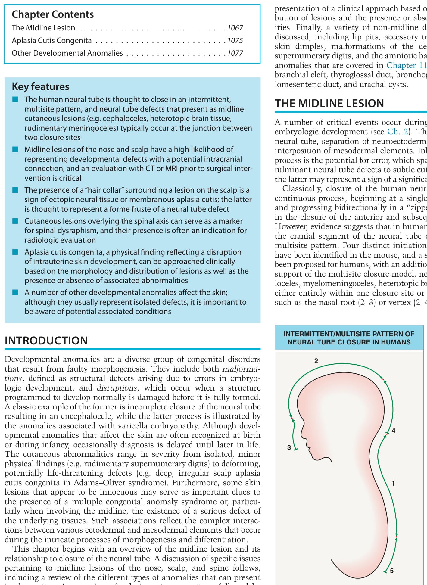
Fig. 64.1 Intermittent/ multisite pattern of neural tube closure in humans. The five separate initiation sites are labeled (in chronological order, 1–5), the direction of neural tube closure is indicated by the arrows, and the junctions (where defects tend to occur) designated with dots. The 2–3 junction corresponds to the location of frontoethmoidal neural tube defects, the 2–4 junction to parietal defects, and the 1–4 junction to occipital defects. Closure in region 4 is thought to occur via membranous growth, proceeding in a superior direction; incomplete closure at this site could therefore result in a membranous defect at the 2–4 junction (i.e. the vertex).
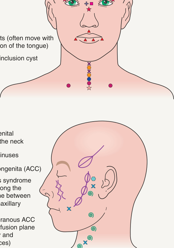
Fig. 64.2 Common sites of developmental anomalies of the face and neck.
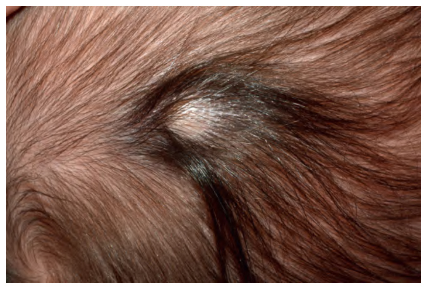
Fig. 64.3 Hair collar sign. Membranous aplasia cutis congenita with a hair collar.
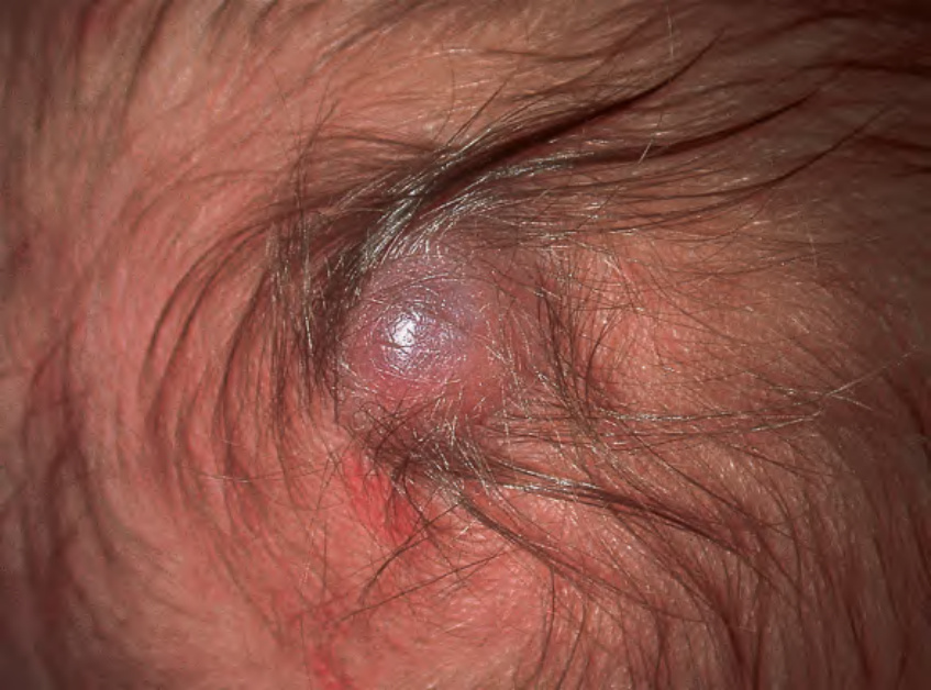
Fig. 64.4 Hair collar sign. Membranous aplasia cutis congenita with a bullous appearance and a surrounding hair collar.
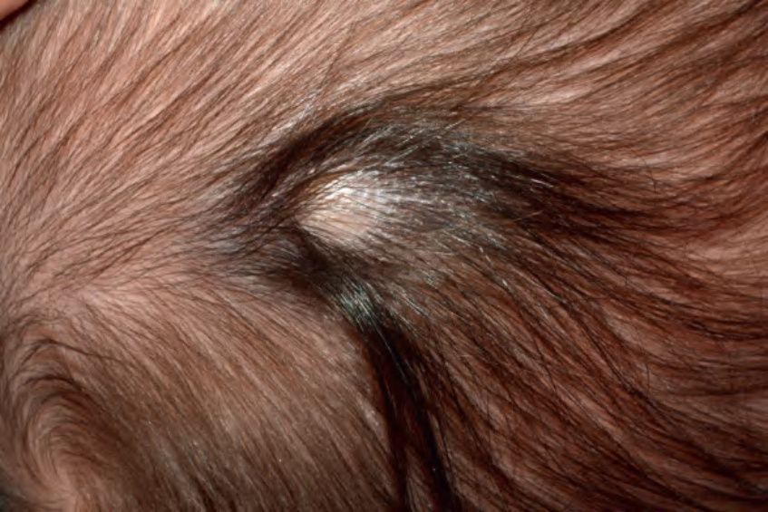
Fig. 64.5 Differential diagnosis and evaluation of a positive hair collar sign. In general, CT is the most accurate method of detecting skull defects. However, if a bony defect is detected by CT, a follow-up evaluation by MRI may be indicated to better determine whether there is extension of soft tissue transcranially.
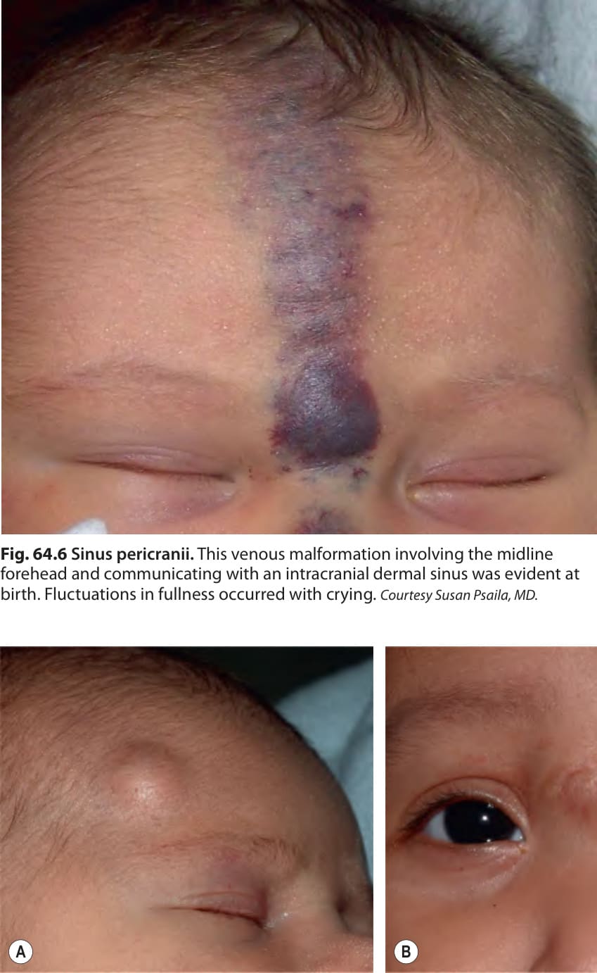
Fig. 64.6 Sinus pericranii. This venous malformation involving the midline forehead and communicating with an intracranial dermal sinus was evident at birth. Fluctuations in fullness occurred with crying. Courtesy Susan Psaila, MD.
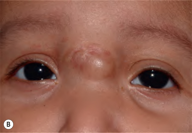
Fig. 64.7 Dermoid cysts.A This dermoid cyst presented in an infant as a firm subcutaneous nodule superior to the lateral right eyebrow. B Congenital firm, skin-colored nodule at the nasal root. Unlike those located in the more common lateral eyebrow location, nasal dermoid cysts have a substantial risk of intra-cranial extension.
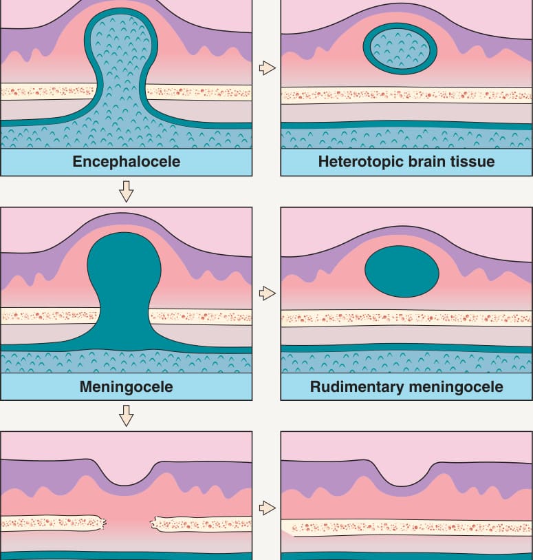
Fig. 64.8 Spectrum of cranial neural tube defects. When there is herniation of both the spinal cord and the meninges, this is referred to as a myelomeningocele.
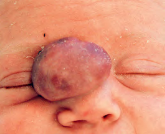
Fig. 64.9 Nasal glial heterotopia (nasal glioma). A mass was noted on prenatal ultrasound, and this reddish, slightly pedunculated, rubbery nodule was evident at birth. Courtesy Mary Chang, MD.
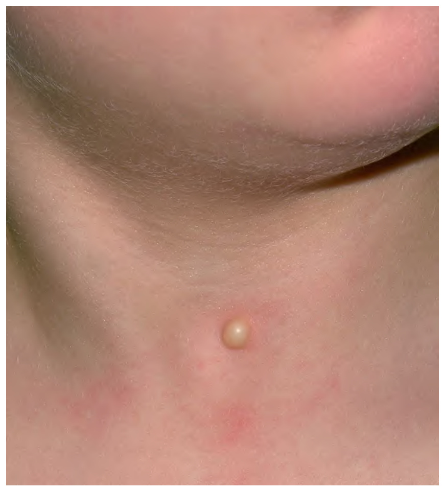
Fig. 64.10 Midline anterior neck inclusion cyst (MANIC) in an infant.Courtesy Antonio Torrelo, MD.
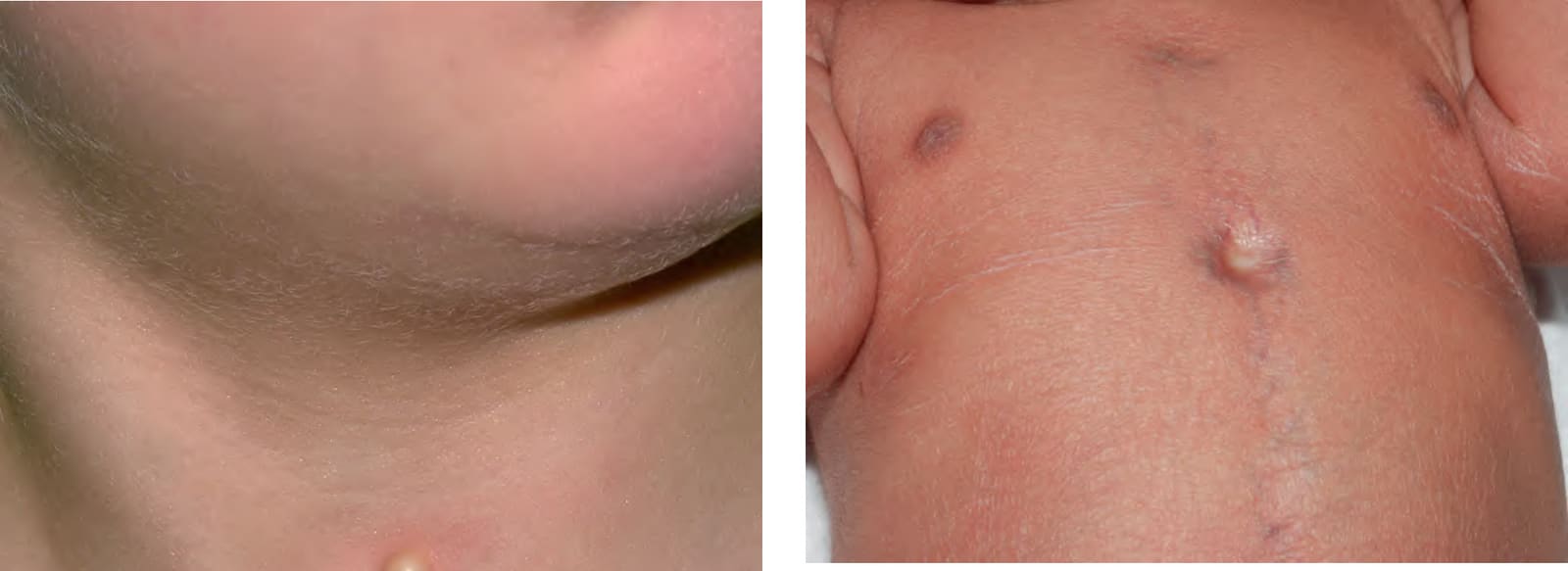
Fig. 64.11 Sternal cleft. Note the atrophic skin overlying the defect and prominent veins in the midline chest. Generalized desquamation is also evident in this 1-day-old post-term neonate. She did not develop an infantile hemangioma and had no cardiac defects or other features of PHACE syndrome.
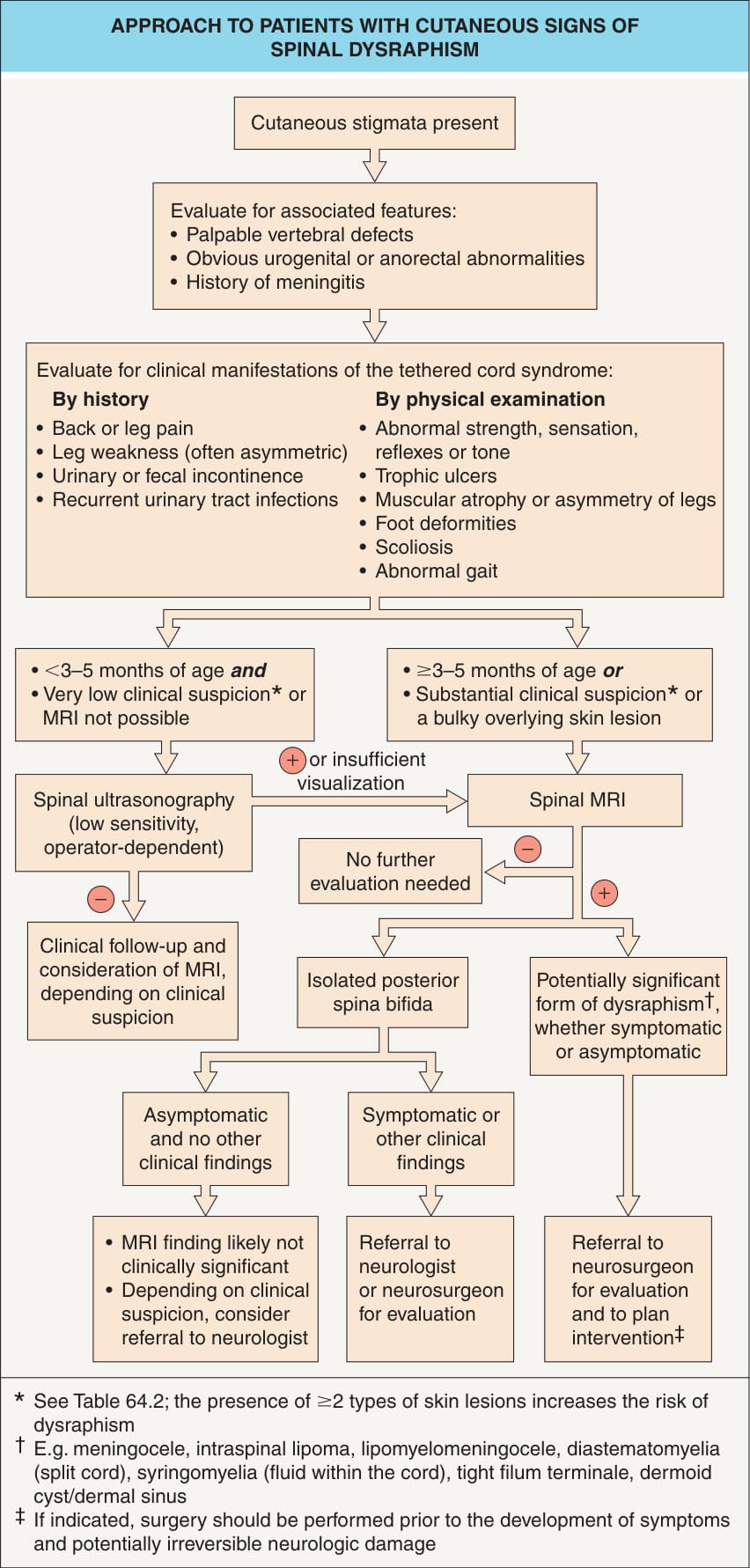
Fig. 64.12 Approach to patients with cutaneous signs of spinal dysraphism.
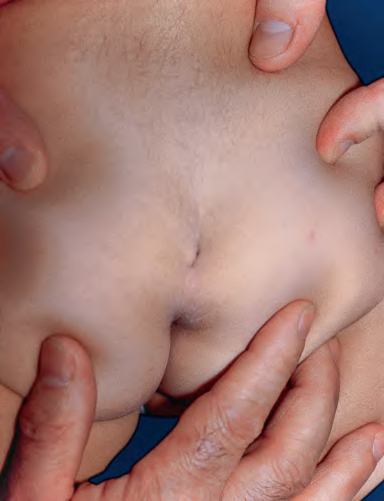
Fig. 64.13 Midline deep sacral dimple. The dimple was located above the gluteal cleft in association with a small tail. In contrast, a shallow dimple within the gluteal cleft is a common finding and not a sign of spinal dysraphism. Courtesy Seth J. Orlow, MD, PhD.
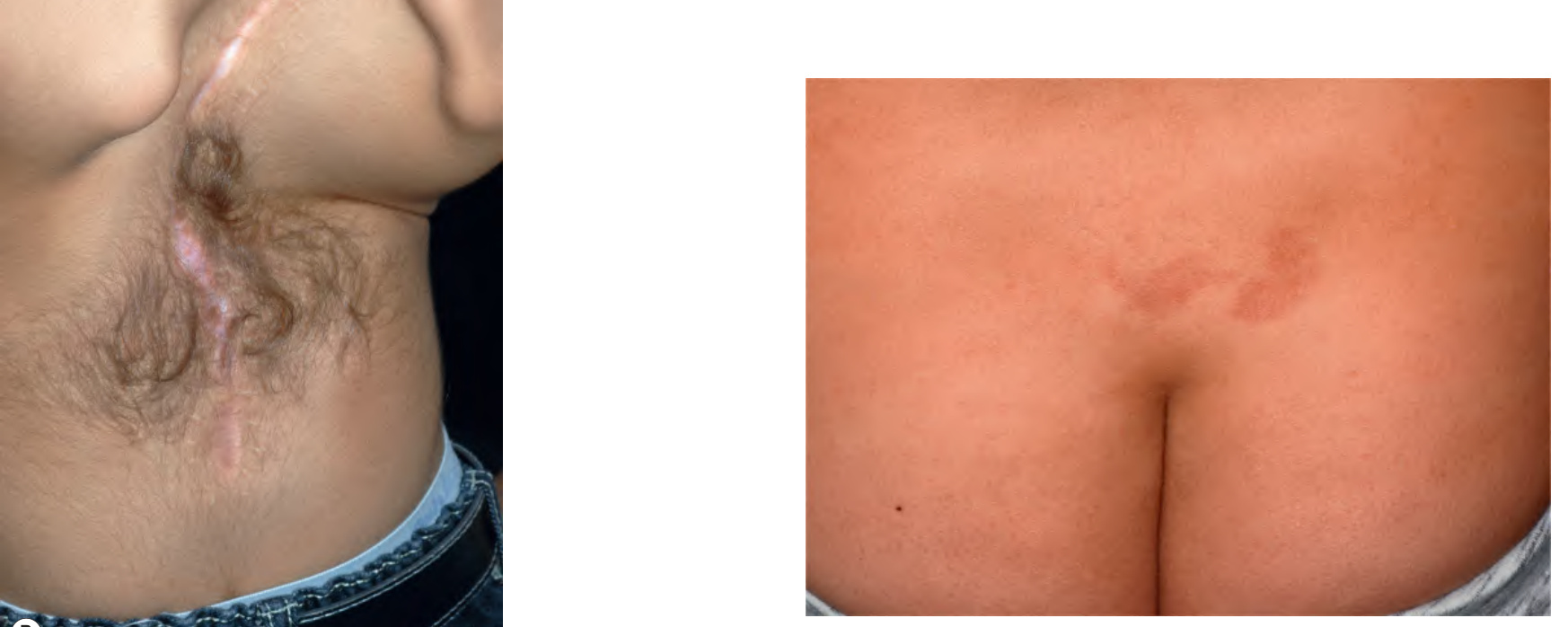
Fig. 64.14 Hypertrichosis associated with spinal dysraphism.
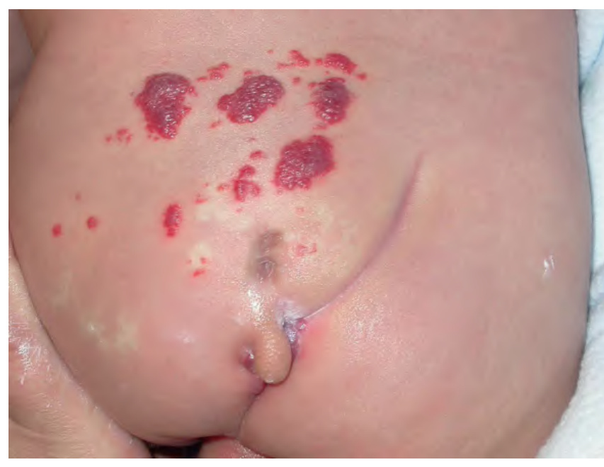
Fig. 64.15 Multiple midline cutaneous stigmata overlying the sacral spine in LUMBAR syndrome. This infant with a segmental infantile hemangioma in the lumbosacral area, a large atypical dimple, a pseudotail, and a deviated gluteal cleft associated with a subcutaneous lipoma had an underlying lipomyelomeningocele.
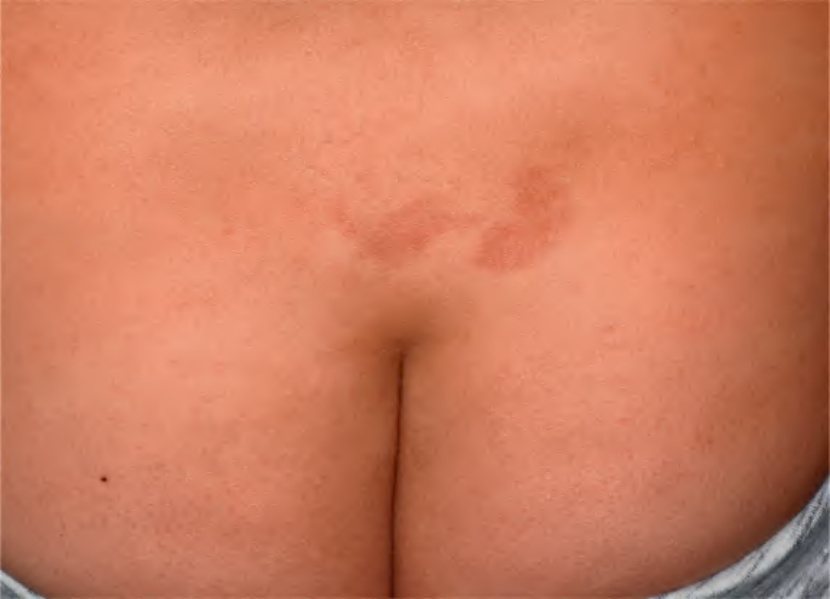
Fig. 64.16 Nevus simplex of the lumbosacral region. Note the characteristic “butterfly-like” shape. The likelihood of spinal dysraphism is extremely low if there are no other local cutaneous findings (e.g. deviated gluteal cleft/lipoma, focal hypertrichosis, dermal sinus/pit), especially if there are additional nevus simplex lesions elsewhere. and (occasionally) upper back. The current consensus is that regardless of spinal level, a nevus simplex without other local skin changes is rarely a sign of dysraphism, especially when there are nevus simplex lesions elsewhere. While additional case series of lumbosacral nevus simplex have supported this expert opinion, i.e. they have not found an association with spinal dysraphism, a prospective study of 3623 neonates found that 1 of the 25 individuals with nevus simplex had diastematomyelia revealed by ultrasonography. These neonates had no additional local skin findings, and it was not noted whether the individual with diastematomyelia had extrasacral vascular lesions.
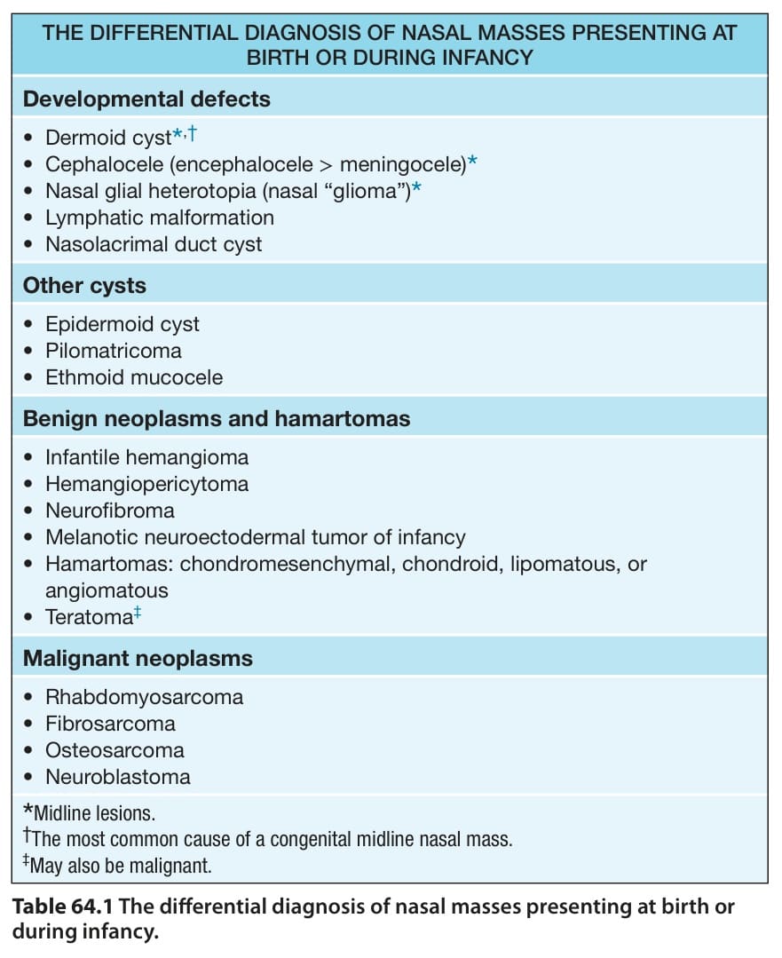
Table 64.1 The differential diagnosis of nasal masses presenting at birth or during infancy.
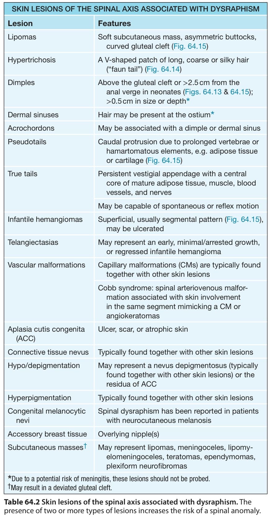
Table 64.2 Skin lesions of the spinal axis associated with dysraphism. The presence of two or more types of lesions increases the risk of a spinal anomaly.