REGULATION OF MELANIN BIOSYNTHESIS
This section begins with a review of the melanin biosynthetic pathway and then examines factors, both external and internal, that can
influence the level of melanin production. The “starting material” for the production of melanin, both the brown–black eumelanin and the yellow–red pheomelanin, is the amino acid tyrosine. The key regulatory enzyme in the pathway is tyrosinase, which controls the initial biochemical reactions in this pathway (Fig. 65.11). It should then come as no surprise that the initial investigations into the molecular basis of OCA focused on the gene that encodes tyrosinase.
In OCA1A, the form of OCA where mutations in both copies of the tyrosinase gene lead to complete loss of enzyme activity, no melanin is found in the hair, skin, or eyes (see Table 65.1). However, in OCA1B, where there is decreased enzyme activity, pheomelanin is produced, especially in the hair as the patient ages. The formation of pheomelanin requires less tyrosinase activity than does the formation of eumelanin and therefore the formation of pheomelanin can be thought of as a default pathway (see Fig. 65.11).
The activity of tyrosinase is enhanced by DOPA and is stabilized by tyrosinase-related protein 1 (TYRP1) (see below). Competitive inhibitors of tyrosinase activity include hydroquinone, which is used to treat disorders of hyperpigmentation such as melasma, and L-phenylalanine. In patients with phenylketonuria (PKU), there is a diffuse pigmentary dilution due to elevated levels of L-phenylalanine resulting from a deficiency in the enzyme L-phenylalanine hydroxylase that converts L-phenylalanine to L-tyrosine. The characteristic blonde hair of PKU can undergo darkening when the patient is on a low-phenylalanine diet. Of note, tyrosinase is a copper-requiring enzyme and it has two copper-binding sites. Rare cases of copper deficiency can lead to diffuse cutaneous pigmentary dilution, and in patients with Menkes disease, where a transmembrane Cu+-transporting ATPase that delivers copper to the trans-Golgi network and melanosomes is dysfunctional, the kinky hair is hypopigmented.
In a test tube, L-DOPA can spontaneously oxidize to form melanin, an insoluble biopolymer. For this reason, it was originally thought that tyrosinase was the sole enzyme involved in melanin biosynthesis. However, by the late 1970s, it was becoming clear that there were additional control points in the pathway (see Fig. 65.11). For example, dopachrome tautomerase, also known as tyrosinase-related protein 2 (TYRP2), which like TYRP1 shares similarities in its amino acid sequence with tyrosinase, converts DOPAchrome to 5,6-dihydroxyindole-2-carboxylic acid (DHICA). In mice and humans, TYRP1 stabilizes tyrosinase and mutations in both copies of TYRP1 lead to OCA3 (see Table 65.1).
Decreased function of yet another transmembrane protein, the P protein, leads to OCA2. Based upon its amino acid sequence, a prediction was made that the P protein was involved in the transport of small molecules across the membrane of the melanosome. Originally, tyrosine, the initial precursor in the melanin biosynthetic pathway, was considered the most likely candidate for transmembrane transport. However, the P protein is now thought to play a role in increasing the internal pH of the melanosome, as acidification can reduce the activity of tyrosinase. P protein may also regulate glutathione levels within the melanocyte’s intracellular compartments.
Melanins represent a group of complex polymers whose functions vary from camouflage to the quenching of oxidative free radicals generated
via exposure to UV radiation (UVR). The level and type of melanin production is a complex interplay of the activity of the various enzymes involved in the biosynthetic pathway as well as the activity of proteins such as the P protein and those that stabilize the activity of tyrosinase (e.g. TYRP1). Several factors are known to influence the activity of these key proteins of melanogenesis, and they include α-MSH, basic fibroblast growth factor (bFGF), endothelin-1, KIT ligand, and UVR (see below).
Among the multiple protein products of the proopiomelanocortin (POMC) gene are adrenocorticotropic hormone (ACTH), β-endorphin, and the three forms of MSH (α, β, and γ) (Fig. 65.12); in humans, α-MSH represents the major biologically active form of MSH. The primary site of expression of POMC is the pituitary gland; however, other sites of expression include the testis, the endothelium, and, of particular importance, epidermal keratinocytes. Although MSH is classically associated with the pigmentary system, this peptide has a wide range of biologic properties, including suppression of inflammation and regulation of body weight. For example, mutations in POMC can result in severe earlyonset obesity, adrenal insufficiency, and red hair. It then follows that mutations in the genes that encode the melanocortin receptors to which the protein products of POMC bind can lead to similar clinical findings; for example, mutations in MC4R are associated with morbid obesity and variant alleles in MC1R are associated with red hair (see below). Of note, subcutaneous afamelanotide (4-norleucyl-7-phenylalanine-α-MSH), which has enhanced binding to MC1R compared to α-MSH, can lead to cutaneous hyperpigmentation. When subcutaneous implants of afamelanotide were administered monthly, patients with erythropoietic protoporphyria (EPP) experienced fewer phototoxic reactions and could tolerate more direct sunlight without pain while patients with vitiligo had more rapid and extensive repigmentation in response to treatment with UVB. More recently, once-daily dersimelagon, a novel oral MC1R agonist, improved sun exposure tolerance in patients with EPP38a.
There are five major melanocortin receptors, all of which have seven transmembrane domains (Table 65.4; Fig. 65.13). Although the MC1R is present on a variety of cells within the skin, from endothelial cells to fibroblasts, the highest density of this receptor is found on melanocytes.
As in the case of the β-adrenergic receptor, the MC1R is a G-proteincoupled receptor, i.e. it uses proteins that bind guanosine triphosphate (GTP) and guanosine diphosphate (GDP) as intermediary messengers (Fig. 65.14). Following the binding of MSH to the MC1R, the protein pair interacts with a complex of G proteins. The GTP-Gsα subunit then activates adenylate cyclase, leading to increased production of cyclic adenosine monophosphate (cAMP) within the melanocyte. An increase in the intracellular concentration of cAMP leads to an increase in tyrosinase activity and eumelanin production via MITF (see Fig. 65.4). If the MC1R is dysfunctional and fails to initiate a significant rise in the intra-cellular level of cAMP, then pheomelanogenesis is favored (Fig. 65.15). Of note, the majority of individuals with red hair are compound heterozygotes or homozygotes for a variant R allele in the gene that encodes the MC1R (see Fig. 65.13). However, the variation in human hair color is complex, with over 120 genes identified.
MC1R also interacts with a protein known as the agouti protein (mouse) or agouti signaling protein (ASIP; human). Agouti is a term used to describe the banding of hairs seen in some mammals, including dogs, foxes and mice, that is due to alternating production of eumelanin and pheomelanin (Fig. 65.16). The production of agouti protein by the cells in the hair follicle papillae is cyclic, and, when the agouti protein is present, it effectively competes with MSH and the formation of pheomelanin within pheomelanosomes is favored (see Fig. 65.15). Compared to eumelanosomes, pheomelanosomes are characterized by a more spherical shape and the presence of an unstructured matrix with vesicular bodies.
An enhancement of pigment production can be seen following exposure of melanocytes to agents that increase intracytoplasmic levels of cAMP such as cholera toxin, forskolin, dibutyryl cAMP, and MSH (see Fig. 65.14). The activation of protein kinase A (PKA) by cAMP leads
to the phosphorylation of various proteins, which can result in their activation. One of the proteins that is phosphorylated by PKA is the cAMP response-element binding protein (CREB), which functions as a transcription factor, regulating the expression of other genes, including MITF (see Fig. 65.4). Patients with the McCune–Albright syndrome (polyostotic fibrous dysplasia) are mosaics for an activating mutation in the gene that encodes the G protein Gsα (see Fig. 65.14). As a result, the cAMP cascade is permanently “turned on” and, with continued transcription of CREB-controlled genes, there is hyperplasia of the bones and endocrine organs, albeit in a mosaic pattern. Presumably within the melanocytes of the segmental café-au-lait macules typical of this disorder, the increase in intracellular cAMP leads to increased tyrosinase activity and eumelanin production.
Exposure of melanocytes to phorbol esters, e.g. tetradecanoyl phorbol acetate (TPA), can lead to increased melanin formation via the activation of protein kinase C (PKC) (Fig. 65.17). Growth factors, including bFGF and KIT ligand, can also increase the pigment content within melanocytes whereas KIT receptor inhibitors (e.g. imatinib) can lead to hypopigmentation. Endothelin-1, a small peptide originally isolated from endothelial cells, is produced by keratinocytes and can lead to an increase in tyrosinase activity followed by an increase in melanin production.
In addition to ultraviolet radiation (see below), inflammation can lead to hyper- or hypopigmentation. Prostaglandin E/F and interleukin (IL)-33, mediators produced by both keratinocytes and fibroblasts, can lead to activation of melanocytes. Inhibitors of pigment production include interferon-γ, tumor necrosis factor (TNF), IL-4, and IL-6.
Multiple genes have been implicated as playing a role in normal pigment variation in humans, including: (1) MC1R and ASIP; (2) several genes that play a role in OCA, e.g. TYR, OCA2, TYRP1, SLC45A2, SLC24A5 (see Table 65.1); (3) KITLG which encodes KIT ligand; (4) TPCN2 which encodes an ion channel transporter; and (5) IRF4 which encodes interferon regulatory factor 4. The presence of a variant (rather than the African ancestral) allele of SLC24A5, whose protein product is a putative cation exchanger in the melanosomal membrane, correlates with lighter skin color. In addition, variants in OCA2 are thought, at least in part, to determine the normal phenotypic variation in human eye color. A haplotype with several polymorphisms in ASIP (and presumably a gain of ASIP function) has been associated with red or blonde hair, freckling, and a tendency to burn, while variants that lead to destabilized ASIP mRNA have been associated with darker skin phototypes. A polymorphism in the regulatory region of KITLG that reduces its responsiveness to the WNT-activated transcription factor LEF1 is associated with blonde hair, and in mice, decreased lef1 leads to light-colored hair. Of note, some loss-of-function mutations in
MC1R confer a risk for developing cutaneous melanoma, independent of pigmentary phenotype.
In humans, graying or whitening of hair is a normal aging phenomenon. There is evidence that reactive oxygen species accumulate within affected hair follicles and lead to oxidative damage of hair follicle melanocytes. Hair bulb melanocytes are thought to undergo apoptosis first, followed by depletion of precursor cells in the bulge region. However, more recent studies of graying hairs have pointed to a reduction in the migration of melanocyte stem cells from the bulge region49a. Hair bulb melanocytes that disappear fail to express TYRP2 and SOX10. The role of SCF production by KROX20+ cells within the hair bulb was discussed previously. Lastly, hyperactivation of sympathetic nerves has been shown to drive depletion of melanocyte stem cells.
Ultraviolet Radiation (UVR)
Following a single exposure to UVR, an increase in the size of melanocytes can be observed, along with an increase in tyrosinase activity.
Repeated exposures to UVR lead to an increase in the number of stage IV melanosomes transferred to keratinocytes, as well as an increase in the number of active melanocytes. When chronically sun-exposed sites (e.g. the upper outer arm) are compared with non-sun-exposed sites (e.g. the upper inner arm), the density of melanocytes is up to two times greater in sun-exposed sites. Melanocytes, like other neural-derived tissues, have a low mitotic rate, and whether this increase in number represents an increase in mitotic rate or an activation of “inactive” melanocytes or melanocyte precursors is not known. Senescent fibroblast-derived GDF15 may play a role in the hyperpigmentation of photoaged skin via β-catenin signaling.
Following exposure to UVA irradiation, an immediate pigmentary darkening can be observed, which occurs within minutes and fades over 20–30 minutes. It is clinically most obvious in darkly pigmented skin and is thought to represent oxidation of pre-existing melanin or melanin precursors. Given its transient nature, it does not provide photoprotection. Delayed tanning is visible within 24–72 hours after exposure to UVB and UVA radiation and represents new pigment production via an increase in tyrosinase activity. In addition to an increase in melanocyte
size and number, tyrosinase activity, and transfer of melanosomes to keratinocytes, the response to PUVA includes an alteration in size and aggregation pattern of melanosomes, i.e. from smaller and grouped to larger and singly dispersed (see Table 65.3).
UVR may work by increasing one or more of the following:
●transcription of the tyrosinase gene (via MITF)
●the number or activity of MC1R on melanocytes
●the expression of POMC and its derivative peptides by keratinocytes and several cell types within the dermis (e.g. endothelial cells, sebocytes, lymphocytes)
●the release of diacylglycerol from the plasma membrane, which activates protein kinase C
●an activation of the nitrous oxide/cGMP pathway
●the production of cytokines and growth factors by keratinocytes (e.g. endothelin-1)
●the induction of an SOS response to UVR-induced DNA damage
●transactivation of the POMC promoter by p53
●the ratio of kinesin to dynein, affecting melanosome transport (see Fig. 65.10).
The inability of the majority of red-haired individuals to develop a tan following exposure to UVR can be explained, at least in part, by dysfunction of their melanocyte MC1R. This phenomenon, along with the production of oxygen radicals following the UV irradiation of pheomelanins, probably contributes to the increased incidence of both cutaneous melanoma and keratinocyte carcinomas in persons with red hair. In addition, chemiexcitation of melanin derivatives induces DNA photoproducts long after UV exposure. Based upon
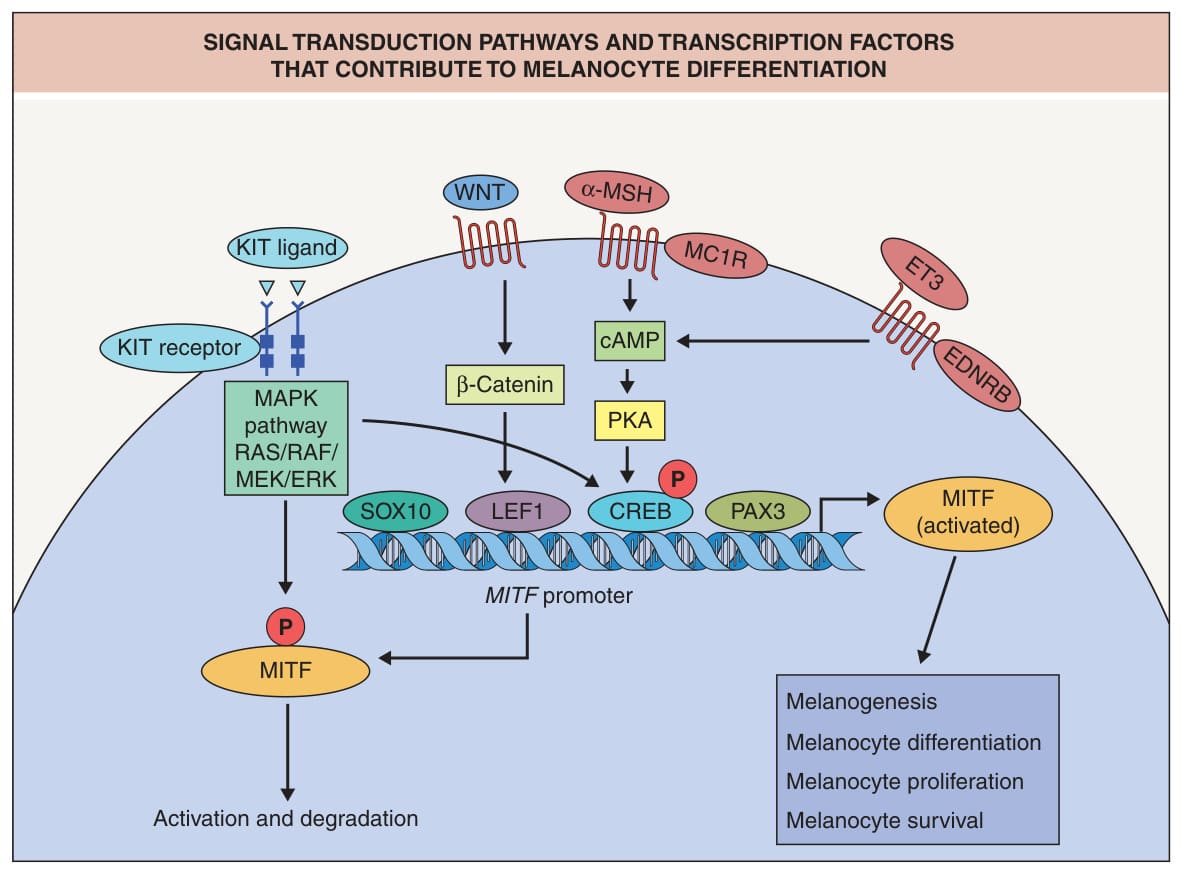
Fig. 65.4 Signal transduction pathways and transcription factors that contribute to melanocyte differentiation. MITF expression is activated early on during the transition from pluripotent neural crest cells to melanoblasts and is required for melanoblast survival; mutations in MITF lead to Waardenburg syndrome, a classic neurocristopathy. MITF also regulates the expression of multiple pigment genes including those that encode tyrosinase, TYRP1, TYRP2, PMEL/PMEL17/gp100, and MART-1/Melan-A. Additional transcriptional targets are CDK2, CDKN2A, and BCL-2 (whose protein product is an inhibitor of apoptosis). Small molecule inhibitors of SIK (salt-inducible kinase) can upregulate MITF, and application of these inhibitors to normal human skin led to an increase in pigmentation. WNT signaling in melanocyte stem cells is critical for hair pigmentation. Details of how activation of G-protein-coupled receptors leads to an increase in intracellular cAMP is shown in Fig. 65.14. Of note, EDNRB interacts with the G proteins GNAQ and GNA11, and activating mutations in the genes that encode these latter two proteins can lead to blue nevi and phakomatosis pigmentovascularis. EDNRB can also activate the MAPK pathway. Additional receptors include ET1 (activates protein kinase C), hepatocyte growth factor receptor, and GM-CSF receptor; the latter two activate the MAPK pathway. cAMP, cyclic adenosine monophosphate; CREB, cAMP response-element binding protein; ET3, endothelin-3; EDNRB, endothelin receptor type B; LEF1, lymphoid enhancer binding factor 1; MAPK, mitogen-activated protein kinase; MC1R, melanocortin 1 receptor; MITF, microphthalmia-associated transcription factor; MSH, melanoctye stimulating hormone; P, phosphorylation; PKA, protein kinase A; SOX10, SRY-box containing gene 10.
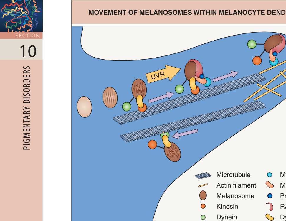
Fig. 65.10 Movement of melanosomes within melanocyte dendrites. As more melanin is deposited within the melanosomes, they migrate into the dendrites along microtubules in preparation for their transfer into neighboring keratinocytes. Kinesin and dynein serve as molecular motors for microtubule-associated anterograde and retrograde melanosomal transport, respectively, and UVR results in augmented anterograde transport via increased kinesin and decreased dynein activity. Myosin Va, which is linked to the melanosomal RAB27A GTPase by melanophilin, captures mature melanosomes when they reach the cell periphery and attaches them to the actin cytoskeleton.
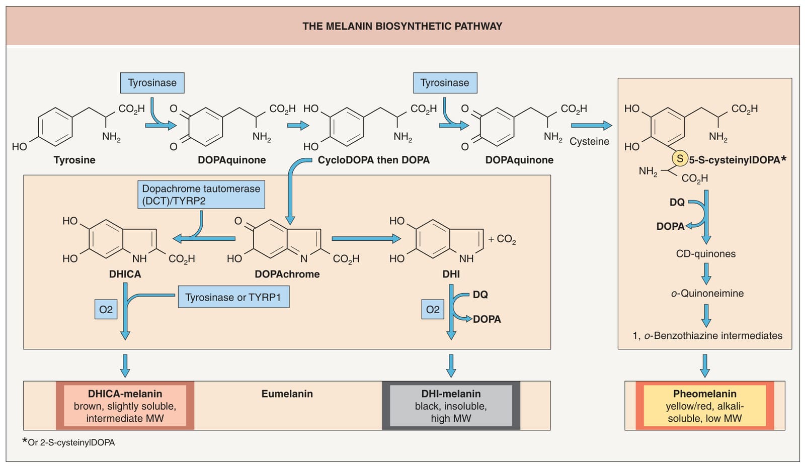
Fig. 65.11 The melanin biosynthetic pathway. The pathway includes the sites of dysfunction in OCA1 (tyrosinase) and OCA3 (TYRP1). The two major forms of melanin in the skin and hair are brown–black eumelanin and yellow–red pheomelanin. The enzymes are transmembrane proteins located within the melanosome. DHI, 5,6-dihydroxyindole; DHICA, 5,6-dihydroxyindole-2-carboxylic acid; DOPA, dihydroxyphenylalanine; MW, molecular weight; TYRP, tyrosinase-related protein. Adapted from Hearing VJ. Determination of melanin synthetic pathways. J Invest Dermatol 2011;131:E8–E11.
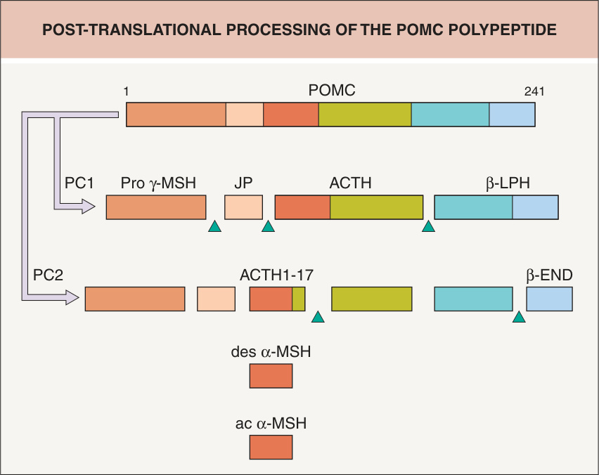
Fig. 65.12 Post-translational processing of the POMC polypeptide. Pituitary hypersecretion of ACTH and/or α-MSH can lead to generalized hyperpigmentation in patients with Addison disease. ac, acetylated; ACTH, adrenocorticotropic hormone; des, desacetyl; END, endorphin; JP, joining peptide; LPH, lipotropic hormone; MSH, melanocyte stimulating hormone; PC, prohormoneconverting enzyme; POMC, proopiomelanocortin.
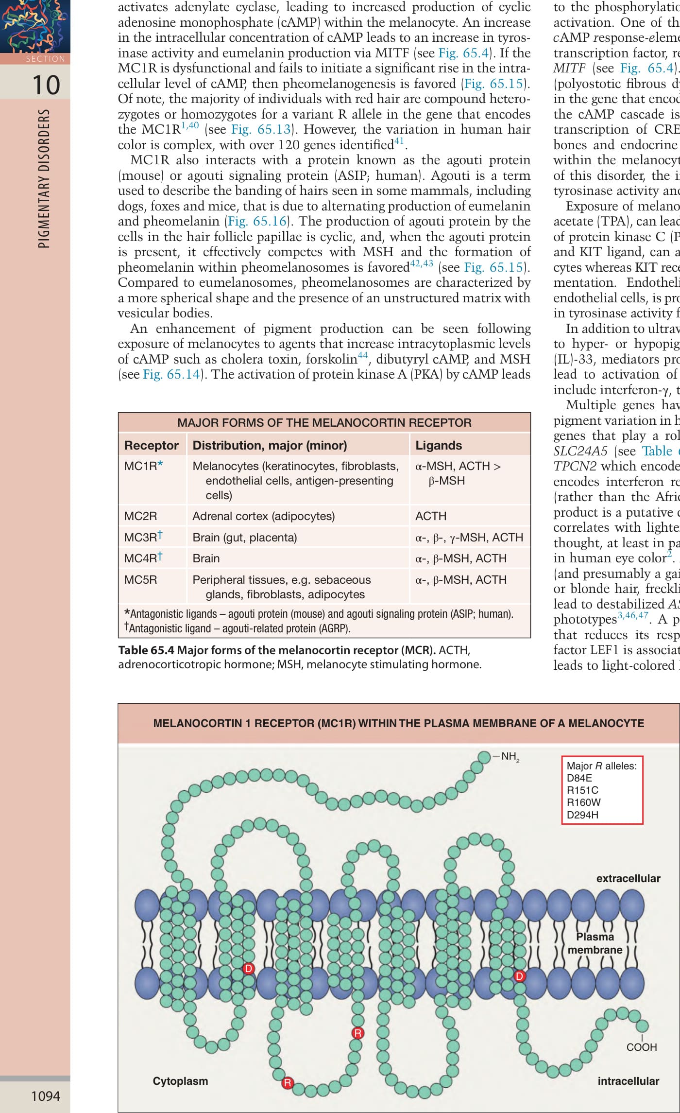
Fig. 65.13 Melanocortin 1 receptor (MC1R) within the plasma membrane of a melanocyte. MC1R has seven transmembrane domains and is a G-coupled receptor (see Fig. 65.14). There are numerous genetic variants of the MC1R. The null or hypomorphic R alleles D84E, R151C, R160W, and D294H have red hair odds ratios of 62, 118, 50, and 94, respectively; the low penetrance r alleles of V60L, V92M, and R163Q have red hair odds ratios of 6, 5, and 2, respectively. In general, R/R individuals have red hair and lightly pigmented skin.
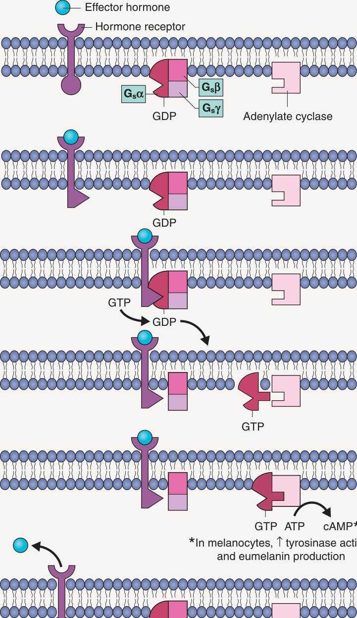
Fig. 65.14 Activation of a G-protein-coupled receptor such as the melano- cortin 1 receptor (MC1R). In the case of the MC1R, the increase in the intra-cellular concentration of cAMP leads to an increase in tyrosinase activity and eumelanin production. GDP, guanosine diphosphate; GTP, guanosine triphosphate; P, phosphate group; S, stimulatory. Adapted from Alberts B. Molecular Biology of the Cell. Garland Publishing; 1989.
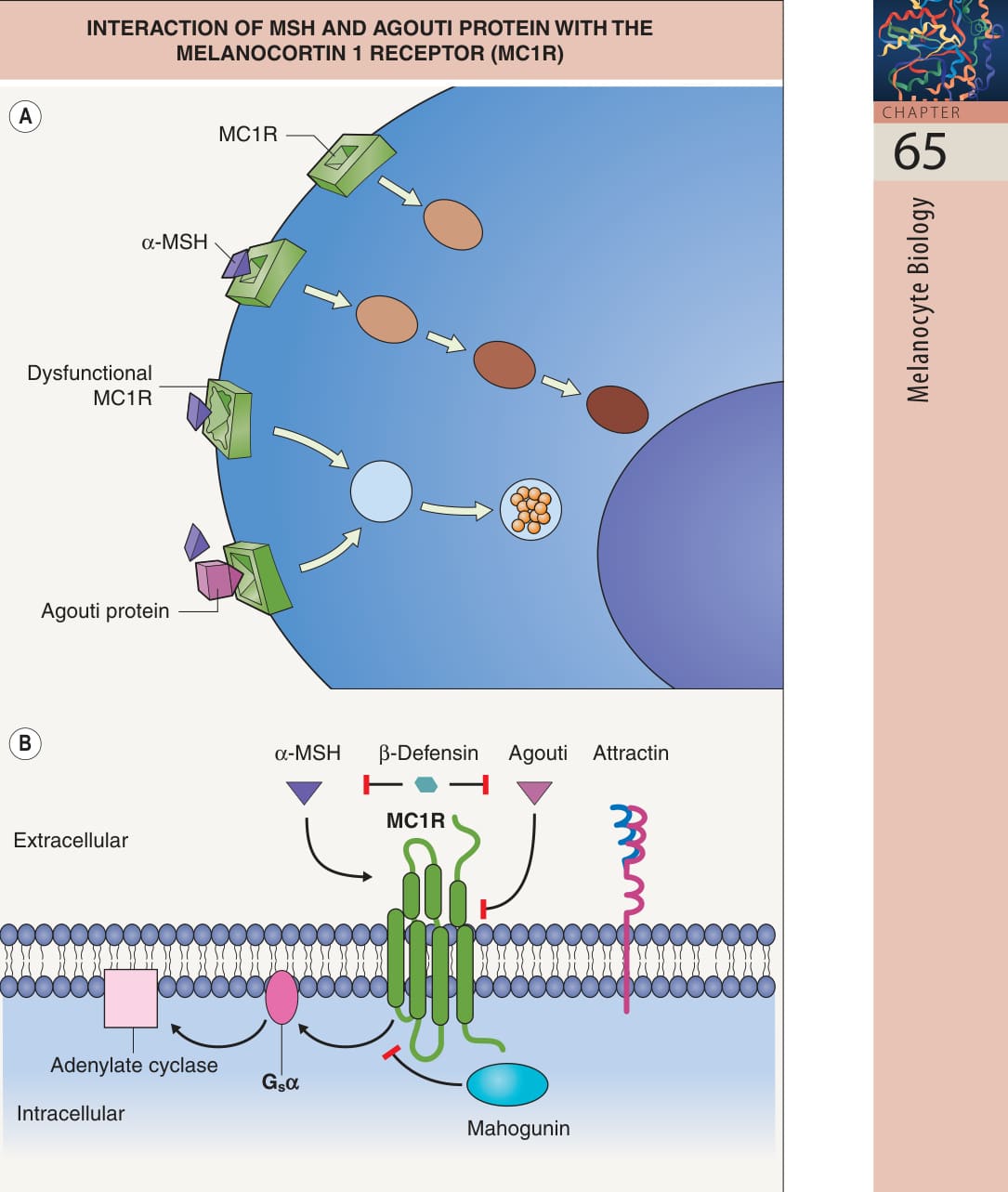
Fig. 65.15 Interaction of melanocyte stimulating hormone (MSH) and agouti protein with the melanocortin 1 receptor (MC1R).A There is baseline activity of the MC1R, enhanced by binding with MSH. Agouti protein represents an antagonist ligand for the MC1R whose binding can lead to pheomelanogenesis. Dysfunction of the MC1R can also lead to pheomelanogenesis, as in the case of humans with red hair. B Interactions are actually more complex, in that to be fully effective, agouti requires attractin and mahogunin; the former aids in the binding of agouti to MC1R while the latter acts on the cytosolic side. There is also a neutral agonist, β-defensin, which interferes with both agonist and antagonist binding. Adapted from Schiaffino MV. Signaling pathways in melanosome biogenesis and pathology. Int J Biochem Cell Biol 2010;42:1094–104.

Fig. 65.16 Formation of agouti hairs – underlying physiology.A Fox hairs with alternating eumelanin and pheomelanin production within individual hairs, a pattern referred to as agouti. B Explanation for the agouti pattern based on different ligands interacting with the melanocortin 1 receptor (MC1R). Both gainof-function mutations at the agouti locus and lossof-function of MC1R can lead to yellow-haired mice. In the CNS, an abundance of agouti protein leads to obesity, and yellow mice with gain-of-function mutations at the agouti locus are also obese.
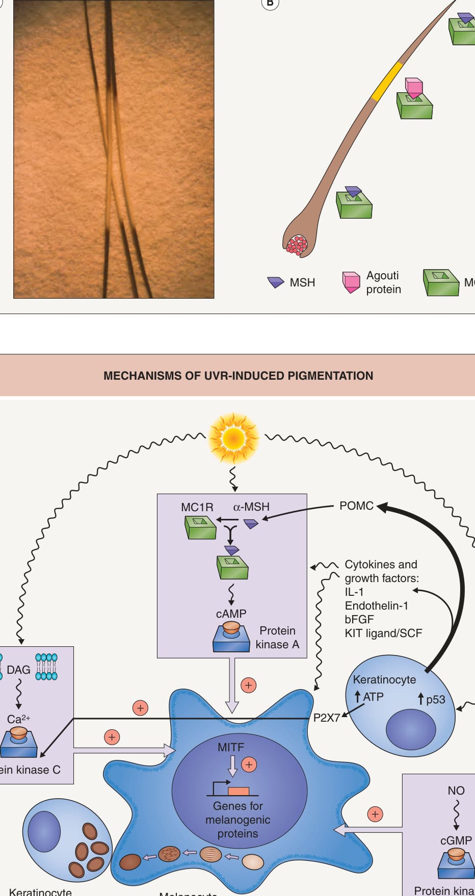
Fig. 65.17 Mechanisms of UVR-induced pigmen- tation. These include an increase in one or more of the following: (1) expression of proopiomelanocortin (POMC) and its derivative peptides by cells within the skin, in particular keratinocytes; (2) the number of melanocortin 1 receptors (MC1R) on melanocytes; (3) the release of diacylglycerol (DAG) from the plasma membrane, which activates protein kinase C; (4) the induction of an SOS response to UVR-induced DNA damage; (5) nitric oxide (NO) production, which activates the cGMP pathway; and (6) production of cytokines and growth factors by keratinocytes. As a result, there is enhanced transcription of the genes that encode microphthalmia-associated transcription factor (MITF) and melanogenic proteins including tyrosinase, tyrosinase-related protein 1 (TYRP1), TYRP2, and PMEL/PMEL17/gp100. In addition, melanocyte dendricity and transfer of melanosomes to keratinocytes is stimulated via increased activity of Rac1 (involved in dendrite formation), the ratio of kinesin to dynein, and expression of proteaseactivated receptor-2 (PAR-2; involved in melanosome transfer). Increased POMC expression in keratinocytes followed by processing to β-endorphin (in addition to α-MSH) may play a role in addiction to ultraviolet light. ATP, adenosine 5′-triphosphate; P2X7, purinergic receptors type 2 X7; TPA, tetradecanoyl phorbol acetate.
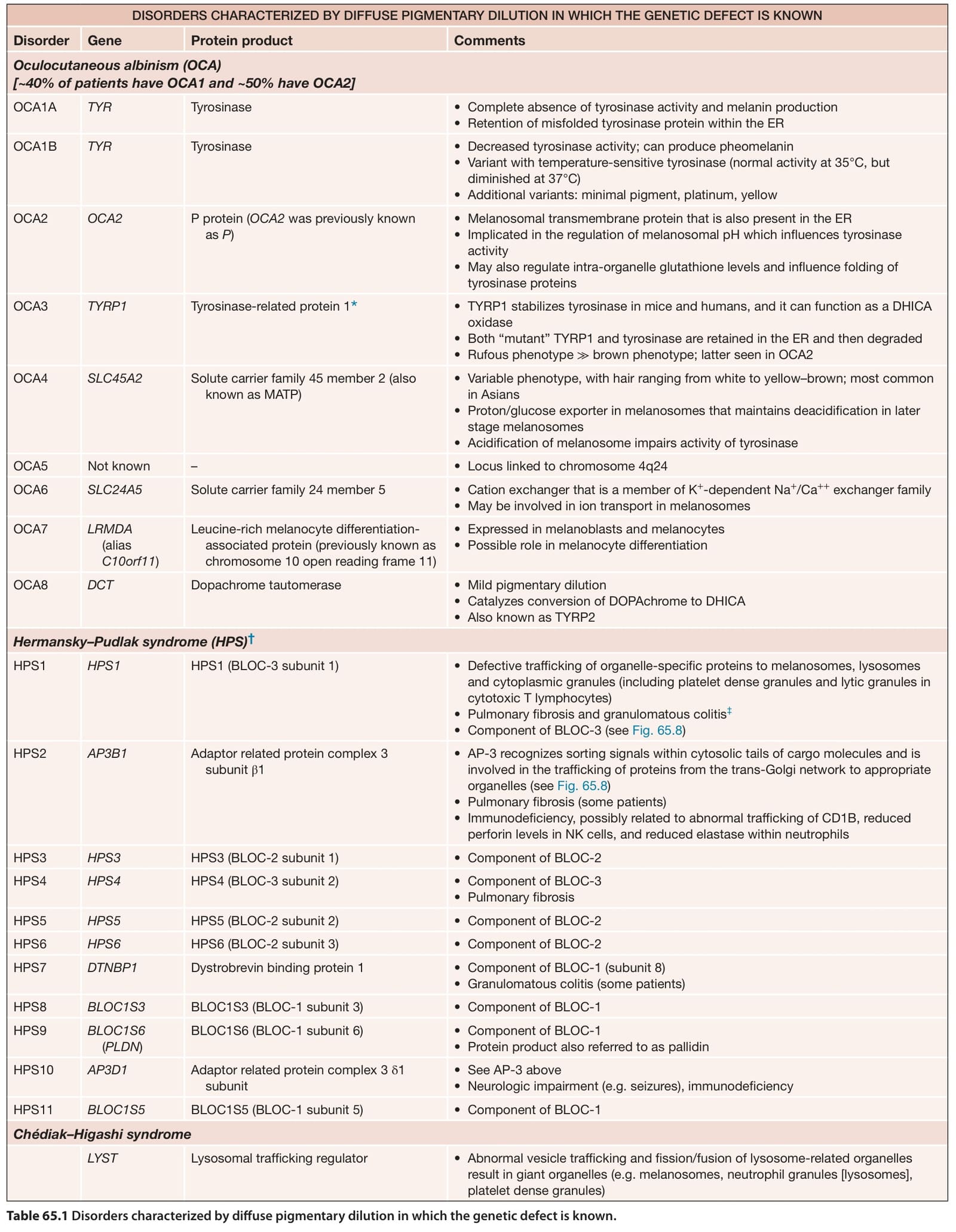
Table 65.1 Disorders characterized by diffuse pigmentary dilution in which the genetic defect is known.
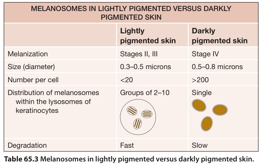
Table 65.3 Melanosomes in lightly pigmented versus darkly pigmented skin.
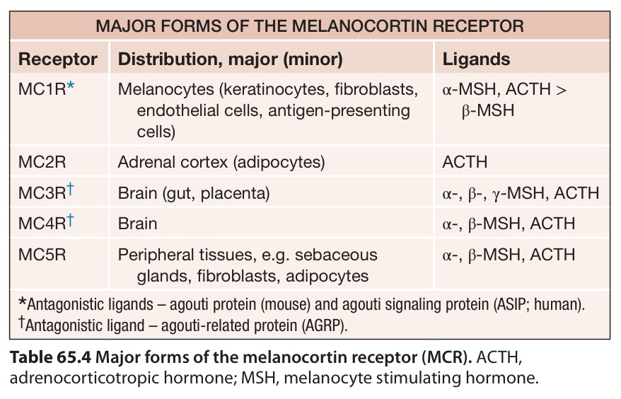
Table 65.4 Major forms of the melanocortin receptor (MCR). ACTH, adrenocorticotropic hormone; MSH, melanocyte stimulating hormone.
a large genome-wide association study in individuals of European ancestry, the ability to tan was associated with 20 loci, including ten that were not previously associated with pigment-related phenotypes.
Additional figure and table on Genes associated with physiologic variation in human pigmentation, available in our eBook (see inside front cover for access code).