TRYPANOSOMIASIS
Synonyms: Chagas disease: American trypanosomiasis, Chagas– Mazza disease African trypanosomiasis: African sleeping sickness
Although the genus Trypanosoma consists of at least 20 species of protozoa, only two are known to cause human disease. Chagas disease (American trypanosomiasis) is caused by T. cruzi infection and transmitted by reduviid bugs, while African trypanosomiasis is caused by T. brucei infection and transmitted by tsetse flies.
Chagas Disease
Key features
An endemic disease in many areas of Central and South America
Transmission of T. cruzi is via infected reduviid bugs and occasionally by contaminated blood
A serious systemic disease with three phases (acute, indeterminate, and chronic). The chronic phase primarily affects the heart and gastrointestinal tract
Following inoculation of the parasite into the skin, localized erythema and edema (chagoma) can develop in conjunction with regional lymphadenopathy
When the conjunctiva is the portal of entry, edema of the palpebral and periocular tissue is seen (Romaña sign)
Epidemiology
T. cruzi and its arthropod vectors (reduviid or kissing bugs) are found in the Americas, from the southern US to southern Argentina (Fig. 83.15). Reduviid bugs feed mainly on human, animal, or avian blood, but they sometimes engage in cannibalism or coprophagia, which may result in vector-to-vector transmission of T. cruzi. A favorite habitat for these bugs is cracks in the walls of mud huts in poor rural areas, and they are known as kissing bugs because of their habit of biting the face of humans. Direct transmission of T. cruzi via transfusion of contaminated blood can occur; since 2007, blood donated in the US has been screened for this organism. In addition, transplacental transmission can result in congenital infection.
Adapted with permission from Nabarro L, Morris-Jones S, Moore DAJ: Peters’ Atlas of Tropical Medicine and Parasitology, 7th edition. London: Elsevier, 2019; and with data from Bern C. Chagas’ Disease. N Engl J Med 2015;373:456–66.
It is estimated that ∼6 million people in the Americas are infected with T. cruzi, including <300 000 individuals in the US, especially immigrants from Latin America living in Texas and California. Chagas disease kills ~10 000 people per year, mostly in rural areas of Latin America; it represents the leading cause of parasitic death in Latin America and the third leading cause in the world. The acute form of the disease (see below) is seen more commonly in children, and, in some areas, 85% of children under the age of 10 years are infected.
Pathogenesis
Blood-sucking reduviid bugs ingest trypomastigotes from the peripheral blood of vertebrate hosts (Fig. 83.16). Once the bug has become infected, it remains so for the rest of its life. The parasites transform into epimastigotes and multiply in the bug’s midgut, later becoming metacyclic trypomastigotes in its hindgut. When the infected bug takes a subsequent blood meal, it usually defecates during or after feeding, depositing infective metacyclic trypomastigotes on the skin. The parasites penetrate the skin (generally via the bite wound) or mucosa (usually the conjunctiva). Following invasion of the host’s cells, amastigotes multiply within the cytoplasm and then develop into trypomastigotes. The latter are released when the cells rupture and then can either infect other cells or enter the bloodstream, where they become the source of infection for new vectors. T. cruzi can also be transmitted transplacentally as well as through blood transfusions, organ transplantation, laboratory accidents, and occasionally contaminated food.
The autonomic nervous system that innervates the heart and gastrointestinal tract appears to be a primary target of T. cruzi infection. The visceral involvement seen in the chronic stage (see below) is thought to reflect neuronal damage during the acute phase. A role for autoimmunity in the chronic phase of the disease has also been hypothesized. In addition, trypomastigotes have the ability to penetrate the smooth muscle of the heart and the wall of the gastrointestinal tract.
Clinical features
Chagas disease has acute and chronic phases with distinct clinical manifestations. Although the acute phase can affect individuals of any age, it is usually observed in children and tends to be more severe in those ≤2 years of age. At the cutaneous site of parasitic entry (see Fig. 83.16), some patients develop a chagoma, which is a localized area of erythema and swelling that is often accompanied by regional lymphadenopathy. When the conjunctiva is the portal of entry, the painless edema of the palpebral and periocular tissues that appears is referred to as the Romaña sign or ophthalmoganglionar complex (Fig. 83.17). The Romaña sign occurs in ~80% of individuals infected by this route. In
Young child with unilateral periorbital edema characteristic of this disease (Romaña sign) when the conjunctiva is the portal of entry. Courtesy Omar P. Sangüeza, MD.
some patients, the swelling is accompanied by preauricular lymphadenopathy (Parinaud sign).
These initial clinical findings can be accompanied by fever, malaise, anorexia, and edema of the face and lower extremities. Some patients develop erythematous macules, hepatosplenomegaly, thrombocytosis, leukocytosis, and/or eosinophilia. The acute phase may last for up to 2 months but usually resolves spontaneously. In addition, inflammation at the sites of parasite entry can persist for several weeks. Occasionally, patients develop life-threatening myocarditis or meningoencephalitis.
After resolution of the acute phase, patients enter the indeterminate phase of Chagas disease. This stage is characterized by asymptomatic parasitemia and positive serology. Then, many years or even decades after the initial infection, 30% of patients develop symptoms of chronic Chagas disease. The heart is the organ most commonly affected and the clinical manifestations include congestive heart failure, arrhythmias, and heart block. The gastrointestinal tract is also frequently involved, leading to megacolon and megaesophagus.
If patients who chronically harbor T. cruzi become immunosuppressed, reactivation of the disease can occur. When this happens, the disease is often more severe than in typical cases of Chagas disease, and patients may develop nodules and plaques that favor the lower extremities.
Pathology
Histologically, at the site of parasite entry (chagoma), there is interstitial edema and a mononuclear cell infiltrate. Within cells in the subcutaneous tissue and muscle, aggregates of amastigotes may be seen. Enlarged lymph nodes are hyperplastic and intracellular amastigotes can be observed. In the chronic stage, trypanosomes are rare in affected organs.
Diagnosis and differential diagnosis
The diagnosis of acute Chagas disease is typically based upon the microscopic identification of either trypomastigotes in the blood (or CSF) or amastigotes within tissue. Because the concentration of parasites is low even in the acute stage, hemoconcentration techniques are used to enhance the yield. PCR currently represents the most sensitive diagnostic method for acute disease, with positive results days to weeks earlier than microscopy; PCR is especially useful for the diagnosis of infections acquired vertically, via organ transplantation, or after laboratory exposure. Cultures of blood, CSF, or tissue are less sensitive for detection of acute T. cruzi infection.
Serologic tests such as ELISAs and immunofluorescent antibody assays (IFAs) are used to establish the diagnosis of chronic Chagas disease. PCR-based detection has variable sensitivity and limited utility for chronic Chagas disease, although it may serve as an indicator of treatment failure or disease reactivation. Xenodiagnosis represents a specific and fairly sensitive (≤50%) method, and it can be used to confirm a serologic diagnosis; however, this technique is expensive and is only available in large laboratories in endemic areas. For xenodiagnosis, trypanosome-free reduviid bugs are allowed to feed on patients and the feces or hindgut contents of the bugs are examined for epimastigotes 10 to 30 days later.
The nonspecific symptoms that may occur during the acute phase of Chagas disease, such as a macular eruption and influenza-like symptoms, make the differential diagnosis broad. When the site of inoculation is the eye, other entities to consider include a reaction to a bite from another type of insect, angioedema, and bacterial cellulitis.
Treatment
Treatment is most effective early in the course of Chagas disease. Benznidazole and nifurtimox (both available through the CDC) can reduce the severity and duration of symptoms associated with the acute phase, with a parasitologic cure in 60%–90% of patients. Because benznidazole is better tolerated, it is favored by most experts. The side effects of these two medications are reviewed in Table 83.8.
Benznidazole, allopurinol, itraconazole, and posaconazole have also been evaluated for the treatment of chronic Chagas disease.
Although these agents can decrease serum parasite load, to date, there is no evidence that pathologic changes or progression of the chronic phase of this disease can be reversed or stopped by any therapy. Thus, chronic disease is managed primarily by supportive care (e.g. antiarrhythmics).
Prevention of the infection involves education about sanitation and the use of residual insecticides in domestic areas. Vaccines are being investigated but are not currently available. Cyanotriazoles, a new class of antiparasitic drugs that inhibit topoisomerase II, may be of utility for the treatment of trypanosomiasis and leishmaniasis.
African Trypanosomiasis
Key features
In West Africa, the disorder is due to Trypanosoma brucei gambiense infection, and in East Africa it is caused by Trypanosoma brucei rhodesiense infection
The vectors are several species of tsetse flies
Cutaneous manifestations include a localized bite reaction
(“trypanosomal chancre”) and an annular erythematous eruption that coincides with fever spikes
Epidemiology
The incidence of African trypanosomiasis decreased from <10 000 cases in 2009 to ~750 in 2021. This disease is almost invariably fatal if left untreated and occurs in two forms that are named based on their separate geographic distributions in Africa (see Fig. 83.15). The West African form, which is due to Trypanosoma brucei gambiense and accounts for >95% of cases, is a chronic anthroponotic disease with primarily neurologic features. Since 2020, elimination of West African trypanosomiasis has been achieved in several countries, including Togo, Côte d’Ivoire, Benin, Uganda, and Equatorial Guinea. The East African form, caused by Trypanosoma brucei rhodesiense, is an acute zoonotic disease (with antelopes as a reservoir) that is often fatal even with treatment. Fortunately, T. b. rhodesiense only occasionally infects humans, mainly farmers, hunters, and tourists.
Both types of T. brucei are transmitted to humans by blood-sucking tsetse flies (genus Glossina), which are found in Africa. The flies become infected by ingesting blood from infected mammalian hosts (Fig. 83.18). Transmission transplacentally or via blood products may also potentially occur.
Pathogenesis
After inoculation, trypanosomes may produce a local chancre before spreading via the lymphatics to regional (and then distant) lymph nodes. The organisms disseminate to the bloodstream and eventually to the CNS, and they multiply by binary fission. They escape significant immunologic response by continuously changing their surface glycoproteins.
Clinical features
An indurated, painful reaction develops at the portal of entry of the organisms, referred to as a “trypanosomal chancre” (Fig. 83.19). This is seen more commonly (70%–80% of cases) in the East African form due to T. b. rhodesiense. The lesion appears 1–2 weeks after the tsetse fly bite and heals spontaneously over several weeks.
The first (hemolymphatic) stage occurs when the trypanosomes enter the lymphatic system and bloodstream 1–3 weeks after infection. Patients develop spiking fevers, malaise, and symmetrical lymphadenopathy. Enlargement of the lymph nodes of the posterior cervical triangle, referred to as the Winterbottom sign, is a classic finding in the West African form due to T. b. gambiense. Patients may have peripheral or facial edema, hepatosplenomegaly, and ascites. A transient eruption (referred to as “trypanids”) of annular and targetoid erythematous patches or urticarial plaques may occur in concert with fever spikes 6–8 weeks after onset of the illness. The eruption favors the trunk and while reported to occur in ∼50% of patients is more easily noted in those with lighter skin phototypes.
The second (meningoencephalitic) stage of the disease is characterized by neurologic manifestations, including irritability, changes in personality, inability to concentrate, and daytime somnolence alternating with nighttime restlessness and insomnia, hence the term sleeping sickness. Deep delayed hyperesthesia after a brisk tap or pinch (Kerandel sign) reflects CNS involvement. CSF examination demonstrates lymphocytic pleocytosis and increased protein content; in some patients, trypanosomes are evident when the fluid is centrifuged.
East African trypanosomiasis tends to follow a more acute course with earlier onset of CNS symptoms, which are often delayed months to years in the West African variant, and the stages are not as well defined. In addition, East African trypanosomiasis may result in heart failure due to myocardial damage, even preceding CNS symptoms.
Pathology and diagnosis
Definitive diagnosis requires identification of the parasites in the skin, blood (especially the buffy coat), lymph node, or CSF. Routine histologic sections from skin lesions demonstrate a superficial perivascular lymphocytic infiltrate and mild spongiosis; trypanosomes are usually
not seen. Serologic tests become positive about 2 weeks after the onset of infection. PCR represents the most sensitive test, and a CRISPR-based assay suitable for point-of-care diagnosis has been developed.
Differential diagnosis
The differential diagnosis of the chancre includes infectious disorders such as extragenital primary syphilis and primary inoculation tuberculosis; entities that lead to an annular erythema are discussed in Chapter 19.
Treatment
According to current WHO guidelines, oral fexinidazole represents the treatment of choice for first and non-severe second (<100 leukocytes/ mcl in CSF) stages of T.b. gambiense infection in patients ≥6 years of age weighing ≥20 kg. In children <6 years of age or weighing <20 kg, pentamidine is recommended for the first stage and nifurtimox-eflornithine combination therapy (NECT) for the second stage. NECT is also indicated for severe second-stage disease (≥100 leukocytes/mcl in CSF) in older children and adults. For T.b. rhodesiense infections, suramin is utilized for the first stage and melarsoprol for the second stage.
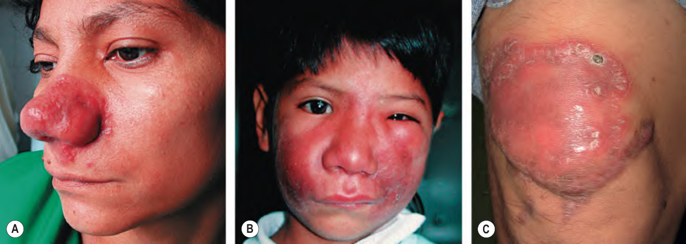
Fig. 83.14 Balamuthia mandrillaris infection.A Indurated plaque on the nose. B Infiltration that has progressed to involve a large portion of the face. C Indurated plaque on the knee, which represents another common site. A, B, with permission from Tyring S, Lupi O, Hengge U (eds). Tropical Dermatology. Churchill Livingstone, 2005.
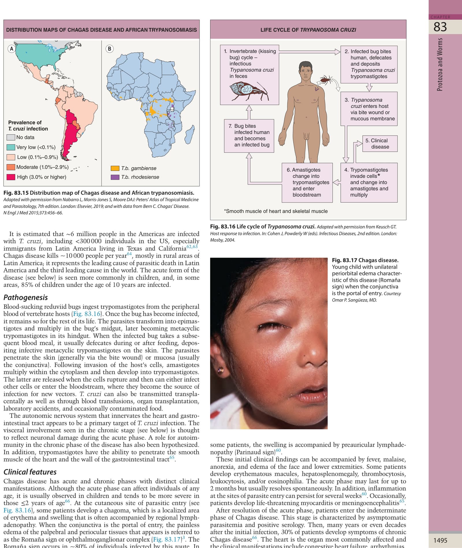
Fig. 83.15 Distribution map of Chagas disease and African trypanosomiasis.
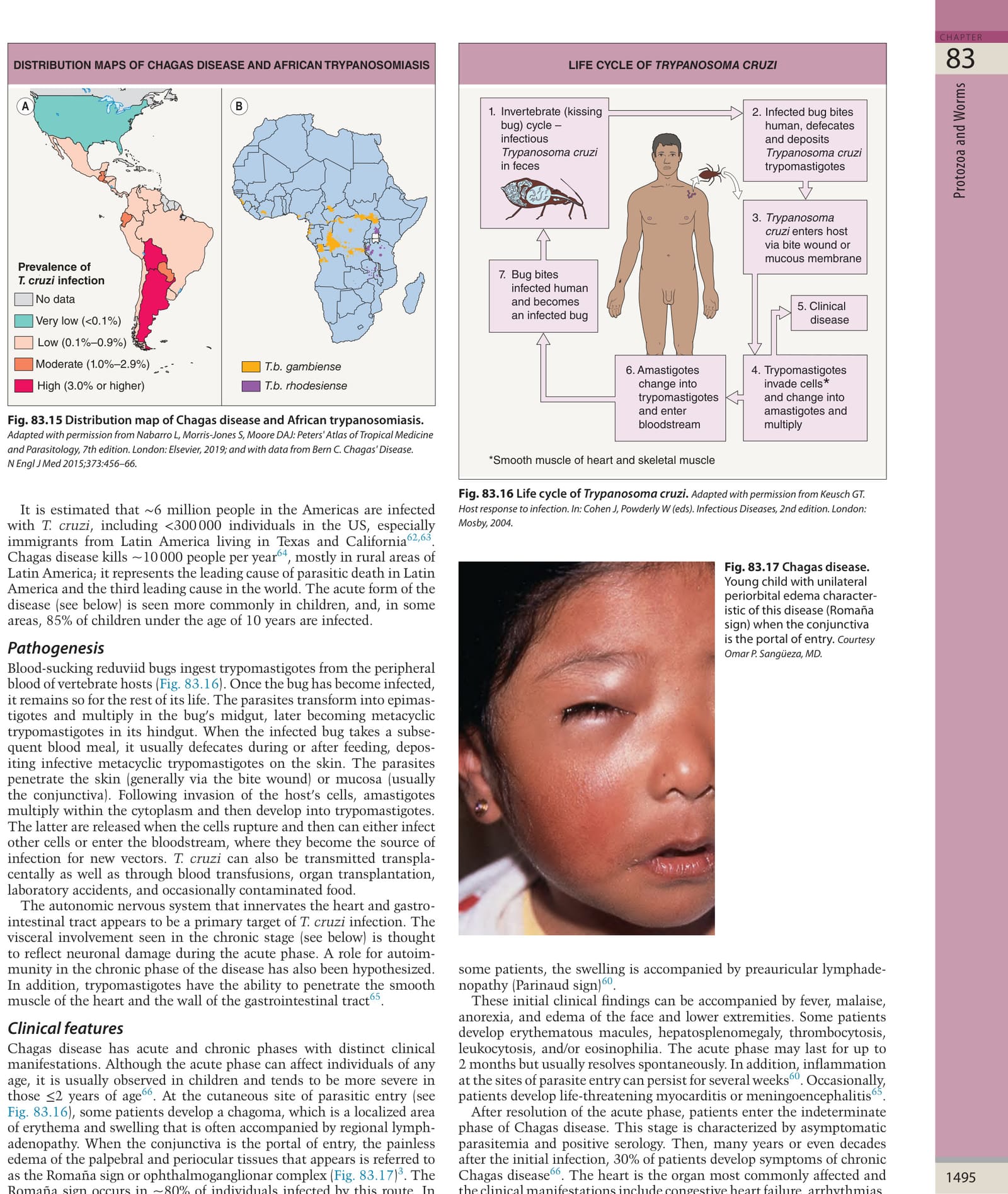
Fig. 83.16 Life cycle of Trypanosoma cruzi.Adapted with permission from Keusch GT. Host response to infection. In: Cohen J, Powderly W (eds). Infectious Diseases, 2nd edition. London: Mosby, 2004.
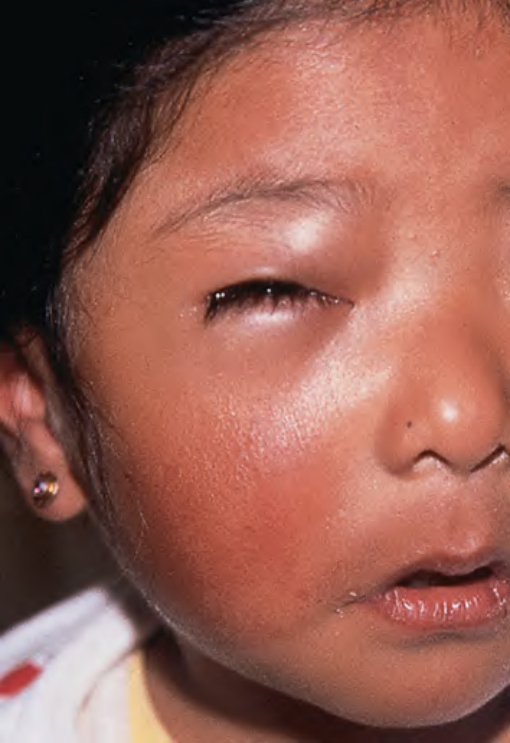
Fig. 83.17 Chagas disease.
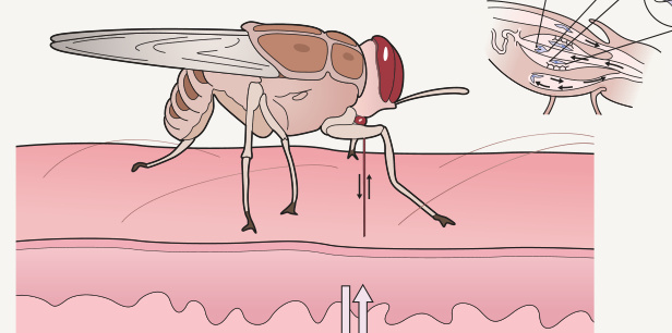
Fig. 83.18 African trypanosomiasis: life cycle of Trypanosoma brucei.
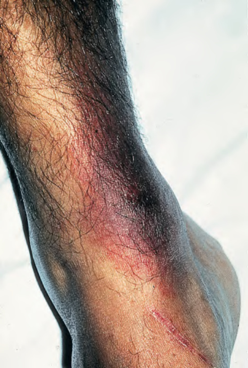
Fig. 83.19 Trypanosomal chancre. The bite reaction, the earliest clinical lesion, is known as a “trypanosomal chancre”. It resembles a boil but is usually painless. Fluid aspirated from the nodule contains actively dividing trypanosomes. This reaction is seen more commonly in T. b. rhodesiense than in T. b. gambiense infection. With permission from Peters W, Pasvol G. Tropical Medicine and Parasitology, 6th edition. London: Mosby, 2007.
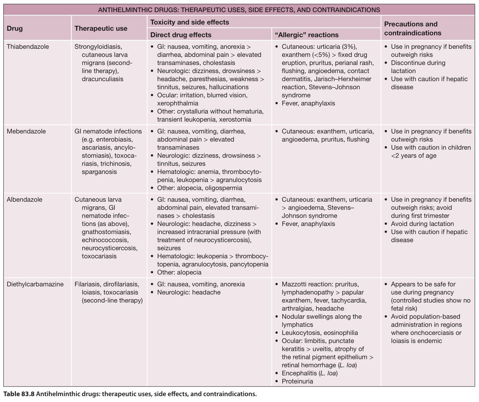
Table 83.8 Antihelminthic drugs: therapeutic uses, side effects, and contraindications.