REPAIR OF UV-INDUCED DNA DAMAGE
The bulky DNA photoproducts (pyrimidine dimers) are usually repaired via the nucleotide excision repair (NER) pathway. In patients with xeroderma pigmentosum (XP), defects in the NER pathway increase UV mutagenesis and this leads to a higher incidence of melanoma and keratinocyte carcinomas (Fig. 86.13). XP includes seven genetic complementation groups (XPA through XPG), which represent different proteins in the NER pathway (Fig. 86.14), in addition to a separate form, XP variant. The latter is due to dysfunction of DNA polymerase-η which normally bypasses T–T dimers with correct insertion of two A residues (see below).
NER involves recognition of DNA damage, incision of the DNA strand containing a lesion, and DNA synthesis and ligation to replace an excised oligonucleotide (see Fig. 86.14). In active genes, the transcribed strand is corrected up to 10 times faster than the non-transcribed strand. Here, recognition of DNA damage is mediated via the RNA polymerase stalled at the site of a pyrimidine dimer plus recruitment of CSA and CSB (protein products of Cockayne syndrome complementation group A and group B genes); this initiates transcription-coupled repair. In non-transcribed genes and non-coding regions, the XPC and XPE proteins bind to UV-damaged DNA, marking it for further processing; this initiates global genome repair. After either form of DNA damage recognition, all subsequent steps of NER are identical. A key intermediate is an open, unwound structure formed around a DNA lesion in a reaction that uses the helicase activities of XPB and XPD. This creates sites for cutting by the endonucleases XPG on the 3′ side of the lesion and the XPF– ERCC1 complex on the 5′ side. A 24- to 32-residue oligonucleotide is released, and the gap is filled by DNA polymerase δ or ε and then sealed by DNA ligase 1.
Defects in NER are found not only in XP, but also in two other photosensitive disorders, Cockayne syndrome and the photosensitive form of the brittle hair syndrome trichothiodystrophy (see Table 87.4). These disorders are not cancer-prone, but sometimes have features that overlap with those of XP. It is remarkable how different mutations in XPB/ERCC3, XPD/ERCC2, and XPG/ERCC5 are associated with dissimilar clinical phenotypes. The transcription/repair syndrome hypothesis has been put forward to explain these various discrepancies. According to this hypothesis, mutations in XPD or XPB could affect
the repair function of transcription factor IIH (TFIIH; see Fig. 86.14), resulting in photosensitivity and cancer development, and/or affect the transcription function of TFIIH, accounting for the typical phenotypes of trichothiodystrophy and Cockayne syndrome.
Cells from patients with XP variant have an intact NER, yet a phenotype that is indistinguishable from the other XP complementation groups. These patients have a deficiency in what they do with unrepaired DNA photoproducts during replication within the S phase of the cell cycle. Replicative DNA polymerases usually stall at unrepaired DNA lesions and detach from the DNA strand. For this situation, cells have several specialized DNA polymerases that are able to bypass different kinds of DNA damage and extend replication forks through damaged sites. Due to a mutation in POLH, patients with XP variant lack the ability of DNA polymerase-η to bypass T–T dimers and correctly insert two A residues. This indicates that NER does not always repair all DNA lesions and that the function of a high-fidelity translesional DNA polymerase is crucial for maintaining genomic stability. In XP variant patients, NER can remove most of the T–T dimers, but because polymerase-η is deficient, any remaining dimers are more likely to be bypassed by translesional DNA polymerases (other than DNA polymerase-η) that insert incorrect residues. This causes a UV-mutator phenotype. If DNA polymerase-η, or any other specialized translesional DNA polymerase, fails to bypass DNA damage during S phase, the cell is faced with a stalled replication fork. In these instances, DNA recombination repair, which utilizes strand invasion from the sister chromatid, can resolve the stalled replication fork.
In contrast to the bulky pyrimidine dimers, which can only be repaired by NER, the non-bulky oxidative DNA base modifications can be processed via base excision repair. As the name implies, the initial step in base excision repair is the removal of a base rather than a nucleotide. This step is carried out by a DNA glycosylase, which removes the damaged DNA base by hydrolytically cleaving the base– deoxyribose glycosyl bond, leaving an apyrimidinic/apurinic site for further processing. This DNA glycosylase has substrate specificity for a particular kind of DNA base damage. The human DNA glycosylase, 8-oxoG DNA glycosylase 1, has specificity for oxidized guanine bases such as 8-oxoG (see Fig. 86.11), and it initiates processing of these lesions via base excision repair.
Lastly, the cytokines IL-12 or -23 can interfere with DNA repair. They are capable of effectively rescuing UV-irradiated cells from apoptosis by triggering DNA repair mechanisms that inhibit immune suppression, most likely due in part to abrogation of induction of regulatory T cells (Tregs). In particular, IL-23 may counteract photocarcinogenesis.
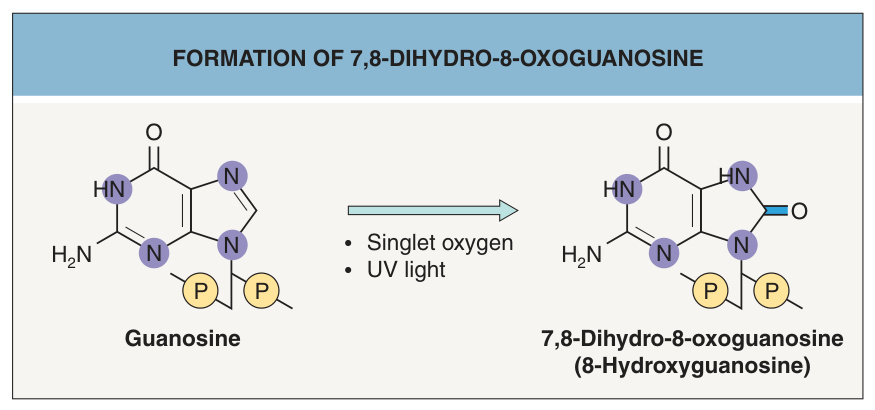
Fig. 86.11 Formation of 7,8-dihydro-8-oxoguanosine. 7,8-Dihydro-8- oxoguanosine (tautomer: 8-hydroxyguanosine) is an oxidation product of guanosine. It is formed by singlet oxygen, which is generated through a photosensitized reaction after excitation of a cellular chromophore by UV light.
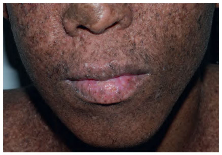
Fig. 86.13 Xeroderma pigmentosum. A 17-year-old with skin phototype V who has hyper- and hypopigmentation admixed with scarring, lentigines, and seborrheic keratoses. Note the squamous cell carcinoma in situ of the lower lip in association with severe actinic cheilitis. Courtesy Julie V. Schaffer, MD.
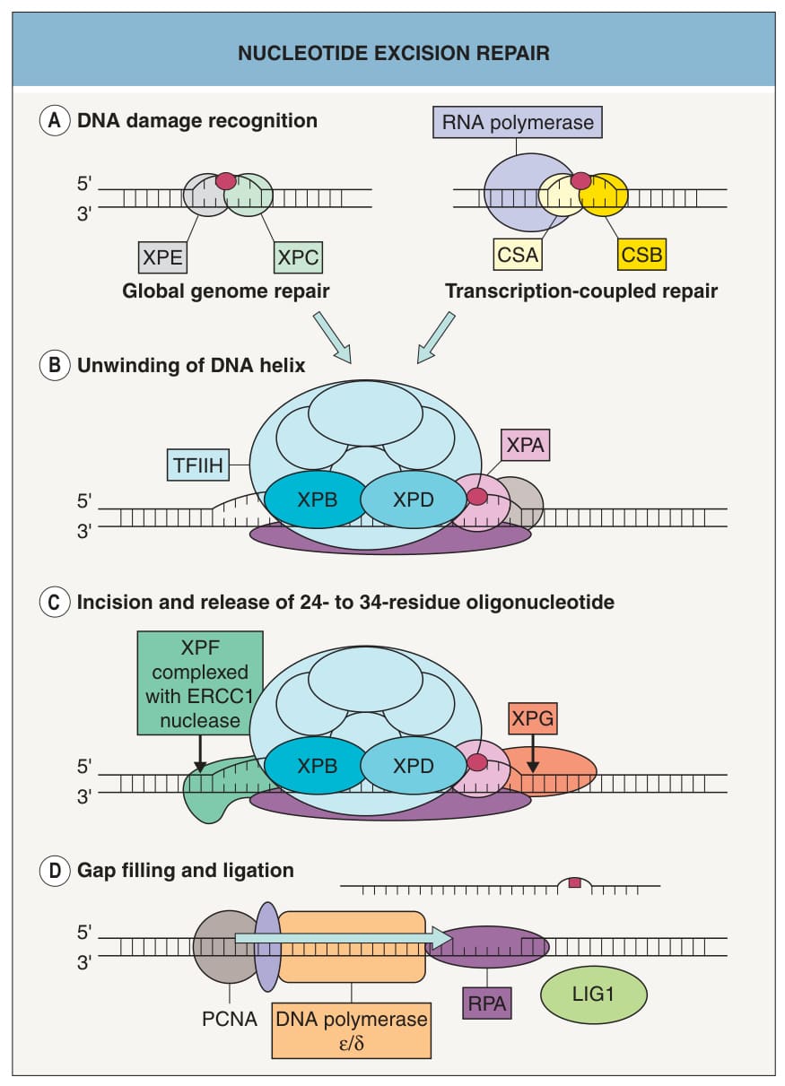
Fig. 86.14 Nucleotide excision repair. This pathway repairs “bulky” DNA lesions, including pyrimidine dimers (red dot). A DNA damage recognition in global genome repair is mediated by XPC and XPE. In transcription-coupled repair, DNA damage recognition is mediated by a stalled RNA polymerase plus recruitment of CSA and CSB. These two types of DNA damage recognition are then followed by identical steps in DNA repair. B Unwinding of the DNA double strand around the lesion by the helicase activity of XPB and XPD. C Incision 5′ and 3′ of the lesion by the endonucleases XPF and XPG. D Repair synthesis and gap closing after release of a 24- to 34-residue oligonucleotide. CSA/CSB, proteins that are dysfunctional in Cockayne syndrome; ERCC1, excision repair cross- complementation group 1; LIG1, DNA ligase 1; PCNA, proliferating cell nuclear antigen; RPA, replication protein A; TFIIH, transcription factor IIH; XP, xeroderma pigmentosum. Adapted with permission from Lindahl T, Wood RD. Quality control by DNA repair. Science 1999;286:1897–905.