INTRODUCTION
The different types of extracellular matrix (ECM) represent specifically organized assemblies of the matrix macromolecules listed in Table 95.1. These macromolecules have characteristic patterns of aggregation into insoluble suprastructures with a high degree of order at successive hierarchic levels. Each of these structures is tissue-specific and adapted to the particular needs of a given tissue. The major constituents are often similar in functionally diverse ECMs. However, various types of less abundant proteins associate with the major ECM components, and differences in their relative composition determine tissue-specific organization of ECM suprastructures. The matrix suprastructures can
be compared to alloys, with each having metallurgic properties that differ from each other and those of the pure metals.
Individual ECM macromolecules are usually oligomers composed of one or several polypeptides. Intimate contacts between the subunits are formed by coiled-coil structures, such as the collagen triple helix or supercoiled α-helices comprised of three or more polypeptides. In addition, large matrix macromolecules can be regarded as linear sequences of structural modules that are similar in a large variety of proteins. The modules can be recognized by several cellular receptors, but receptor clustering is determined in a tissue-specific manner, and the response may be different in different tissues.
Our knowledge of matrix macromolecules has increased dramatically due to advances in molecular genetics and proteomics. Numerous molecules have been characterized and their expression, regulation, tissue specificity, and functions discerned. The assembled ECM structures are generally adhesive, enabling the attachment of tissue-specific cells, leukocytes, tumor cells, and even microorganisms (Fig. 95.1). Through integrin-mediated interactions with cells, matrix molecules control cell proliferation, differentiation, and migration, especially during development and regenerative processes. Without contact with the ECM, many cells undergo a form of apoptosis known as anoikis. Furthermore, the ECM can function as a reservoir of information; certain proteoglycans and proteins bind growth factors (e.g. transforming growth factor [TGF]-β), releasing and activating them as needed to control cellular functions. To date, mutations in >50 different genes encoding ECM molecules have been found to cause heritable disorders in humans and mice.
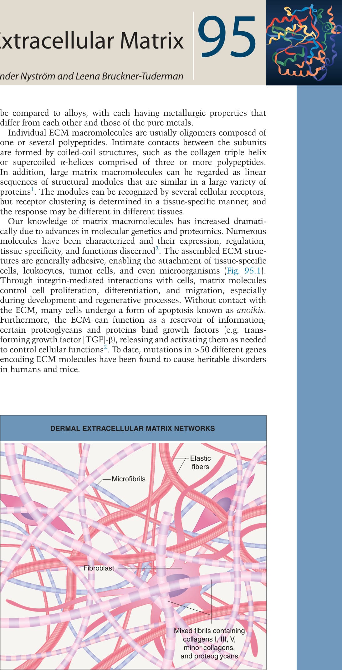
Fig. 95.1 Dermal extracellular matrix networks. Different molecules polymerize into distinct fibril networks and within the mesh of the networks, cells are embedded in the amorphous extrafibrillar matrix. The fibril networks interact with each other, with the extrafibrillar matrix and with the cells. These networks have a dual function: support of the tissue and regulation of cellular functions.
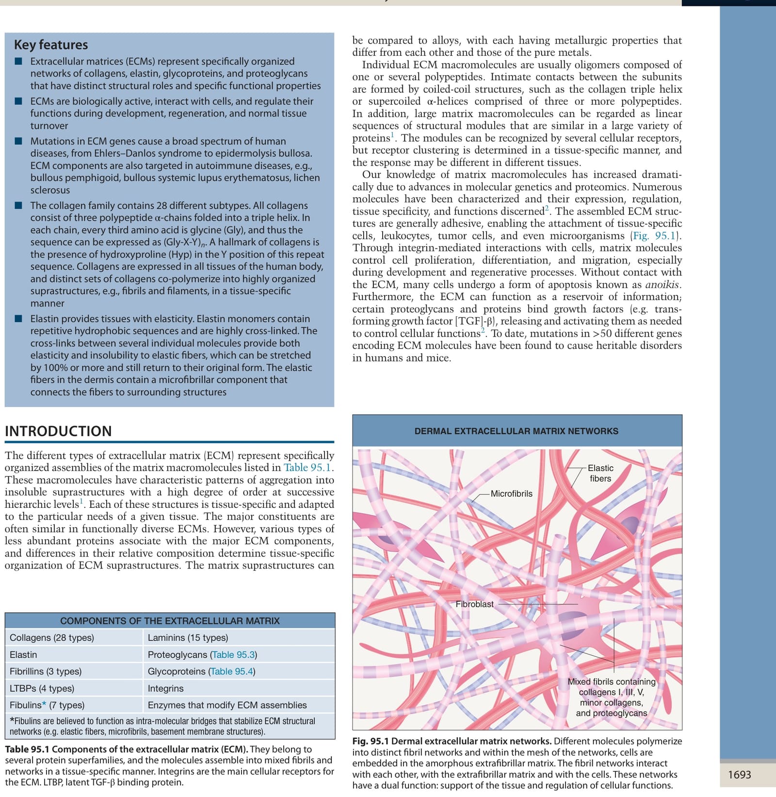
Table 95.1 Components of the extracellular matrix (ECM). They belong to several protein superfamilies, and the molecules assemble into mixed fibrils and networks in a tissue-specific manner. Integrins are the main cellular receptors for the ECM. LTBP, latent TGF-β binding protein.