EPIDERMAL DEVELOPMENT
The ectoderm that covers the developing embryo after gastrulation is a single-layered epithelium (Fig. 2.2A). The first step in epidermal development occurs when cells of the surface ectoderm adopt an epidermal fate. Although this process does not result in major morphologic changes, it is marked by dramatic alterations in gene expression that result in the formation of the embryonic epidermis, which initially consists of a simple epithelium (Fig. 2.2B). Primitive keratinocytes subsequently generate cells of the periderm, a single cell layer that covers the developing epidermis until the cornified cell layer is formed (Figs. 2.2C–F & 2.3A,B). The periderm is believed to exchange substances across fetal skin and to protect the developing epidermis from forming interepithelial adhesions.
The embryonic epidermis begins to stratify at approximately 8 weeks’ estimated gestational age (EGA). At this time, basic organogenesis is complete and bone marrow hematopoiesis commences, marking the transition from embryo to fetus. Of note, expression of TP63 is required for epidermal stratification. During the first stage of stratification, an intermediate cell layer is formed between the basal layer and periderm (see Figs. 2.2D & 2.3B). Unlike suprabasal keratinocytes in the postnatal epidermis, the intermediate layer consists of actively proliferating cells. As a consequence, it is able to expand to accommodate the rapid growth of the embryo as well as create additional layers of intermediate cells over the next several weeks (see Fig. 2.2E). However, the intermediate cell layer is ultimately replaced by post-mitotic keratinocytes under-going terminal differentiation.
Terminal differentiation, the process resulting in the formation of mature keratinizing epidermal cells, begins during the second trimester. Early cornification can be observed within the hair canal at approximately 15 weeks’ EGA, but it does not commence in the interfollicular epidermis until 22–24 weeks’ EGA, occurring first in the skin on the head, palms, and soles. The process begins when cells in the intermediate layer permanently withdraw from the cell cycle and differentiate into spinous and granular cells (see Fig. 2.2F). The cornified cell layer, which is composed of “dead” keratinocytes (corneocytes) held together by a matrix of proteins and lipids (see Chs. 56 & 124), subsequently starts to form and is several cells thick by 24–26 weeks’ EGA. The corneocytes are a reflection of the closely regulated process of terminal differentiation that is required for normal functioning of the skin. At the time of keratinization, the periderm detaches from the underlying epidermis and is sloughed off into the amniotic fluid, with remnants contributing to the vernix caseosa that coats newborns. During the third trimester, the number of keratohyalin and lamellar granules as well as stratum corneum layers increases. By the mid third trimester, the epidermis is morphologically similar to adult skin (Figs. 2.2G & 2.3C), although it does not acquire full barrier function until a few weeks after birth.
Clinical Relevance
Genetic abnormalities affecting various stages of epidermal morphogenesis have been found to underlie inherited skin disorders in humans. However, generalized abnormalities in epidermal specification, the process through which the surface ectoderm adopts an epidermal fate, have not been identified. It is likely that generalized epidermal defects would be incompatible with survival past the first trimester. Mosaic skin conditions that result in abnormalities of the epidermis and/or its appendages often have a distribution pattern that follows the lines of Blaschko, which are thought to represent the migration pathways of epidermal cells during embryonic development. Classic examples include epidermal nevi (due to postzygotic mutations in genes encoding
fibroblast growth factor receptors, the phosphoinositide-3-kinase alpha subunit, Ras family members, and keratins) and sebaceous nevi (due to postzygotic mutations in HRAS>KRAS) (see Ch. 62).
There are a number of genetic disorders that result in abnormal epidermal differentiation and barrier formation. One clinical presentation of such conditions is a “collodion baby” born encased in a taut, shiny, transparent membrane that is formed by aberrant stratum corneum. After shedding the membrane, most of these infants manifest with lamellar ichthyosis or congenital ichthyosiform erythroderma, two forms of autosomal recessive congenital ichthyosis that exist on a spectrum. However, following a collodion membrane, some patients develop normal or almost normal appearing skin. The “self-improving” collodion baby is an example of a dynamic epidermal phenotype that depends on environmental conditions. All of these outcomes can result from mutations in the same set of genes that encode proteins essential for formation of the epidermal barrier, including transglutaminase-1 (an enzyme that cross-links lipids to the cornified cell envelope; TGM1), lipid processing enzymes (ALOXE3, ALOX12B), and lipid transporters (ABCA12) (see Ch. 57).
More deleterious mutations in ABCA12 cause harlequin ichthyosis (HI), an especially severe disorder of cornification characterized by aberrant epidermal maturation. Patients with HI are born with a tremendously thick, armor-like shell of hyperkeratosis, severe ectropion and eclabium, and underdevelopment of the nose and ears. The extreme phenotype of HI highlights the importance of lipid transport into lamellar bodies for epidermal formation and function.
Abnormalities in the stratum corneum are present not only in infants with ichthyosis, but also in premature infants, especially those born before 28 weeks’ EGA. The immaturity of the stratum corneum results in impaired barrier function, which leads to an increased risk of infection, dehydration, and excessive absorption of topical medications or chemicals. Even healthy full-term infants do not attain full skin barrier function until 3 weeks of age. The structural features of premature skin and adult skin are summarized in Table 2.1.
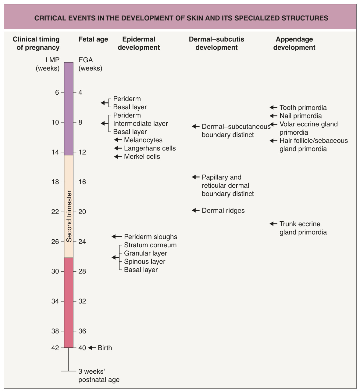
Fig. 2.1 Critical events in the development of the skin and its specialized structures. The timeline indicates the time of initiation defined by estimated gestational age (EGA) and duration of pregnancy (by last menstrual period [LMP]). Refers to skin on the back unless otherwise noted.
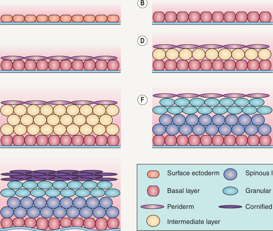
Fig. 2.2 Development of the epidermis.A The epidermis develops from the surface ectoderm, a single-layered epithelium that initially covers the developing embryo. B Through changes in gene expression, cells of the surface ectoderm adopt an epidermal fate. C Epidermal cells subsequently give rise to the periderm, a cell layer that covers the developing epidermis until cornification occurs. D Epidermal stratification begins with the formation of a highly proliferative intermediate layer between the basal layer and the periderm. E The intermediate layer becomes several cells thick over the next few weeks. F Intermediate cells ultimately withdraw from the cell cycle and differentiate into spinous and granular keratinocytes. G The periderm is replaced by the cornified cell layer.
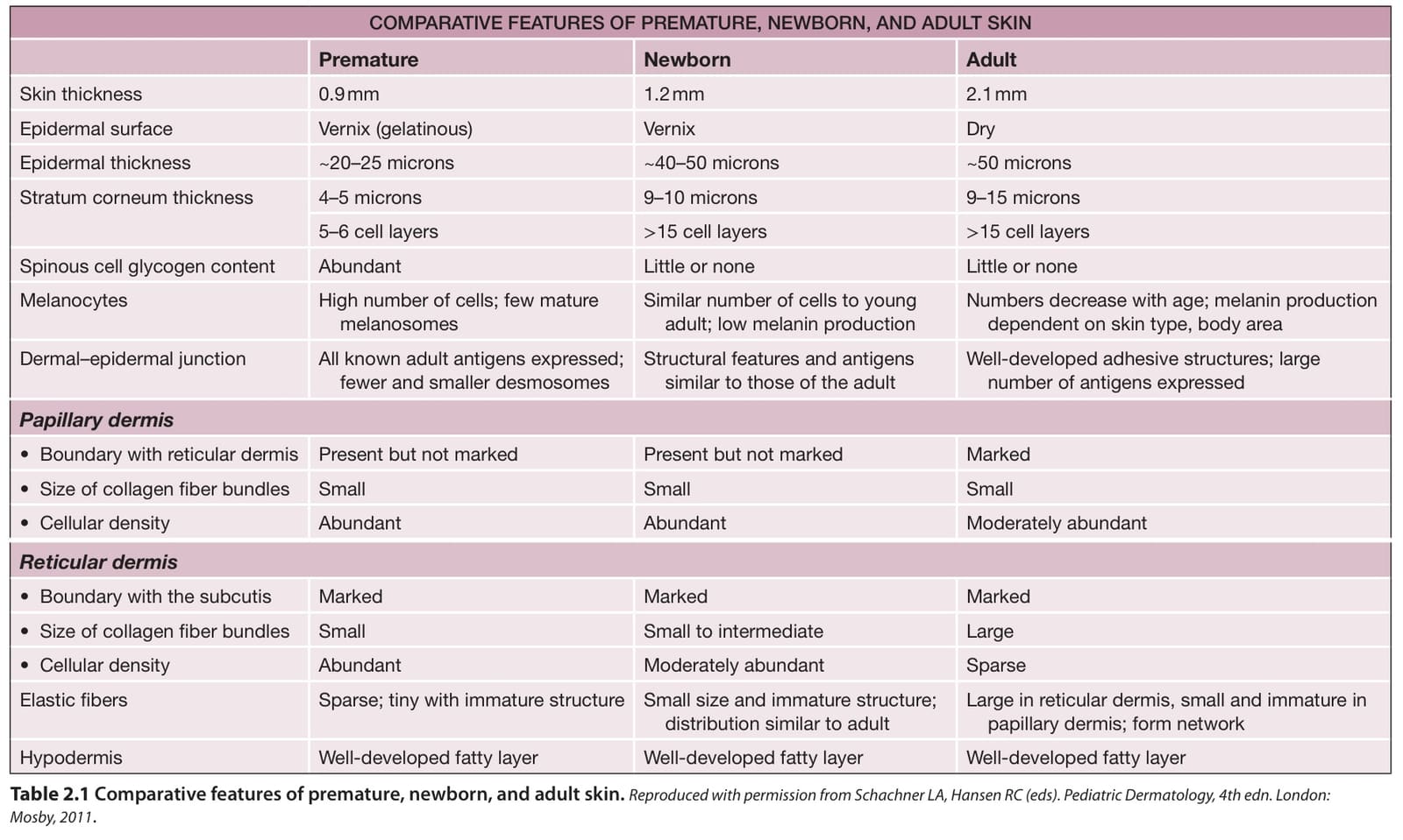
Table 2.1 Comparative features of premature, newborn, and adult skin.Reproduced with permission from Schachner LA, Hansen RC (eds). Pediatric Dermatology, 4th edn. London: Mosby, 2011.