EXPERIMENTAL TECHNIQUES
Tissue Processing
Dermatologists are accustomed to placing a biopsy specimen in formalin followed by paraffin embedding and then staining tissue sections with hematoxylin and eosin. However, this is just one of several possible starting points for tissue processing (Fig. 3.1). By placing the sample in
formalin (for light microscopy), glutaraldehyde (for electron microscopy), or freezing a fresh sample before processing and sectioning, the sample can be analyzed histologically in various ways, including by routine staining, immunohistochemistry, or in situ hybridization.
Cryosections from frozen samples have lower quality tissue architecture than sections from formalin-fixed, paraffin-embedded tissues, but the former preserve DNA, RNA, and protein better than other fixation methods. Fresh or frozen tissue can also be processed directly with buffers and reagents to extract DNA, RNA, and protein from the whole tissue. In addition, fresh tissue can be used to prepare a suspension of single cells for single-cell analysis of DNA, RNA, or protein. The desired lineage of cells (e.g., keratinocytes, fibroblasts, immunocytes, endothelial cells) can be propagated in vitro by using selective culture media and isolation techniques.
One method for isolating a particular cell population from a tissue section is laser microdissection, which allows selection of microanatomic regions for analysis (Fig. 3.2). Alternatively, once tissue is dissociated and living cells are in a single cell suspension, flow cytometry can be used to identify populations of cells (Fig. 3.3). Individual cells pass through in a stream and through a set of lasers and electronic detectors that can measure multiple parameters for each cell. Fluorescence-activated cell sorting (FACS) utilizes an electrostatic deflection system and it can be used to obtain pure cell populations with specific desired characteristics, e.g. cells tagged by lineage-specific antibodies. The field of single-cell molecular biology is rapidly evolving, and it has become possible to analyze 40 or more parameters for each cell by combining the principles of flow cytometry and elemental mass spectrometry in a technique called mass cytometry.
The Foundations of Molecular Techniques for Analyzing DNA, RNA, and Protein
The concepts behind molecular biology are simple and unifying. In general, they consist of extracting the molecules of interest, amplifying them to measurable amounts, and then detecting them. Polymerase chain reaction (PCR) is a standard technique for amplifying DNA (Table 3.1; Fig. 3.4). To perform PCR, short sequences of synthetic DNA (primers) are used to hybridize to a DNA region of interest, which is then replicated through multiple cycles. The PCR-amplified DNA, typically 50 to 2000 base pairs in size depending on the primers designed, can be detected in a gel using an intercalating dye that fluoresces with ultraviolet light. The nucleotide sequence can then be determined via fluorescence sequencing techniques. This simple and relatively inexpensive approach, also called Sanger sequencing, is still widely used.
Nowadays, however, massively parallel sequencing, also known as next-generation sequencing, offers the opportunity for reading millions of DNA fragments in a single run at a marginally higher cost (Table 3.2; Fig. 3.5). The advent of massively parallel sequencing not only allows access to DNA sequences, but it also allows one to infer DNA copy number changes. This is because the final amplified DNA is proportional to the number of DNA copies in the original reaction, allowing inference of copy number variation of specific genes and genomic regions. One limitation of massively parallel sequencing is that its increased power and reading depth requires bioinformatics and sophisticated data analyses, which are challenging to standardize in clinical practice.
RNA is also easy to purify, but is more unstable and degrades much faster than DNA. Therefore, a typical first step in the analysis of RNA is to convert it into DNA using reverse transcription (RT; Table 3.3; Fig. 3.6). Following RT, the generated complementary DNA (cDNA) can be amplified by PCR, as described above. The technique of RT-PCR has also been modified to allow accurate quantitation of
very low levels of mRNA. Because the amount of PCR product is monitored during each amplification cycle, this technique is called “real-time” quantitative PCR.
The amount of protein in a cell is a complex balance of synthesis and degradation controlled at multiple steps, including efficiency of protein translation and post-translational modifications that affect protein stability. One method for measuring protein levels is the Western blot, which is also called an immunoblot because an antibody is used to detect the protein of interest (Table 3.4; Fig. 3.7). In addition to measuring protein levels, Western blot analysis can determine protein size and whether there are different forms of the protein. Another common method for measuring protein levels is an enzyme-linked immunosorbent assay (ELISA; see Table 3.4), which can provide accurate quantifications of proteins. In multiplex bead-based immunoassays, up to 100 analytes can be detected simultaneously (see Fig. 40.2). For this, distinct fluorophore-colored microbeads, coupled to specific antibodies, are incubated with a sample; this mixture is then exposed to secondary detection antibodies and reporters, followed by exposure to laser excitation. The technique allows for a quantitative interpretation of the amount of analyte bound to the bead.
Measuring Cells Within Tissues
For populations of cells, PCR, RT-PCR, and Western blots are useful for measuring DNA, RNA, and protein, respectively. However, to identify features at a cellular level, tissue sections can be analyzed via various assays. For example, fluorescence in situ hybridization (FISH) is employed to assess the presence or expression of DNA or RNA. This technique relies on the use of fluorescently labeled nucleic acid probes that are complementary to the DNA or RNA sequences of interest in the tissue sections (Table 3.5; Fig. 3.8). The probes are hybridized to the target sequences in the tissue, and the slide is then examined using
Immunohistochemistry is employed to assess the expression of proteins in tissue sections. An antibody is added to a tissue section where it specifically binds to the protein of interest. The bound antibody is usually detected based on an enzymatic reaction that generates a colored product at the binding site. Immunohistochemistry can be used to determine the locations and expression level of a protein within a tissue, including proteins that mark specific cell lineages or cell states such as differentiation, activation, proliferation, or apoptosis. Of note, immunohistochemistry has evolved beyond quantitative visualization of a single protein – it can also detect specific sequences of messenger RNA (mRNA) in a tissue. In addition, revolutionary visualization assays allow assessment with multiple antibodies, utilizing the same principles of antibody-bound color reagents (multiplex IHC and FISH).
Measuring the Transcriptome and Proteome
There are several approaches to quantitatively analyze the expression levels of nearly all cellular genes, i.e. the transcriptome. RNA-seq utilizes massively parallel sequencing to evaluate gene expression levels, gene splicing, non-coding RNA, microRNA, gene fusions, and mutations. Nowadays it is also possible to perform RNA-seq on single cells, an approach that is providing new insights into the complexity of human skin. Additional methods are based on the hybridization of labeled sample mRNA to known oligonucleotide or cDNA sequences
(representing thousands of genes) placed on chips, beads, or glass slides (arrays). The level of hybridization to the nucleotide sequence of a given gene indicates how much mRNA of that gene is present.
The proteome represents all cellular proteins expressed under specific conditions. Mass spectrometry provides extremely accurate measurements of protein mass by ionizing the protein and measuring the “time-of-flight” through a tube to a detector on the opposite end (Table 3.6; Fig. 3.9). In most instances, the proteins are separated by two-dimensional gel electrophoresis or capillary electrophoresis before mass spectrometry. Quantification is accomplished by labeling proteins with stable isotopes to provide internal standards or by labelfree shotgun proteomic approaches that allow a faster analysis at lower costs. Another technique is the use of reverse-phase protein arrays, in which whole protein lysates are spotted onto slides and probed using antibodies to specific proteins. The field of proteomics promises to provide a tremendous amount of information regarding cell biology and disease states.
Mouse Models of Skin Disease
Transgenic mice are commonly used as experimental models for elucidating the in vivo functions of genes. The traditional approach is to microinject the gene of interest into a fertilized mouse egg, where it randomly integrates into the mouse genome (Table 3.7; Fig. 3.10). Although the gene is present in every cell as the egg develops into a mouse, expression of the gene can be limited or targeted to specific tissues or cell types by using a promoter that is only expressed in a
particular cell linage. Promoters initiate mRNA expression and are located upstream of the gene’s coding region. For example, a promoter that ensures that a gene is expressed only in melanocytes would be a promoter of a gene involved in melanin biosynthesis, such as tyrosinase. Although every mouse cell will contain a randomly integrated copy of this new gene, it is expressed only in melanocytes, because of the melanocyte-specific promoter. This principle can be applied to other cell lineages.
The functional loss of a gene can also be studied by creating a “knockout” transgenic mouse (Table 3.8; Fig. 3.11). It is also possible to selectively delete a gene (e.g. a tumor suppressor gene) in a specific cell type or tissue rather than deleting the gene from all cells in the mouse (Fig. 3.12). A further refinement is to trigger deletion of the gene at a particular point in time, including during development. One advantage of these inducible systems over gene deletion in all mouse cells is that they may more faithfully mimic how gene deletion occurs in a specific tissue of a patient, faithfully recapitulating the hallmarks of diseases such as cancer. Inducible models can also circumvent problems that arise from gene deletion in all mouse cells, which often impact normal mouse development. In addition to using tamoxifen to control Cre expression (see Fig. 3.12), the “Tet-On/Off” system can be used to address the problem of embryonic death due to gene deletion.
The discovery of CRISPR-Cas9 genome editing tools has revolutionized and accelerated mouse model engineering, allowing for the precise introduction of mutations at targeted DNA sites (Fig. 3.13). The CRISPR-Cas9 system facilitates RNA-guided, site-specific cleavage of DNA, which results in the deletion of a DNA region following DNA repair (knockout); alternatively, exogenous DNA is used as a template during the repair process and this leads to a new genetic code (knock-in). These new techniques simplify the engineering of models for skin diseases and hold the potential to accelerate therapy development.
Gene-Based Therapy for Skin Diseases
Gene-based therapy is a therapeutic approach that holds promise as a treatment for skin diseases. In gene-based therapy, DNA or RNA is manipulated to add or correct a gene in the cells of a target tissue in order to achieve a therapeutic effect (Table 3.9). Although this therapy
could be administered directly to the patient’s skin in vivo, there are concerns regarding safety and the potential for genetic alteration of the germline. Therefore, ex vivo techniques have been developed in which cells are grown from a skin sample, treated in vitro, and grafted back onto the patient (see Fig. 2.7).
Because genetic manipulation of stem cells may result in long-term engraftment and stable benefits, they are often preferred. However, the number of stem cells that can be obtained from adult tissues is limited, so there is considerable interest in gene therapy utilizing induced pluripotent stem (iPS) cells (see Ch. 2). Fibroblasts grown from human skin can be reprogrammed into iPS cells via exposure to transcription factors; these iPS cells are pluripotent and can be propagated in vitro indefinitely. iPS cells from a patient may be genetically corrected and then differentiated into the desired cell type, such as keratinocytes, for grafting back to the patient (see Fig. 2.7).
In gene therapy, there are several ways to manipulate DNA. To compensate for the loss-of-function of a gene, a new copy of the gene may be introduced into cells utilizing either non-viral or viral vectors. Sleeping Beauty is a non-viral, cut-and-paste DNA transposon that improves long-term gene expression compared to that obtained when only DNA from the gene of interest is used. However, the DNA transposon poses risks for insertional mutagenesis because it may randomly insert near a proto-oncogene that then becomes activated. Viral vectors can also be used to introduce DNA, including self-inactivating lentiviral and gamma retroviral vectors. These vectors integrate into the genome and have less risk of insertional mutagenesis compared to earlier retroviral vectors. However, viral vectors also have disadvantages such as immunogenicity, broad tropism, and limited DNA packaging capacity.
It may be possible to avoid the complication of insertional mutagenesis by correcting the endogenous gene rather than inserting a gene. Methods for targeted gene correction include the use of zinc
finger nucleases (ZFNs), transcription activator-like effector nucleases (TALENs), and, most importantly, CRISPR-Cas9 (see Fig. 3.13). These nucleases have sequence-specific DNA-binding modules that are linked to a DNA cleavage domain and this allows them to induce a DNA double-strand break at a specified site. The double-strand break increases the frequency of homology-directed repair so that a co-delivered plasmid with extended homology arms can correct the existing gene. Correction of endogenous genes holds promise for treating genetically based diseases.
Another therapeutic approach is to target specific mRNAs for degradation. Double-stranded RNAs with a length of 21–23 nucleotides and complementary to the mRNA of interest are designed. These synthesized antisense oligodeoxynucleotides (AS-ODNs) are delivered to the cell, where they mediate sequence-specific mRNA degradation using an endogenous complex of proteins called the RNA-induced silencing complex (RISC). However, AS-ODN techniques are being replaced by synthetic vectors that deliver short interfering RNA (siRNA) and microRNA (miRNA) (Fig. 3.14). These synthetic delivery vectors include lipid-based or polymer-based nanoparticles, as well as covalent delivery systems such as dynamic polyconjugates and GalNAc conjugates. These approaches have been used successfully in the laboratory and are being tested in clinical trials.
Fig. 3.1 Tissue processing. A tissue sample can be processed in various ways for the analysis of DNA, RNA, or protein. FACS, fluorescence-activated cell sorting.
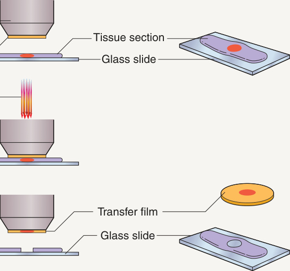
Fig. 3.2 Laser capture microdissection. Laser capture microdissection is one method of microdissection used to selectively procure individual cells or clusters of cells from tissue sections. A cap coated with a thermoplastic transfer membrane is placed directly over the tissue section. The operator visually identifies the cells of interest and triggers a low-energy infrared laser to melt the transfer film onto cells of interest. The cap is then lifted from the section to separate the selected cells from the remainder of the tissue section.
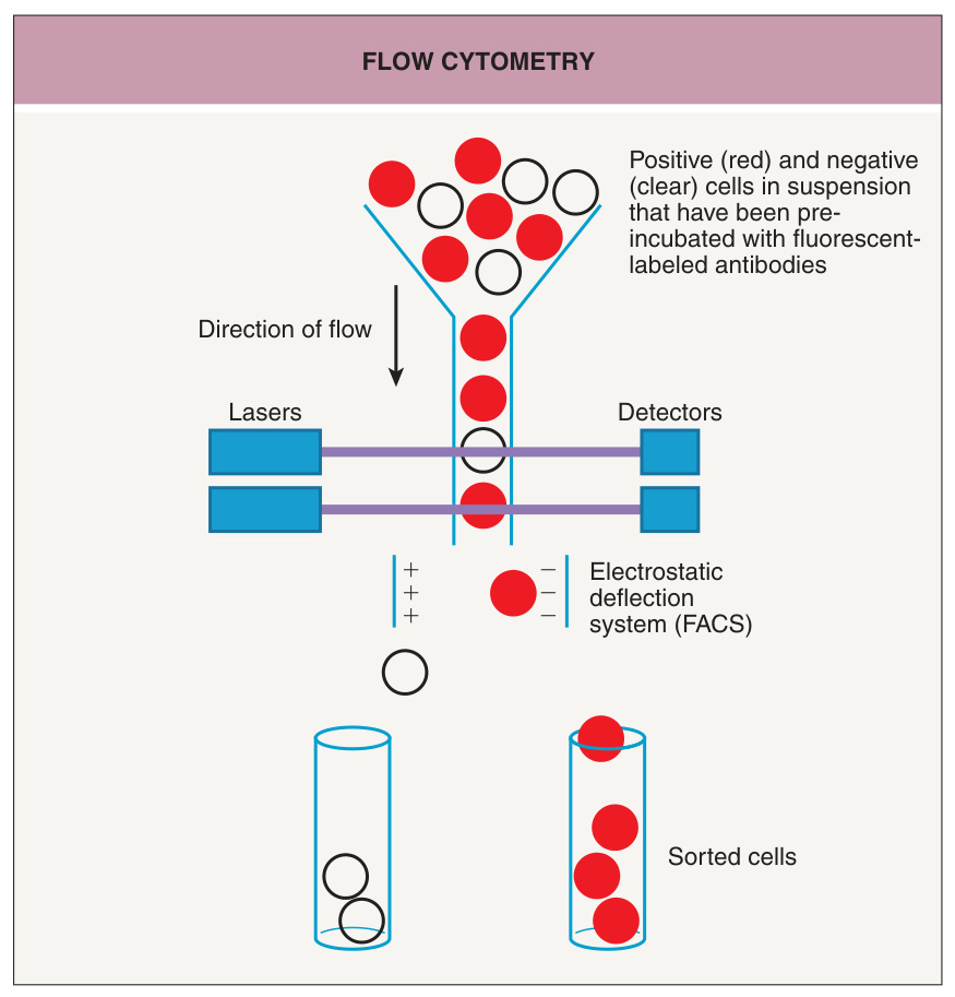
Fig. 3.3 Flow cytometry. Cells in suspension, pre-incubated with fluorescentlabeled antibodies, flow single file in a liquid stream through lasers. Detectors measure the fluorescence intensities of each cell. In fluorescence-activated cell sorting (FACS), the single cells are divided into charged droplets that are separated into different tubes based on the marker(s) of interest as they pass through an electrostatic deflection system.
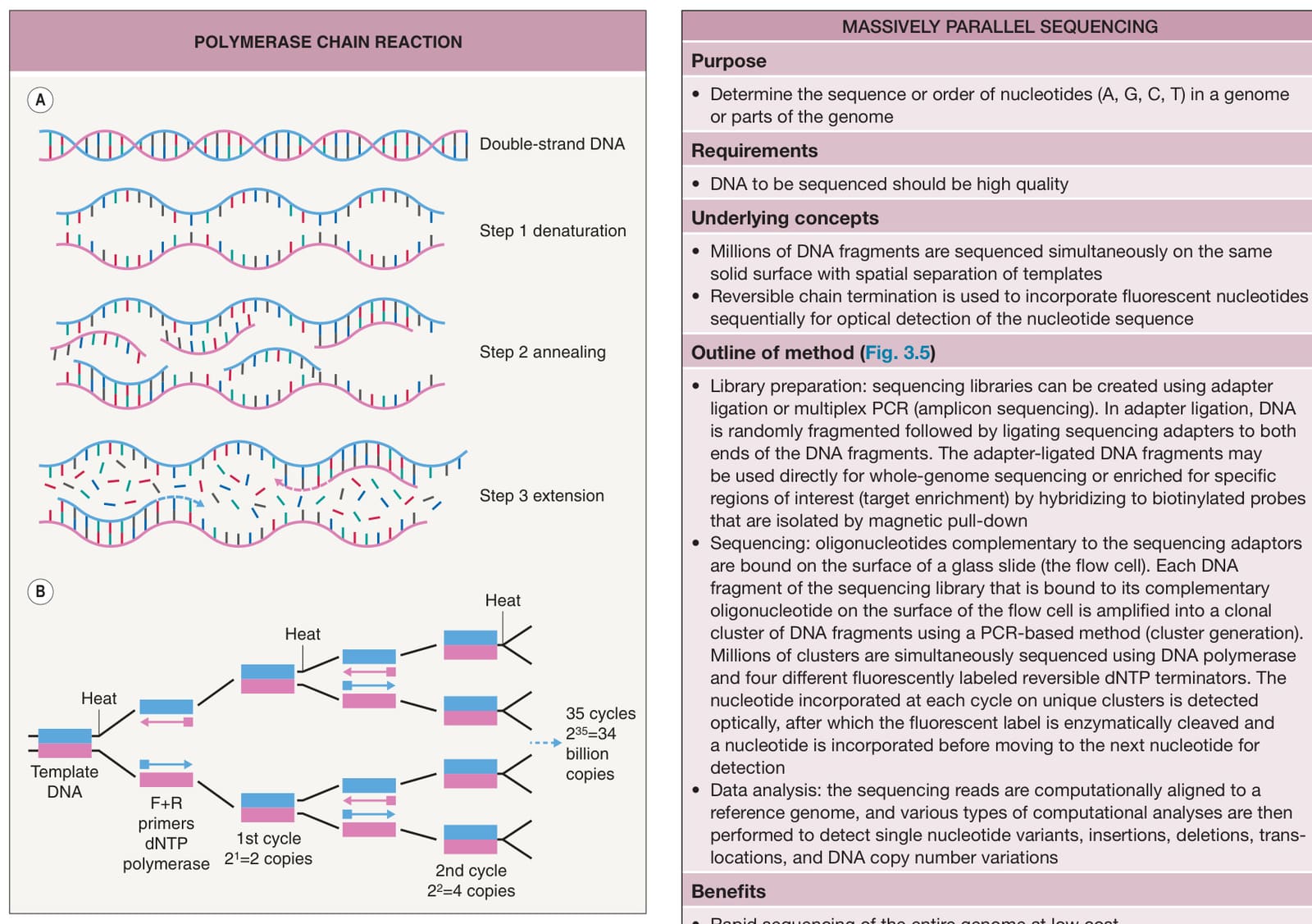
Fig. 3.4 Polymerase chain reaction. A Each cycle contains the following steps: (1) denaturation – separate the two strands of DNA by heating to >90°C; (2) primer annealing or primer hybridization – allow the oligonucleotide primers to bind to the template DNA by cooling to 50–65°C; (3) primer extension – DNA polymerase catalyzes the addition of nucleotides (A, G, C, T) that are complementary to the DNA template, beginning with the primer and extending 3′ at the optimal temperature of 72°C; and (4) repeat the complete cycle 30–40 times. B Each cycle increases the number of PCR products twofold. The total number of PCR products after n cycles will be 2n times the original amount. F+R, forward and reverse.
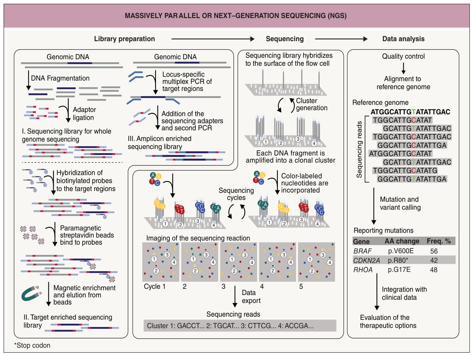
Fig. 3.5 Massively parallel or next generation sequencing (NGS). The technology used for massively parallel sequencing varies among different platforms. The workflow on Illumina platforms can be divided into three steps: (1) library preparation – the test DNA is flanked with sequencing adaptors either by ligation (adaptor ligation) or PCR (amplicon sequencing); (2) sequencing – the adaptor-flanked test DNA binds to oligonucleotides on the surface of the flow cell and DNA polymerases catalyze the incorporation of fluorescently labeled nucleotides, which are identified by imaging; after enzymatic removal of the fluorophores, a new fluorescently labeled nucleotide is incorporated and read out by imaging; (3) data analysis – after quality controls and alignment of the sequencing reads to reference genomes, the genetic aberrations are called and annotated using computational algorithms. Freq, frequency fluorescence microscopy. As a diagnostic tool, FISH can be used to detect genomic aberrations in single cancer cells (see Fig. 117.19).
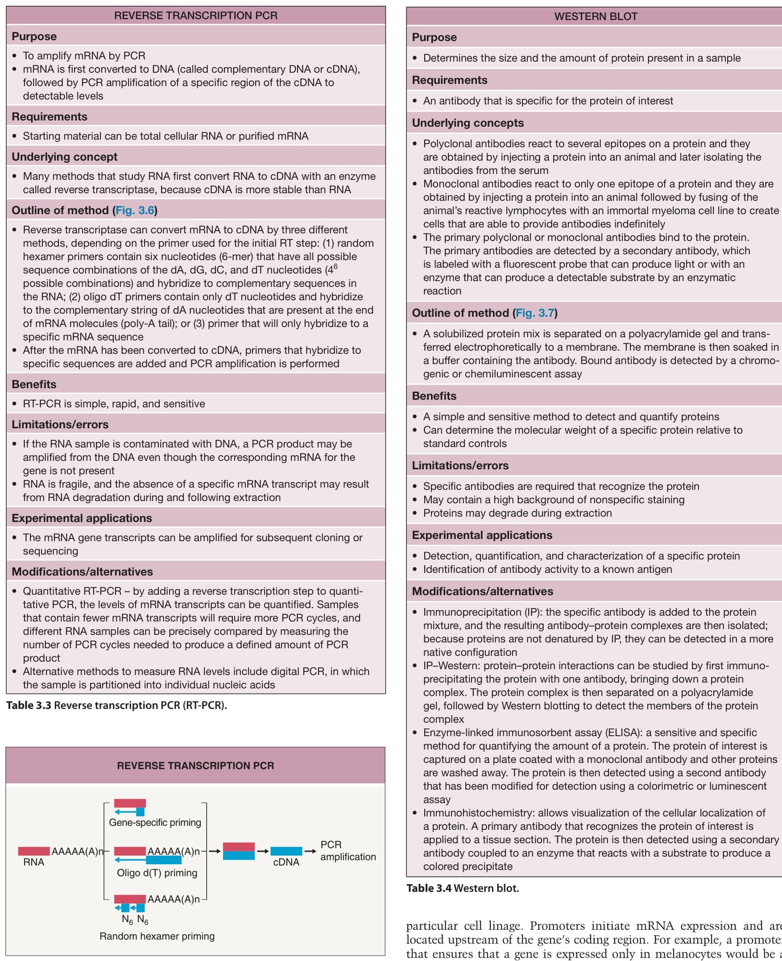
Fig. 3.6 Reverse transcription PCR (RT-PCR). Reverse transcriptase can convert mRNA to cDNA in three different ways, depending on the primer used: (1) random hexamer primers; (2) oligo dT primers; and (3) gene-specific primers. After the mRNA has been converted to cDNA, primers that can hybridize to specific nucleotide sequences are added and PCR is performed.
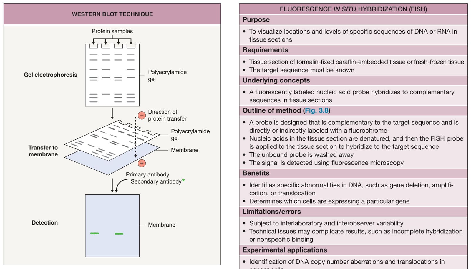
Fig. 3.7 Western blot technique. A solubilized protein mix is separated on a polyacrylamide gel and electrophoretically transferred to a membrane. The membrane is soaked in an antibody-containing buffer, and the bound antibody is detected by a chromogenic or chemiluminescent assay utilizing a labeled secondary antibody (green asterisk).
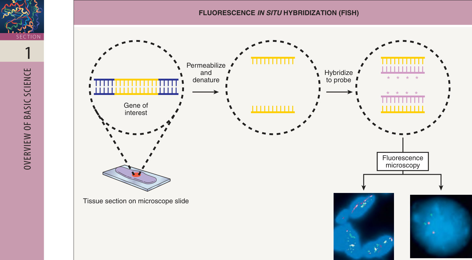
Fig. 3.8 Fluorescence in situ hybridization (FISH). A tissue section on a microscope slide is fixed and permeabilized. The nucleotides on the section (yellow) and the fluorescently labeled (asterisks) probes (dark pink) are denatured; then the probes are added to the section to hybridize with complementary sequences. The unbound probe is washed away, and the tissue is examined by fluorescence microscopy. Normally, two fluorescent signals appear in each nucleus for an autosomal gene, but in cancer cells, there may be only one signal, indicating a deletion, or three or more signals, indicating an amplification. Here, a normal nucleus contains two green signals and two dark pink signals for two separate autosomal genes, whereas a tissue section from an angiosarcoma shows multiple separate signals using a MYC break-apart probe (the probe consists of two flanking sequences in which variable breakpoints have been observed) instead of only two copies of the two signals attached to one another. FFPE, formalinfixed, paraffin-embedded.
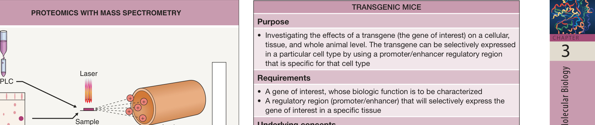
Fig. 3.9 Proteomics with mass spectrometry. The first step is to reduce the complexity of the mixture of cellular proteins or peptides to be analyzed. The two main methods to separate proteins/peptides from each other are high-performance liquid chromatography (HPLC) columns or two-dimensional (2D) gel electrophoresis. Mass spectrometry analysis is then performed by first ionizing the separated proteins/peptides into positively charged ions using lasers. Based on the time-of-flight of the charged ion, mass spectrometry can measure, record, and print out the mass/charge ratio of every peptide along with signal intensity.
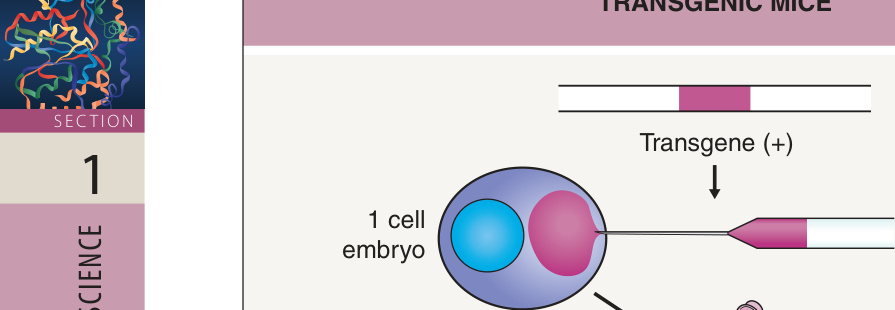
Fig. 3.10 Transgenic mice. A transgene construct, defined as the transgene and regulatory region (promoter/enhancer), is prepared for injection. The transgene is microinjected into fertilized eggs (single-cell stage) and the transgene integrates into the genome, usually at a single site. These injected eggs are then implanted into a recipient mother, who then gives birth to a heterozygous “founder” mouse. The founder mice are bred with normal non-transgenic mice of the same strain, and then two heterozygous transgenic mice are mated.
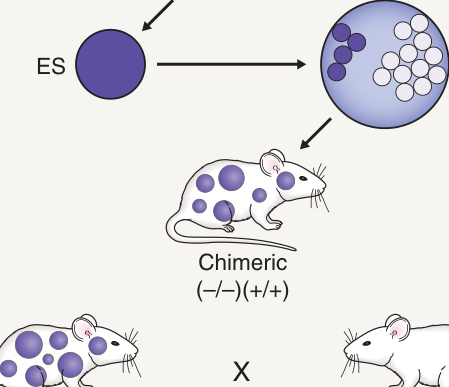
Fig. 3.11 Knockout transgenic mice. A targeting vector that contains some sequences of the gene to be targeted is created and introduced into cells. This targeting vector hybridizes to one of the endogenous alleles of a gene and modifies or deletes the endogenous gene so that no normal protein is produced. The embryonic stem (ES) cells containing the “knocked out” gene are introduced into early mouse embryos, which are then implanted into recipient mothers. The progeny will be chimeric mice, which are then mated to normal mice. To obtain completely knocked out mice, two heterozygous mice are mated.
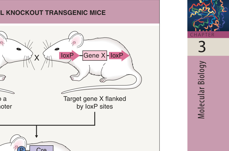
Fig. 3.12 Conditional knockout transgenic mice. In the conditional knockout model using the Cre-lox system, a target gene can be selectively deleted in a particular organ or tissue. Cre is a site-specific DNA recombinase that catalyzes recombination between two loxP sites, resulting in cleavage, exchange, and ligation; this leads to excision (and subsequent degradation) of the DNA between the loxP sites. A transgenic mouse with the Cre gene linked to a tissuespecific promoter is mated to a transgenic mouse with two loxP sites flanking the target gene. Progeny inherit both of these constructs, and the target gene is knocked out only in cells that activate the tissue-specific promoter. By adding a ligand-binding domain to the Cre recombinase, such as one that binds tamoxifen, investigators can achieve temporal control of Cre expression in these cells. The target gene can be temporally deleted, inverted, or translocated, depending on the orientation of the loxP sites.
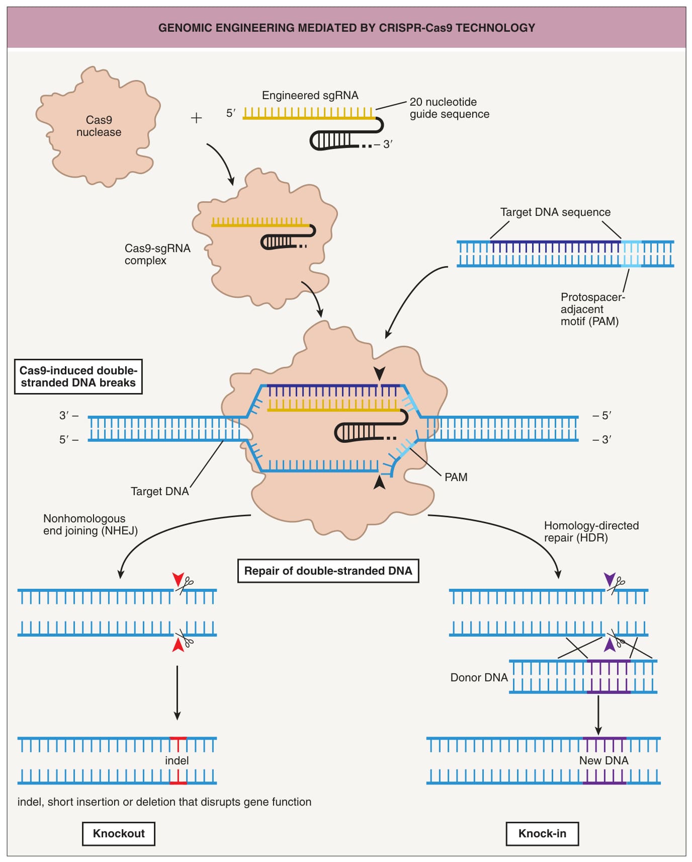
Fig. 3.13 Genomic engineering mediated by CRISPR-Cas9 technology. A single-guide RNA (sgRNA) is engineered to target genomic DNA adjacent to a protospacer adjacent motif (PAM) utilizing a 20-nucleotide guide sequence. The hairpin structure recruits Cas9 to form a complex that binds to the target sequence. Cas9 catalyzes the cleavage of both strands of DNA three nucleotides upstream of the PAM. A double-strand break may be repaired by non-homologous end-joining (NHEJ) that leads to the formation of short insertions or deletions that disrupts gene function. In the presence of a DNA-repair template, homology-directed repair (HDR) may occur to introduce exogenous DNA at the site of the break.
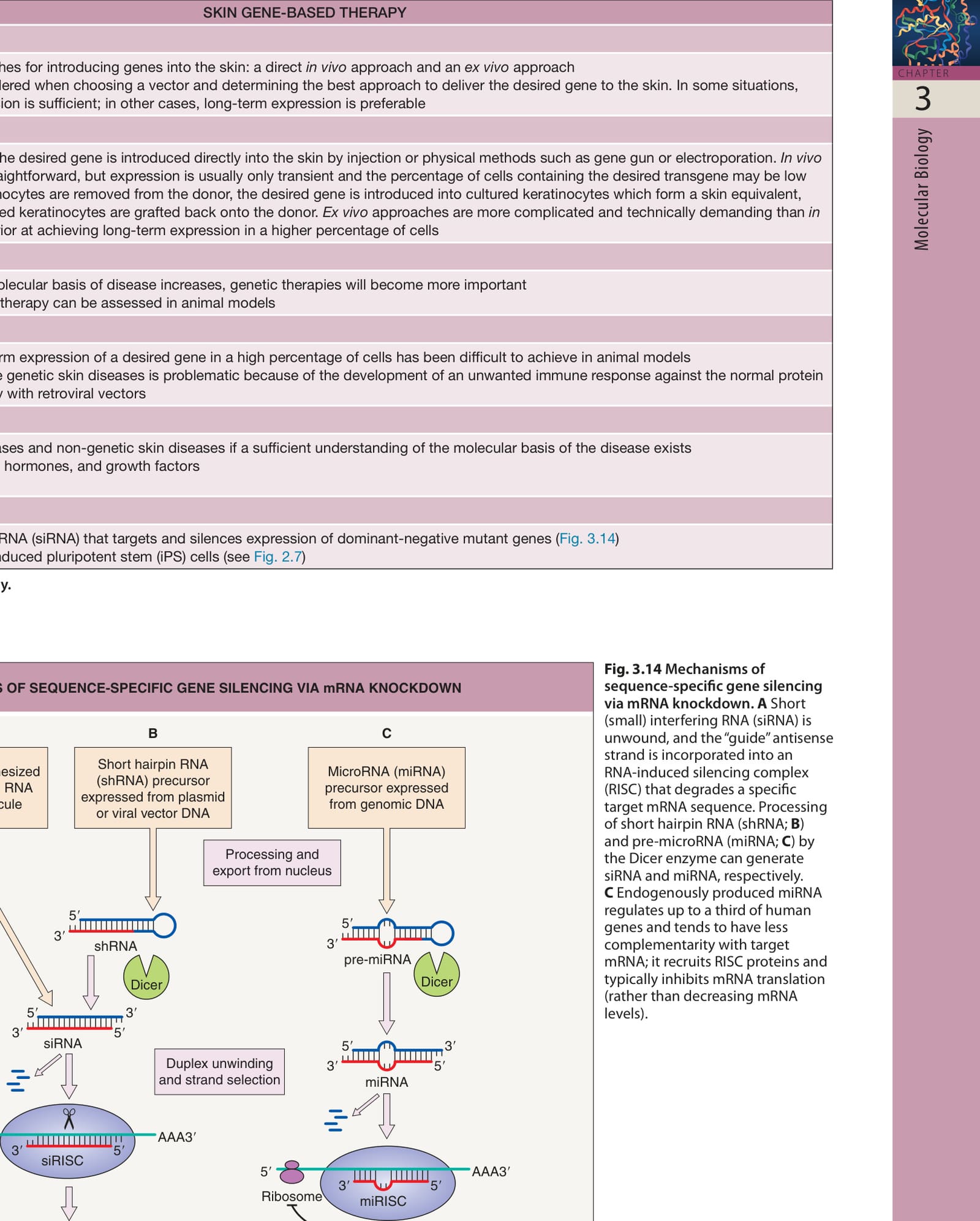
Fig. 3.14 Mechanisms of sequence-specific gene silencing via mRNA knockdown. A Short (small) interfering RNA (siRNA) is unwound, and the “guide” antisense strand is incorporated into an RNA-induced silencing complex (RISC) that degrades a specific target mRNA sequence. Processing of short hairpin RNA (shRNA; B) and pre-microRNA (miRNA; C) by the Dicer enzyme can generate siRNA and miRNA, respectively. C Endogenously produced miRNA regulates up to a third of human genes and tends to have less complementarity with target mRNA; it recruits RISC proteins and typically inhibits mRNA translation (rather than decreasing mRNA levels).
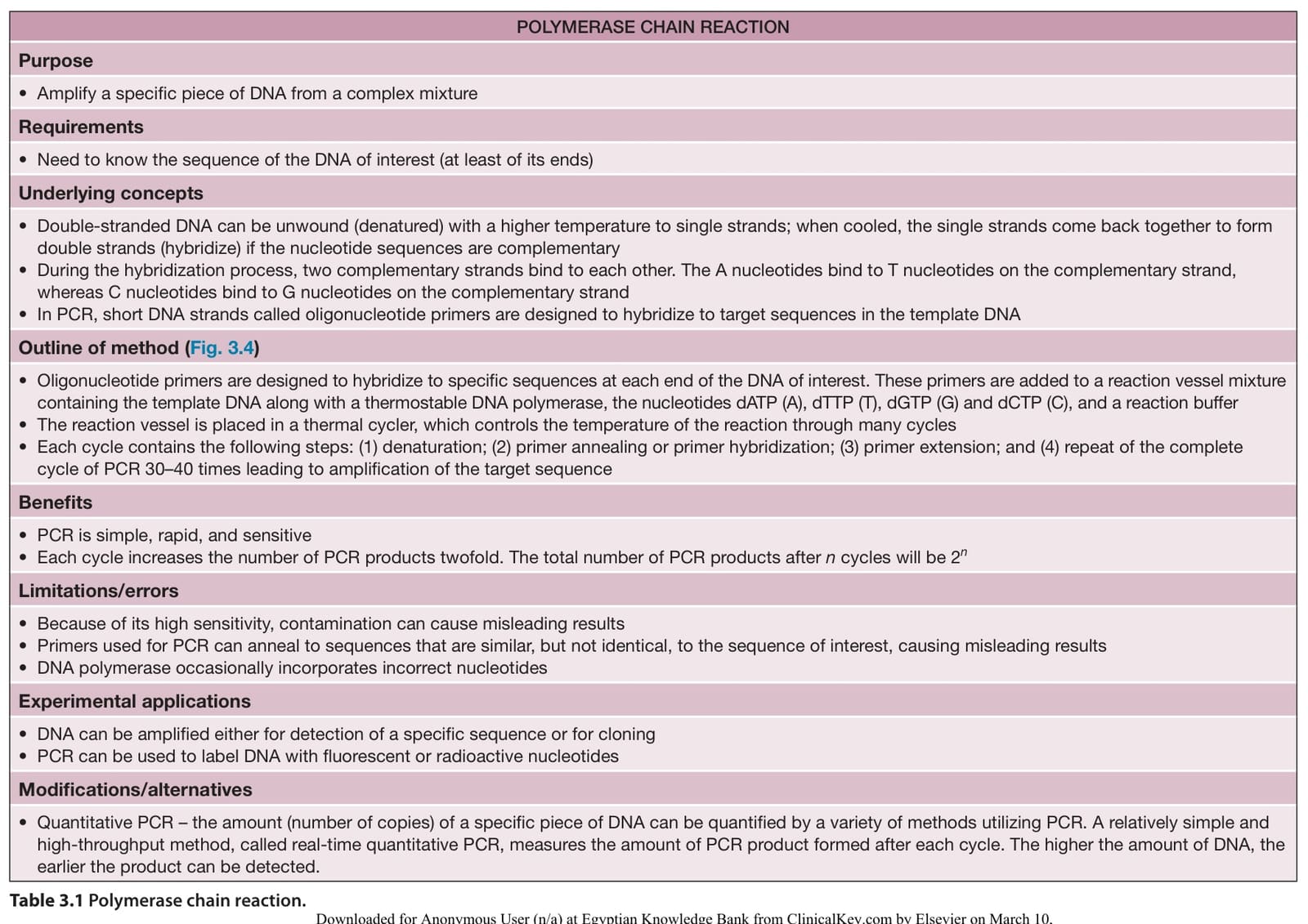
Table 3.1 Polymerase chain reaction.
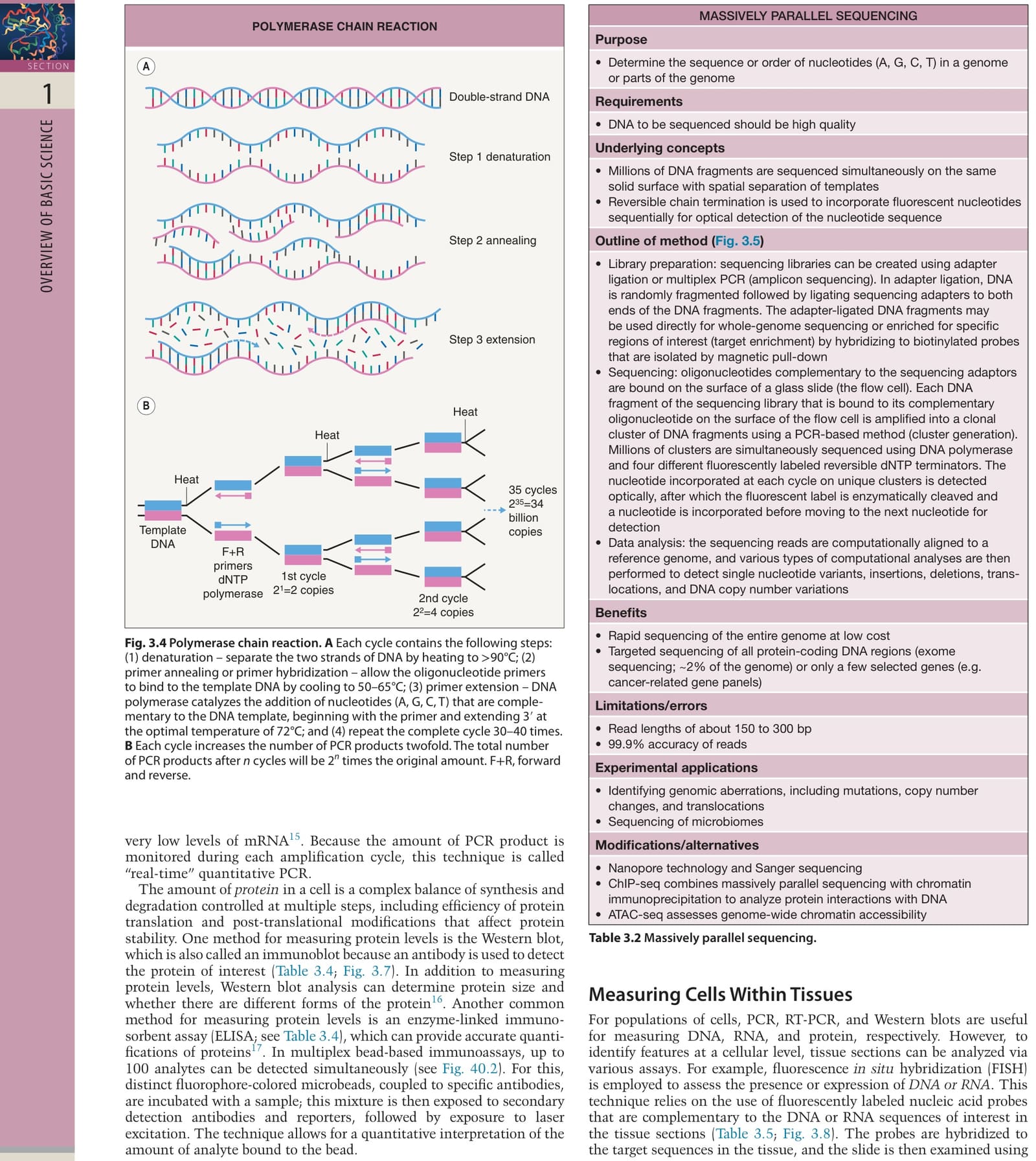
Table 3.2 Massively parallel sequencing.
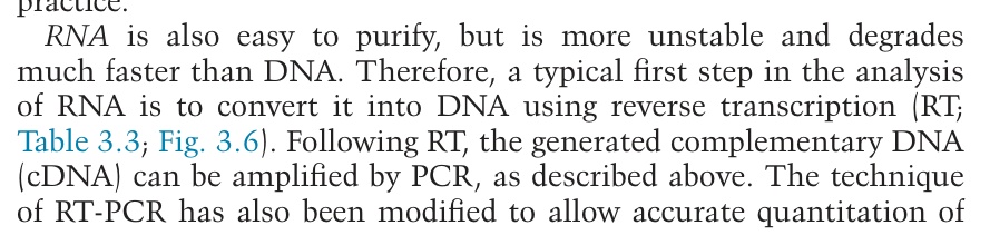
Table 3.3 Reverse transcription PCR (RT-PCR).
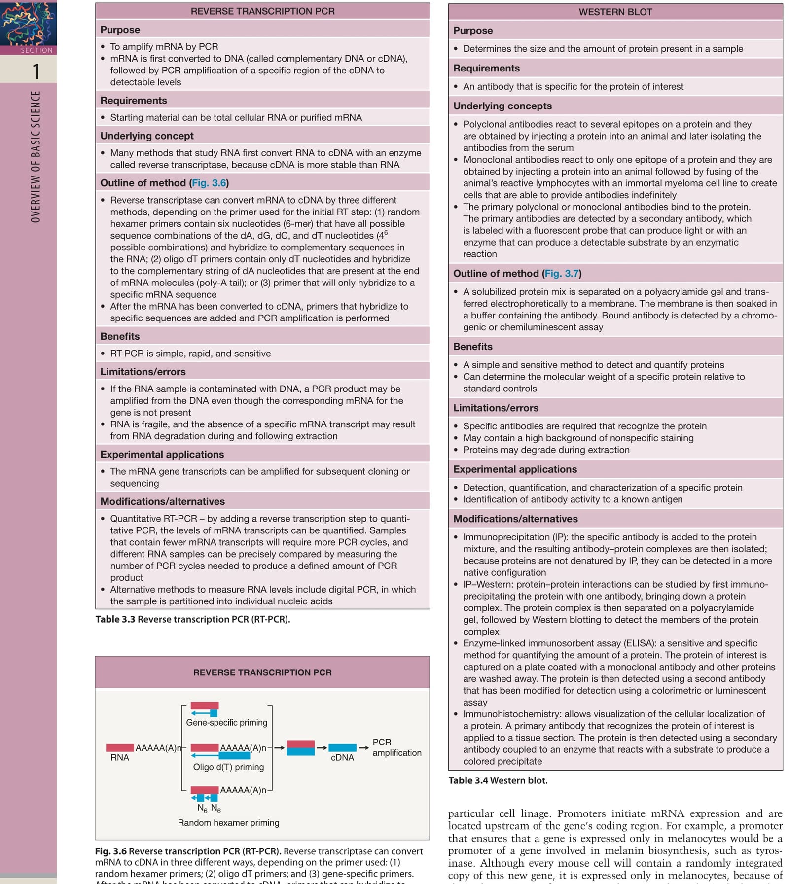
Table 3.4 Western blot.
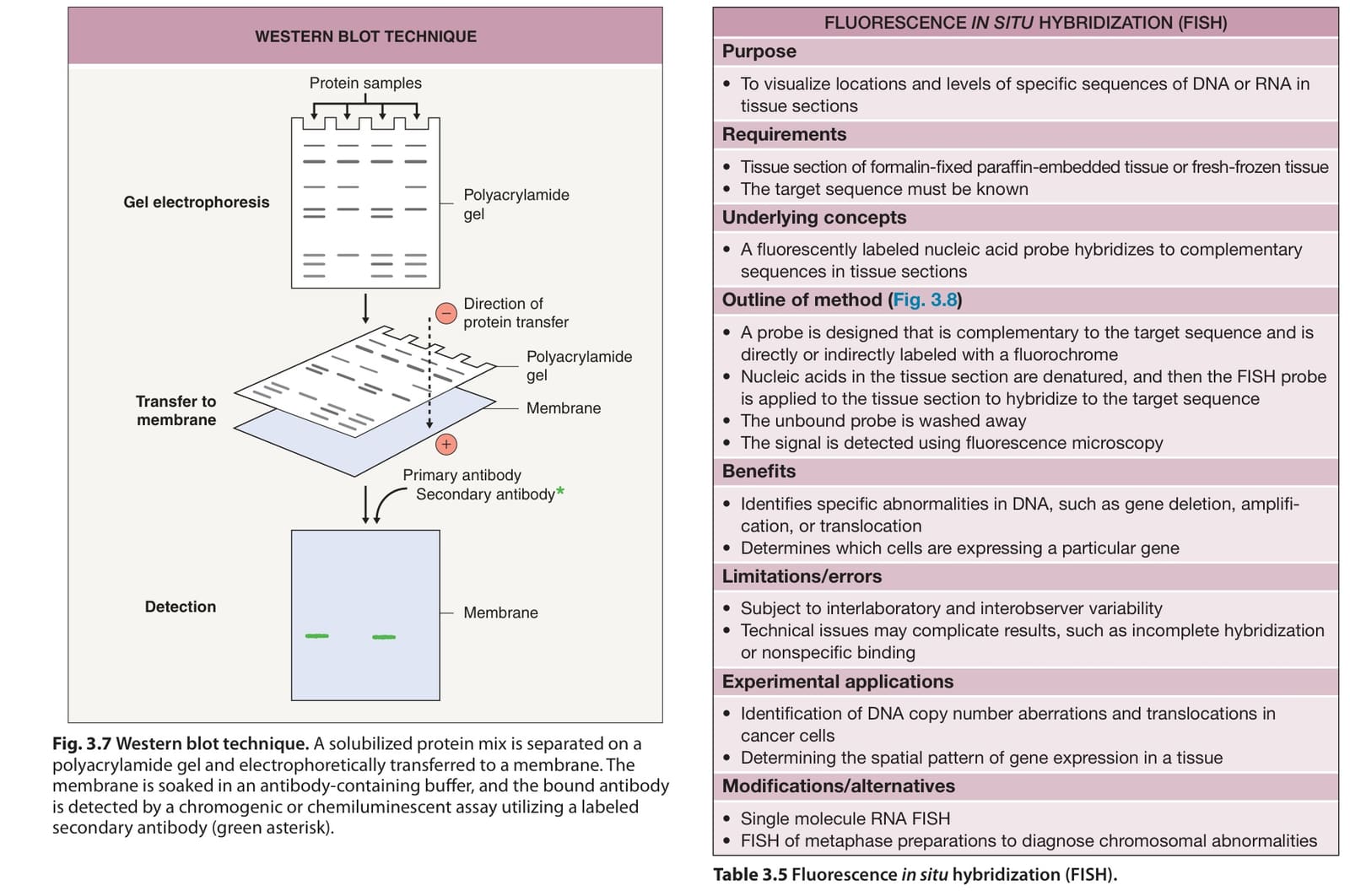
Table 3.5 Fluorescence in situ hybridization (FISH).
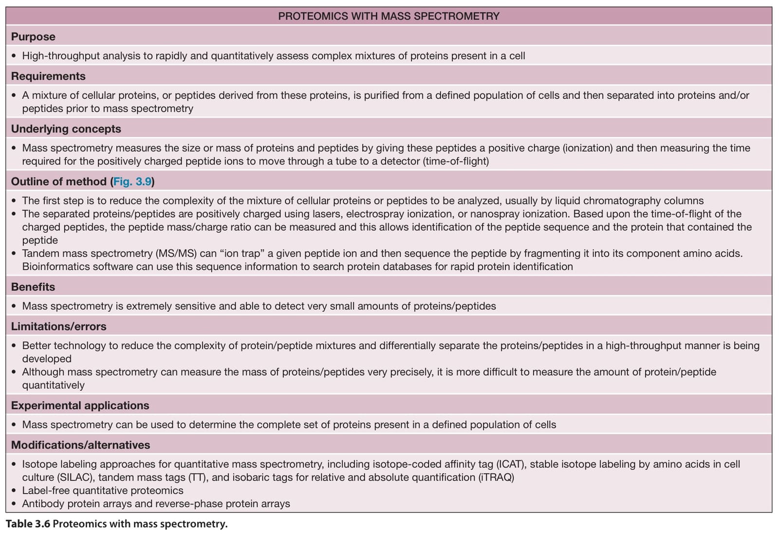
Table 3.6 Proteomics with mass spectrometry.
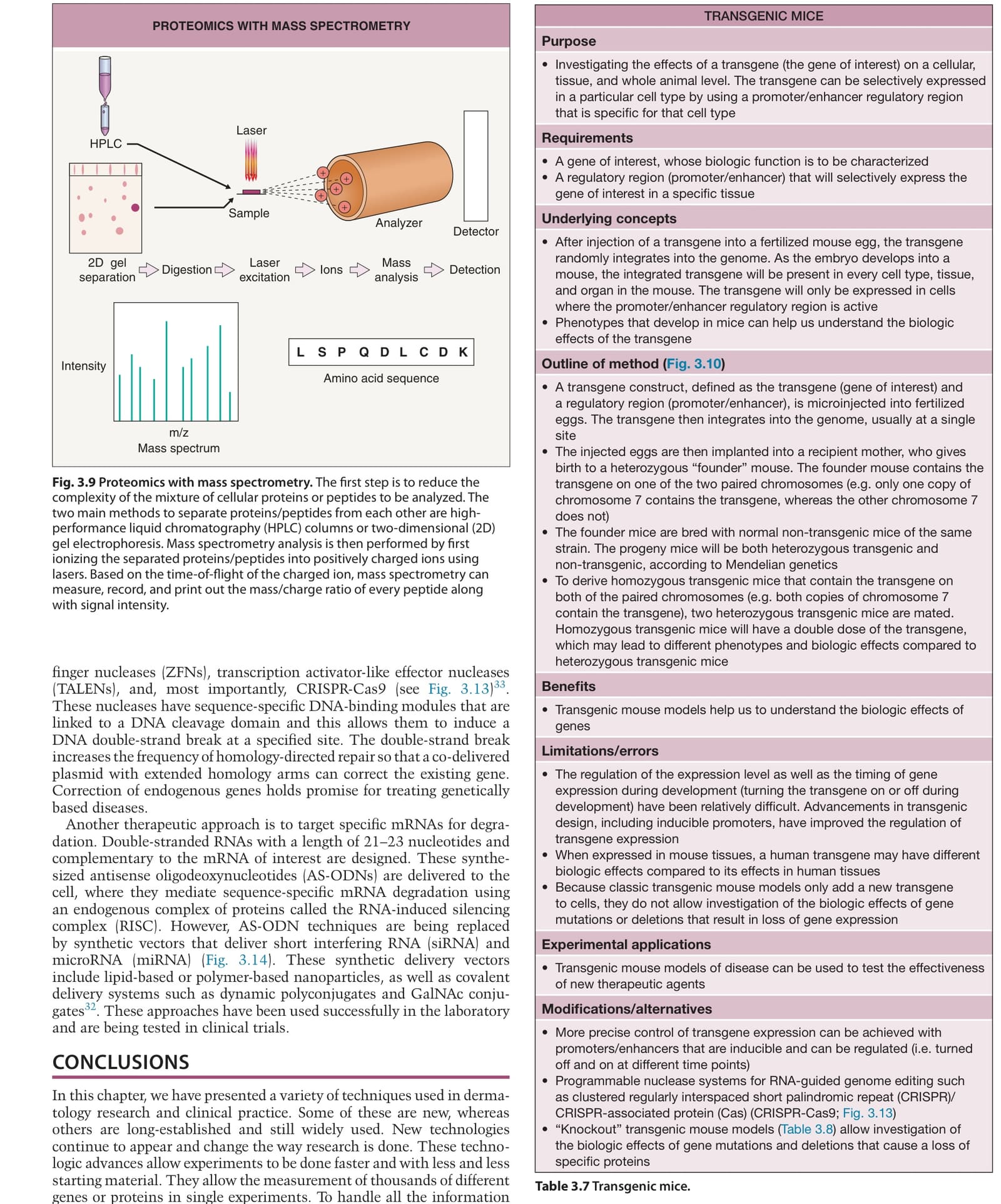
Table 3.7 Transgenic mice.
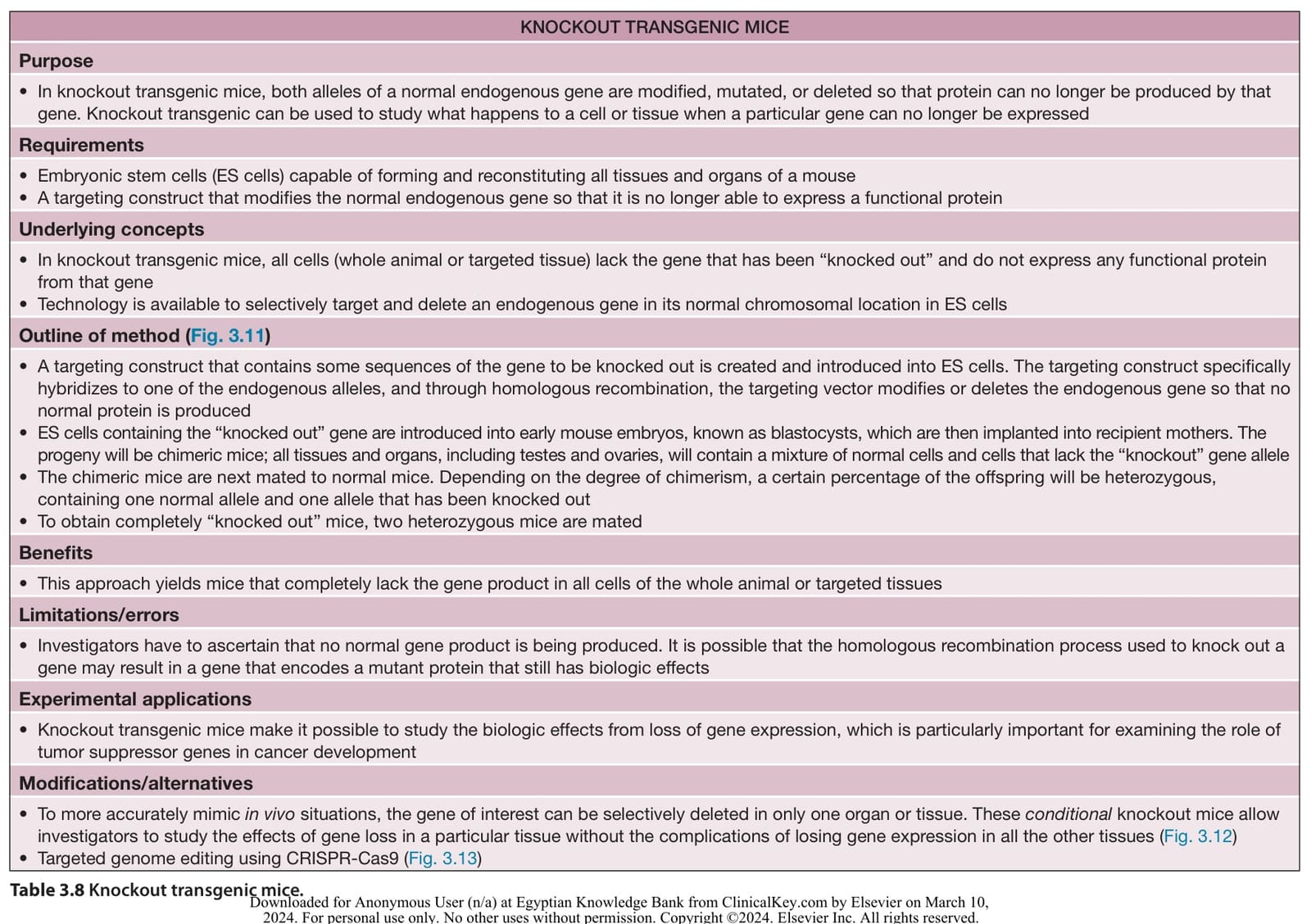
表 3-8
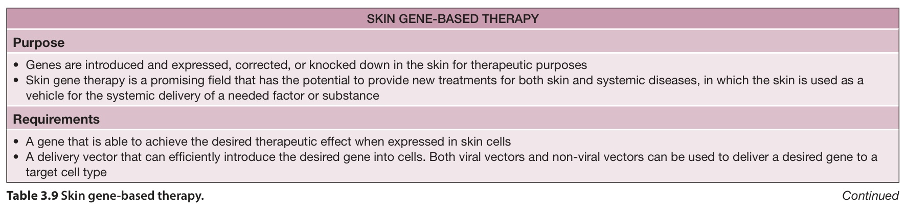
Table 3.9 Skin gene-based therapy. Continued