INNATE IMMUNE RESPONSE
Essential components of the cutaneous innate immune system include tissue-resident cells such as mast cells, macrophages, dendritic cells (DCs), non-classical T cells, natural killer (NK) cells, and innate lymphoid cells. In addition, circulating cells, including neutrophils and eosinophils, can be rapidly recruited from the bloodstream into the skin. Innate immune cells are characterized by their capacity to immediately respond to harmful signals by virtue of expression of danger sensing receptors such as Toll-like receptors (TLRs). Soluble mediators, including cytokines and complement factors, represent effector molecules of the innate immune response. In addition, microbiota as well as antimicrobial peptides produced by the epithelium and other cells support these responses against invading pathogens. Innate immune reactions are less complicated than adaptive responses and developed earlier in evolution. Nevertheless, failures in these “primitive” immune responses may be associated with severe, even fatal, health problems.
Complement
The complement system plays an important role in innate immunity. It consists of at least 20 serum glycoproteins that are activated by an enzymatic amplifying cascade (see Ch. 60). Complement proteins sense and transmit internal danger signals originating from tissue disruption (damage-associated molecular patterns, DAMPs) or products released from microbes (microorganism-associated molecular patterns, MAMPs). Three pathways can trigger the proteolytic cascade which results in protein fragments that activate complement receptors. The classical pathway is stimulated by antigen–antibody complexes, the alternative pathway by polysaccharides derived from microbial cell walls, and the later identified lectin pathway by the interaction of microbial carbohydrates with mannose-binding proteins. All three pathways lead to activation of the central C3 component and, finally, the generation of a number of immunologically active substances. For example, C3b, the cleavage product of C3, binds to the surface of microbes. Since phagocytic cells express receptors for C3b, phagocytosis of the microorganisms is enhanced. In addition, complement components bind to antigen– antibody immune complexes, which help complement receptor-bearing antigen-presenting cells target these immune complexes.
C5a is a powerful attractant for neutrophils. C3a, C4a, and C5a, also called anaphylatoxins, induce the release of inflammatory mediators from mast cells. This increases vascular permeability, thereby enabling proteins (e.g. antibodies) to enter the tissue. Assembly of the complement components C5b, C6, C7, C8, and C9 forms the membrane-attack complex (MAC), which generates pores in cell membranes and leads to death by osmotic lysis. Human cells are much less susceptible to killing by complement than microbes, since the former express certain complement receptors that can inhibit cell lysis. For example, human cells express complement receptor type 1 (CR1, CD35), decay- accelerating factor (DAF, CD55), and membrane cofactor protein (MCP, CD46), which inhibit C3 convertase and thereby block progression of the complement cascade. In addition, CD59 is a protein that binds to C8 and inhibits insertion of C9 into the cell membrane.
Complement fragments can also bind to complement receptors expressed on immune cells, such as NK cells, neutrophils, monocytes, macrophages, DCs, and, although still controversial, B and T cells. Complement receptor engagement induces and modifies several effector functions including phagocytosis of apoptotic cells and immune complexes, cell migration and adhesion, surface receptor and cytokine expression, tissue repair, and modification of cell signaling responses to pattern recognition receptors. Furthermore, components of the complement system can crosstalk and modulate TLR signaling cascades. These synergistic interactions potentially boost immune system responses directed against invading pathogens.
Toll-Like Receptors
Several families of pattern recognition receptors (PRRs) mediate responses to pathogen-associated molecular patterns (PAMPs) that are conserved among microorganisms. TLRs, the mammalian homologs of the Toll receptors identified in Drosophila, are one such family of PRRs. To date, ten TLRs have been identified in humans (Fig. 4.1), and those localized to the cell surface have the following specificities: TLR2 (in association with either TLR1 or TLR6) – recognition of lipoproteins and peptidoglycans; TLR4 – lipopolysaccharide; and TLR5 – flagellin (a component of bacterial flagella). TLR3, TLR7, TLR8, and TLR9 reside within endosomal vesicles and recognize nucleic acids, with TLR9 recognizing bacterial CpG DNA sequences and the other intracellular TLRs involved in the recognition of viral RNA.
The signaling pathway of TLRs is highly homologous to that of the receptor for interleukin (IL)-1. Upon interaction with myeloid differentiation factor 88 (MyD88), IL-1 receptor-associated kinase (IRAK) is recruited, ultimately leading to activation of the transcription factor NF-κB (see Fig. 4.1). Activation of TLRs can also result in the release of interferons (IFNs) via activation of interferon regulatory factor 3 (IRF3).
DCs express several types of TLRs. Upon activation of these receptors by microbial components, DCs mature and migrate to the lymph nodes, where they present pathogen-derived antigens to naive T cells and induce an adaptive immune response. TLRs thereby bridge the gap between the innate and adaptive immune systems. These two systems constantly interact within the skin, and therefore the innate immune system represents a potential target for modulating adaptive immune responses. Different PAMPs and danger signals polarize DCs, giving them the ability to produce certain cytokines and to induce T cells to differentiate into particular subtypes.
Inflammasomes
Closely connected with the rapid recognition of danger signals are inflammasomes – innate multi-protein complexes that sense intracellular DAMPs or PAMPs. Four types of inflammasomes have been identified: Aim2 (absent in melanoma 2), the pyrin domain-containing NLRP1 (nucleotide-binding domain leucine-rich repeat-containing receptor 1, also called NALP), NLRP3, and NLRC4 (Nod-like receptor CARD domain-containing 4). Aim2 is activated by double-stranded DNA resulting from either viral or bacterial intracellular pathogens, NLRP1 by muramyl dipeptide, and NLRC4 by flagellin. NLRP3 is triggered by numerous PAMPs and DAMPs and is thus the most important inflammasome (Fig. 4.2). The ultimate result of activation of inflammasomes is the cleavage of pro-IL-1β into active IL-1β, a highly potent inflammatory mediator. NLRPs are large backbone proteins of the complex which, upon assembly of the inflammasome complex, bind to ASC (apoptosisassociated speck-like protein containing a caspase recruitment domain [CARD]). The latter interacts with caspase-1, resulting in its activation. Active caspase-1 cleaves and activates pro-IL-1β, as well as the proinflammatory cytokine pro-IL-18. Dysregulation of inflammasomes is the cause of a group of inherited autoinflammatory diseases called cryopyrin-associated periodic syndromes (CAPS) which are associated with recurrent episodes of fever, urticarial skin lesions, arthritis, and systemic inflammation (see Ch. 45). These disorders respond very well to IL-1 blocking drugs (see Fig. 4.2).
The Skin Microbiota and Antimicrobial Peptides
The skin microbiota is comprised of billions of bacteria, archaea, viruses, and fungi that are in symbiosis with the host. Extensive research over the past decade has shown that the commensal microorganisms on the skin are essential for training cells of the cutaneous
immune system as well as for protecting against invasion of pathogenic microbes. Re-evaluation of the composition of the skin microbiota via next generation sequencing (see Fig. 3.5) has expanded the microbiome kingdom landscape beyond bacteria. The diversity of bacterial species and strains (see Fig. 74.1), viruses, and fungi is strongly dependent on host genetics, body site, skin pH, sebum content, moisture, and UV radiation exposure.
On healthy skin, microbiota outcompete pathogenic microorganisms by building a physical and biological barrier which contributes to the first line of defense against pathogen invasion. Some commensal bacteria can produce antimicrobial substances (e.g. antibiotics, serine proteases) while others interact with and train immune and skin cells to secrete interleukins, complement components, and antimicrobial peptides (AMPs). A change in the equilibrium of commensal microbiota and pathogens is an important factor in the pathogenesis of several skin disorders, most notably atopic dermatitis in which there is an overgrowth of Staphylococcus aureus.
To cope with pathogenic invaders, plants and invertebrates produce a variety of highly effective AMPs. Human epithelia, including the epidermis, also secrete AMPs as a mechanism of innate defense. The first AMP to be isolated from human skin was human β-defensin-2 (hBD-2). A number of other AMPs have subsequently been described (Table 4.1). In addition to antibacterial properties, some of these
Upon release of autoinhibition, cryopyrin interacts with apoptosisassociated speck-like protein with a CARD domain (ASC) and CARD-inhibitor of NF-κB-activating ligand (CARDINAL) within an inflammasome, leading to activation of caspase-1 and generation of mature interleukin (IL)-1β. Pyrin can competitively bind ASC and (pro)-caspase-1, thereby preventing them from being incorporated into the NLRP3 inflammasome. Proline–serine– threonine phosphatase-interacting protein 1 (PSTPIP1) binds (and perhaps inhibits) pyrin, and this interaction is strengthened by the PSTPIP1 mutations that underlie PAPA syndrome. CAPS, cryopyrinassociated periodic syndromes; CARD, caspase-recruitment domain; CINCA, chronic infantile neurologic, cutaneous and articular syndrome; IL-1R, interleukin-1 receptor; NF-κB, nuclear factor-κB; NLRP3, NOD-like receptor pyrin domain-containing protein 3; NOD, nucleotide-binding oligomerization domain; NOMID, neonatalonset multisystem inflammatory disease; PAPA, pyogenic arthritis, pyoderma gangrenosum and acne; TLR, Toll-like receptor; TNF, tumor necrosis factor. Courtesy Julie V. Schaffer, MD.
peptides possess antimycotic and, likely, antiviral activity. As demonstrated for psoriasin, which prevents Escherichia coli infection, these peptides may protect the skin from pathogenic bacterial infections.
Expression of AMPs can be induced by bacteria, bacterial products, or proinflammatory cytokines via TLRs and other mechanisms. Enhanced and reduced production of these peptides in psoriasis and atopic dermatitis, respectively, may explain why superinfections are so rare in the former disease and common in the latter. However, enhanced expression of AMPs in atopic skin has also been reported, which may reflect disruption of the epidermal barrier. Ultraviolet (UV) B radiation has been shown to induce expression of AMPs, potentially explaining the paucity of UVB-related bacterial infections despite its immunosuppressive effects (see Ch. 86). The role of microbiota in the response of the epidermis and the immune system to UV radiation has been shown experimentally in a mouse model under germ-free conditions.
Beta-defensins can also attract immature DCs and memory T cells via the chemokine receptor (CCR)-6, illustrating another link between innate epithelial defense and adaptive immunity. The antimicrobial peptide LL-37, also known as cathelicidin antimicrobial peptide (CAMP), mediates DC activation in psoriasis by binding self-DNA and forming structures that stimulate TLR9 and thereby induce IFN production (see Fig. 4.1).
Cytokines
Cytokines represent a large, heterogeneous family of low-molecularweight messenger substances that play a crucial role in intercellular communication. Cytokines can be secreted by almost any cell type, and they may act in an autocrine, paracrine, or endocrine manner. Cytokines exert their biologic activities by binding to specific cell surface receptors. Although the vast majority of cytokines occur in a soluble form, some can be membrane-bound. In addition, soluble cytokines can be sequestered by extracellular matrix molecules thereby becoming immobilized. The latter limits diffusion, aids in the build-up of cytokine concentration gradients within tissues, and results in increased force loads being transferred onto the cellular cytoskeleton. These intracellular forces can enhance stimulatory signals following cytokine binding resulting in increased cellular responses.
Cytokines influence the proliferation, differentiation, and activation of cells. Each cytokine exhibits multiple activities, a fact that can complicate strict categorization. Cytokines that are produced by leukocytes and exert effects preferentially on other white blood cells are called interleukins (ILs). Colony-stimulating factors (CSFs) are mediators that induce differentiation and proliferation of hematopoietic progenitor cells while IFNs interfere with viral replication. Cytokines that have chemoattractant activity are termed chemokines, and they play a crucial role in leukocyte migration. The main subgroups of chemokines are differentiated according to the position of two cysteine (C) residues compared with the other amino acid residues (X), CXC (α-chemokines) and CC (β-chemokines). Chemokines that recruit leukocytes are termed inflammatory chemokines, whereas those that regulate trafficking within lymphoid tissues are called lymphoid chemokines.
Early innate immune responses are dominated by cytokines with inflammatory (e.g. IL-1, IL-6, IL-10, IL-12, IL-18, tumor necrosis factor [TNF], inflammatory chemokines) and antiviral (e.g. IFN-α, IFN-β) capacities. Induction of adaptive immune responses is critically dependent on cytokines with immunomodulatory capacities (e.g. IL-2, IL-4, IL-5, IL-12, IL-13, IL-17, IL-22, IL-23, IFN-γ and TGF-β). However, since most of these mediators exhibit multiple and sometimes overlapping activities, a strict separation into inflammatory and immunomodulatory cytokines is not possible. Due to structural similarities, some cytokines are grouped into families, e.g. the IL-6 family (IL-6, IL-11, oncostatin M, leukemia inhibitory factor [LIF]), the IL-10 family (IL-10, IL-19, IL-20, IL-22, IL-24, IL-26), or the IL-12 family (IL-12, IL-23, IL-27).
Monocytes and Macrophages
Monocytes are short-lived mononuclear cells with proinflammatory and regulatory activities. Two main subtypes of monocytes and one intermediate stage with distinct functions have been described in human blood: classical CD14++CD16−; intermediate/transitional CD14+CD16+; and non-classical CD14dimCD16++. Monocytes are derived from a common myeloid progenitor in the bone marrow, circulate in the bloodstream, and migrate into tissues to respond to inflammation and infection. Within tissue, they can give rise to inflammatory DCs and macrophages whose functions are defined by the tissue environment and tissue type. Although residing in different niches, these cell types have similar functions: phagocytosis, antigen presentation to T cells, and production of cytokines.
Monocyte-derived macrophages express receptors for carbohydrates that are usually not expressed on vertebrate cells (e.g. mannose). Through this mechanism, macrophages can discriminate between “foreign” and “self” molecules. Furthermore, macrophages possess receptors for antibodies and complement. Hence, microorganisms that are coated with antibodies and/or complement are more readily phagocytosed. After phagocytosis, the microorganisms are exposed to a variety of toxic intracellular molecules, including superoxide anions, hydroxyl radicals, hypochlorous acid, nitric oxide, lysozyme, and antimicrobial cationic proteins. Macrophages can also present processed antigens to T and B cells. However, their T cell stimulatory capacity is less effective than that of DCs.
Activated macrophages release granulocyte colony-stimulating factor (G-CSF) and granulocyte–macrophage colony-stimulating factor (GM-CSF). These two cytokines induce the division of myeloid precursors in the bone marrow, releasing millions of neutrophils into the circulation.
Neutrophils
Under normal conditions, neutrophils circulate in the bloodstream, some rolling along the vascular endothelium. To enter the site of an infection, neutrophils utilize a complex process that involves adhesion molecules (e.g. integrins), chemoattractants, and other surface receptors (see Chs. 25 & 102). The recruited neutrophils phagocytose organisms and kill them within phagolysosomes by using oxygen-dependent and oxygen-independent mechanisms. The former, called the respiratory burst, involves the production of hydrogen peroxide, hydroxyl radicals, and singlet oxygen. The oxygen-independent mechanism utilizes highly toxic cationic proteins and enzymes, such as myeloperoxidase (MPO) and lysozyme. Organisms that are coated with antibodies or complement components bind to Fc and complement receptors, respectively, on neutrophils (as well as macrophages) and are more effectively phagocytosed and killed.
Ectosomes are vesicles released from the plasma membrane of neutrophils upon their stimulation (e.g. by bacteria). They carry membrane surface receptors (e.g. CD15, L-selectin), phosphatidylserine, and granule proteins (e.g. CD66b, CD87, MPO, elastase, proteinase 3, defensins, lactoferrin, collagenase I). Depending on the composition, ectosomes can induce a variety of responses in their target cells which include endothelial cells, platelets, natural killer cells, and DCs.
In addition, neutrophils can release neutrophilic extracellular traps (NETs) and thereby develop into anuclear cytoplasts. NETs are composed of extracellular strands of DNA bound to neutrophil-derived AMPs and proteins. Because NETs can entrap bacteria, fungi, and viruses, they may offer some antimicrobial protection. On the other hand, NETs can induce autoimmunity by stimulating immune responses against the NET-associated nuclear antigens (see Fig. 41.1). Whether formation of NETs contributes to the exacerbation of autoimmune diseases by infections remains to be determined.
Eosinophils
The major function of eosinophils is most likely to protect the host from infections by parasites, particularly nematodes. Infections with these organisms are associated with the production of antigen-specific IgE antibodies that coat the parasite. Via their low-affinity receptors (FcεRII, CD23), eosinophils bind to IgE antibodies and become activated. In contrast to macrophages and neutrophils, eosinophils are only weakly phagocytic. They harbor large granules that contain major basic protein, eosinophilic cationic protein, eosinophil peroxidase, and eosinophil-derived neurotoxin (see Fig. 26.1). Upon activation, eosinophils release these toxic products, which can kill parasites, together with prostaglandins, leukotrienes, and various cytokines. Eosinophils also play an important role in the pathogenesis of allergic reactions.
Basophils and Mast Cells
Basophils (found in the blood) and mast cells (located within tissues) exhibit similar functional and morphologic characteristics. At least two populations of mast cells exist, which can be differentiated by the enzymes they contain and by their tissue location. Mucosal mast cells contain only trypsin, while connective tissue mast cells contain both trypsin and chymotrypsin (see Ch. 118). In contrast to pulmonary, uterine, and tonsillar mast cells, cutaneous mast cells express the receptor for C5a (CD88), which implies that triggering of mast cells through the anaphylatoxin C5a results in cutaneous, but not systemic, reactions. Further, skin mast cells have been shown to express CD117 (c-Kit), CD26 (dipeptidyl peptidase IV), and the neural cell adhesion molecule L1. Both basophils and mast cells express high-affinity receptors for IgE (FcεRI) that avidly bind IgE (see Ch. 18).
When a specific antigen binds to mast cell-bound IgE, the FcεRI becomes activated, which leads to degranulation and release of preformed mediators, including histamine and serotonin. Other mediators such as prostaglandins, leukotrienes (B, C, D and E), and platelet-activating factor are also released, and they enhance vascular permeability, bronchoconstriction, and induction of an inflammatory response (see Ch. 18). Hence, basophils and mast cells play an important role in immediate-type allergic reactions such as urticaria and angioedema. There is also evidence that mast cells are involved in contact hypersensitivity reactions.
Innate Lymphoid Cells
Innate lymphoid cells (ILCs) are lymphoid progenitor-derived leukocytes which lack T and B cell receptors and do not express conventional hematopoietic lineage markers. ILCs require the transcription factor Id2 and the cytokine IL-7 for their development. Three subgroups have been defined based upon their cytokine and transcription factor expression profiles. ILC1s rely on the T-bet transcription factor and secrete type 1 cytokines such as IFN-γ and TNF in response to intra-cellular pathogens. ILC2s are under the control of GATA3 and ROR-α transcription factors and produce type 2 cytokines (e.g. IL-4, IL-5, IL-9, IL-13) in response to extracellular parasite infections. ILC3s are under the control of the ROR-γt transcription factor and produce IL-17, IL-22, and GM-CSF. There may be some plasticity between different ILC subgroups, but this is still under investigation.
The exact role of ILC1s in physiologic and pathologic processes within the skin still remains to be determined. In contrast, there is mounting evidence to support the role of ILC2s in allergic skin diseases such as atopic dermatitis. ILC2s activate B cells to secrete IgE via class switching, increase mast cell degranulation, and induce eosinophilia. ILC3s may play a role in triggering psoriatic flares since an increase in NKp44+ILC3s that release IL-17 and IL-22 have been found in the circulation and skin of psoriatic patients. Their potential pathogenic role is also supported by a significant reduction in NKp44+ILC3s after successful treatment with adalimumab. These ILC subsets have distinct gene expression profiles and localize to different skin compartments. For example, when they reside in compartments close to hair follicles and sebaceous glands, they negatively regulate sebaceous gland function in a TNF- and lymphotoxin-dependent manner; absence of ILCs results in overgrowth of sebaceous glands and increased fatty acid production. By controlling the production of antimicrobial lipids these cells positively influence microbiota diversity.
Natural Killer Cells
The major task of natural killer (NK) cells is to eliminate virally infected or malignant cells. NK cells can recognize their targets in two ways. Since they express Fc receptors that bind IgG (FcγRIII, CD16), NK cells can adhere to and kill target cells that are coated with IgG. This killing process is referred to as antibody-dependent cellular cytotoxicity (ADCC).
The second recognition system involves killer-activating and killerinhibitory receptors. Killer-activating receptors recognize molecules that are expressed by nucleated cells. This provides a signal for the NK cell to kill the target cell by secretion of perforins, which make holes in the cell membrane through which granzymes are injected. Granzymes lyse target cells by activating the apoptotic caspase cascade. Killer-inhibitory receptors (KIR) on NK cells recognize major histocompatibility complex (MHC) class I molecules. KIRs shut off the killer signal and thus prevent autolysis of the host. Tumor cells and viruses often downregulate MHC class I molecules to escape recognition by cytotoxic T cells, but this renders “MHC class I-low” cells susceptible to recognition by NK cells.
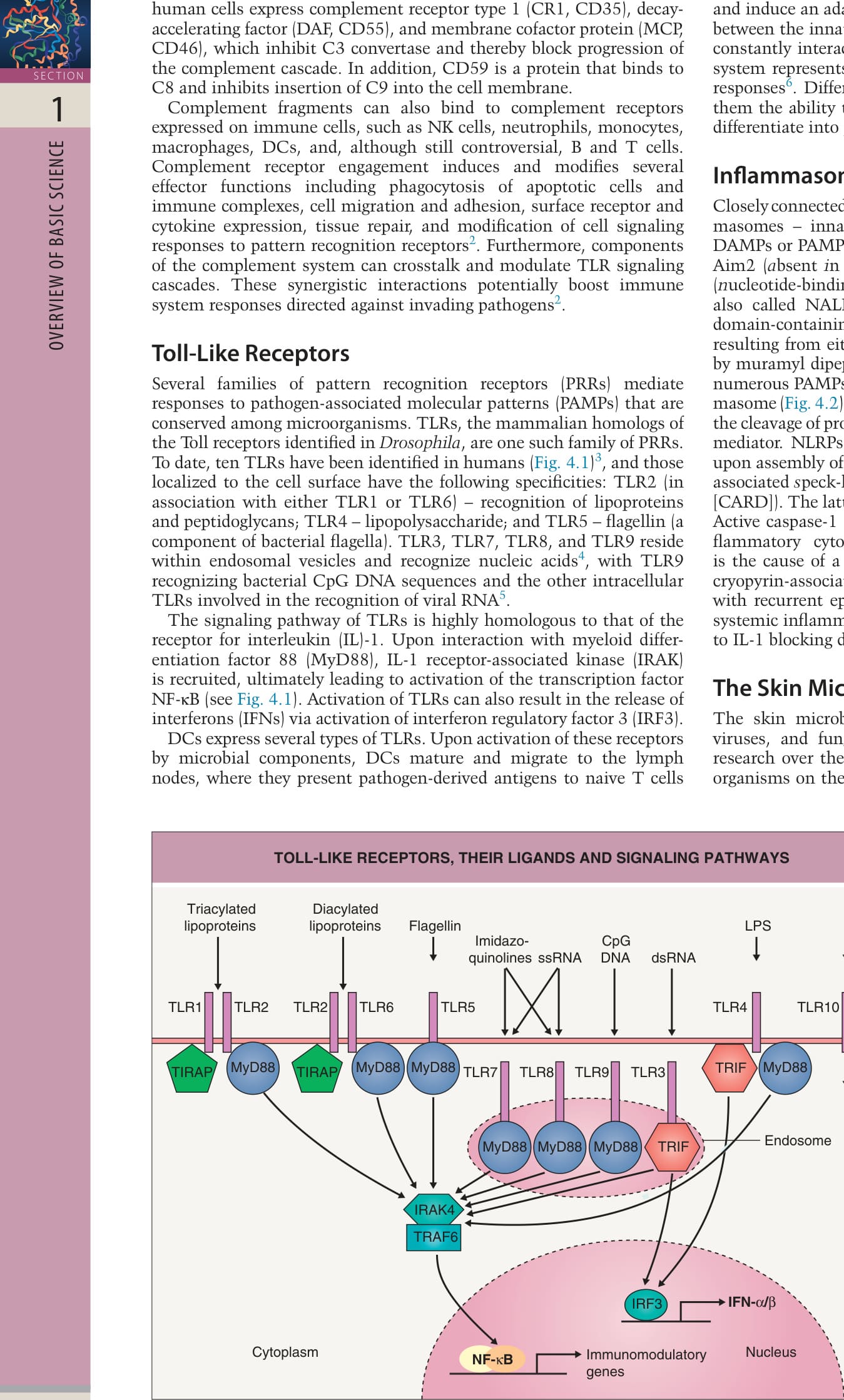
Fig. 4.1 Toll-like receptors, their ligands, and signaling pathways. Toll-like receptors (TLRs) recognize pathogen-associated molecular patterns and/or synthetic compounds in a specific fashion. A TLR2/TLR1 dimer recognizes triacylated lipoproteins, and a TLR2/TRL6 dimer interacts with diacylated lipoproteins. TLR5 recognizes flagellin and TLR4 recognizes lipopolysaccharide (LPS). These TLRs are located on the cell membrane and are internalized upon ligand interaction. TLR3, TLR7, TLR8, and TLR9 are located on intracellular membranes of endosomes and lysosomes. TLR3 recognizes viral double-stranded RNA (dsRNA); TLR7 and TLR8 viral single-stranded RNA (ssRNA); and TLR9 bacterial and viral hypomethylated DNA (CpG [5′-C-phosphate-G-3′] motifs). TLR7 and TLR8 also bind to synthetic compounds (imidazoquinolones). All TLRs except TLR3 utilize myeloid differentiation factor 88 (MyD88) for signaling, while TLR2 and TLR6 also require Toll-interleukin-1 receptor domaincontaining adaptor protein (TIRAP). MyD88 signaling via IL-1 receptor-associated kinase-4 (IRAK-4) and tumor necrosis factor receptor-activated factor-6 (TRAF-6) mostly results in activation of nuclear factor κB (NF-κB), ultimately inducing transcription of genes encoding immunomodulatory and proinflammatory molecules. TLR3 and TLR4 signal via TIR-domaincontaining adapter-inducing interferon-β (TRIF). The TRIF pathway induces the production of interferons (IFNs) via interferon regulatory factor-3 (IRF3). Both the ligand and signaling pathway of TLR10 are still unknown. Adapted from Miller LS. Toll-like receptors in skin. Adv Dermatol 2008;24:71–87 and from McInturff JE, Modlin RL, Kim J. The role of toll-like receptors in the pathogenesis and treatment of dermatological disease. J Invest Dermatol 2005;125:1–8.
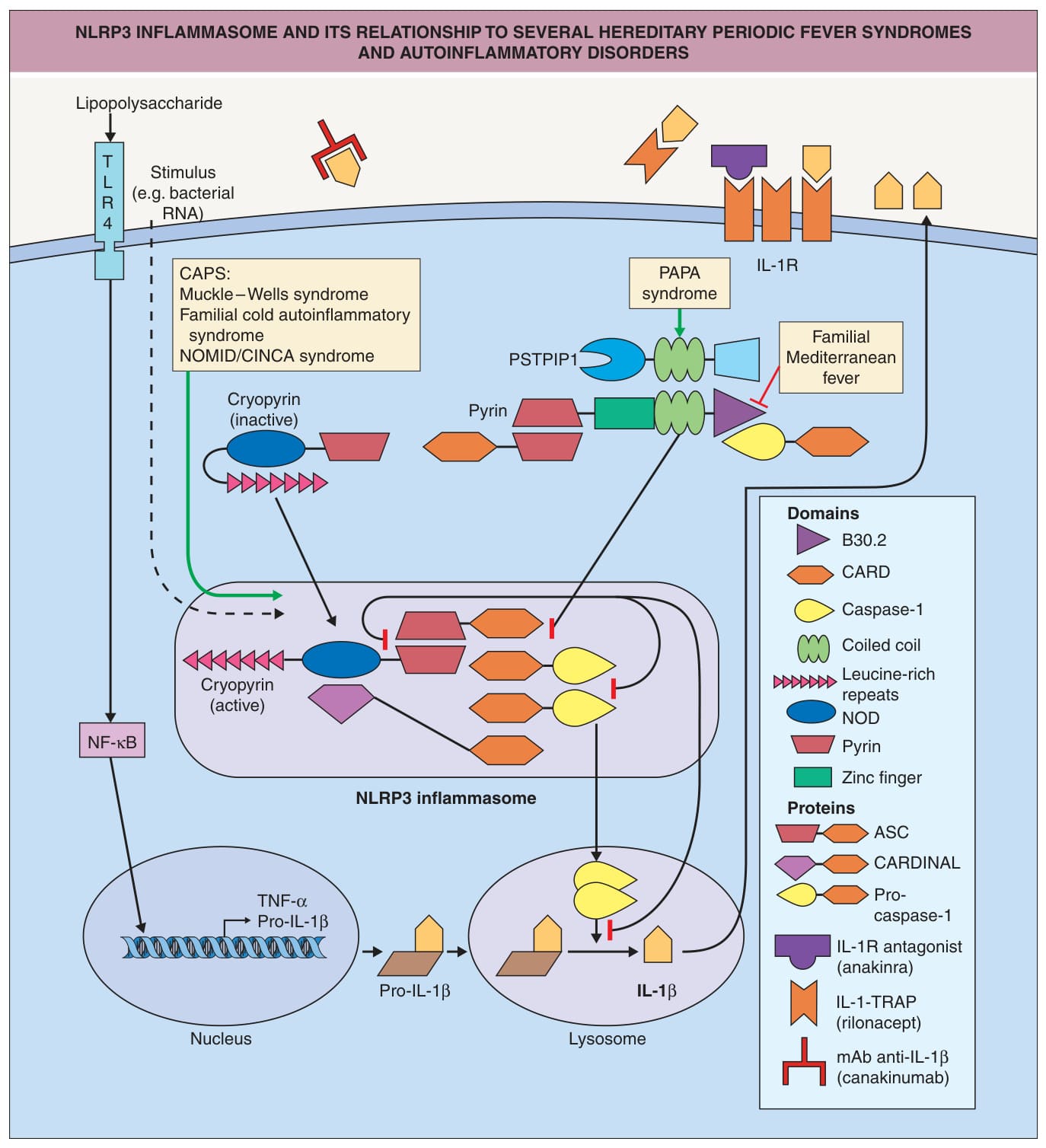
Fig. 4.2 NLRP3 inflammasome and its relationship to several hereditary periodic fever syndromes and autoinflammatory disorders.
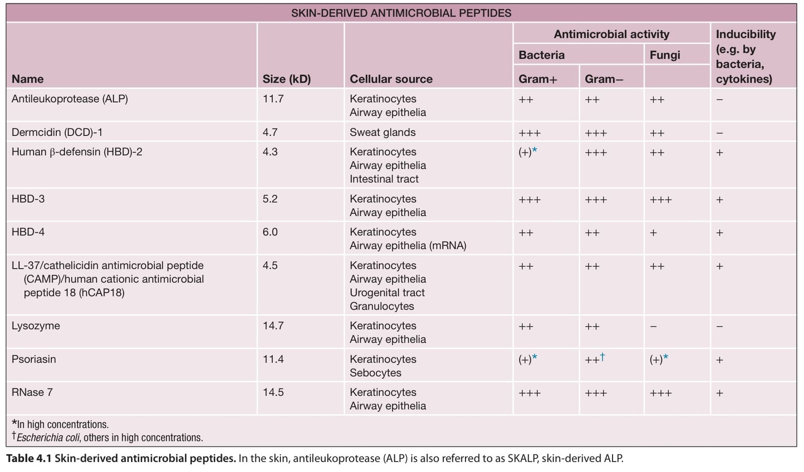
Table 4.1 Skin-derived antimicrobial peptides. In the skin, antileukoprotease (ALP) is also referred to as SKALP, skin-derived ALP.