ADAPTIVE IMMUNE RESPONSE
The characteristic features of an adaptive immune response are its specificity and enhancement with each successive antigen encounter owing to the accumulation of cellular “memory”. A crucial event during the generation of an adaptive immune response is antigen presentation.
Antigen-Presenting Cells
Various cells can present antigens, depending on how and where the antigen first encounters cells of the immune system. Professional antigen-presenting cells (APCs), such as DCs and B cells, are specialized in capturing and displaying antigens to lymphocytes. Within the epidermis, Langerhans cells (LCs) are key APCs.
Langerhans cells
In 1868, Paul Langerhans first described dendritic cells that localized within the epidermis. Because of their shape, Langerhans thought that these cells might be of neural origin. More than 100 years later, it was shown that LCs are derived from bone marrow. LCs share ontogeny with tissue-resident macrophages, but functionally resemble DCs. Formation of LCs depends on transforming growth factor-β1 (TGF-β1) and macrophage colony-stimulating factor receptor (M-CSFR) ligands (M-CSF, IL-34), which are provided in an autocrine fashion.
LCs are intimately associated with nerve fibers that extend from the dermis into the epidermis and they can modulate LC function via the release of neuropeptides such as calcitonin gene-related peptide (CGRP). Psychological stress has been shown to result in a reduction of LCs and to influence local neuropeptide expression in human skin, suggesting a connection between environmental influences, mental health, and skin immune homeostasis. In humans, the number of LCs is reduced on the palms and soles, genitalia, and buccal mucosa. Of note, their density decreases with age and is reduced in chronically UV-exposed skin.
Ultrastructurally, LCs have rod-shaped organelles termed Birbeck granules (see Fig. 91.5). A Ca+-dependent lectin with mannose-binding specificity called langerin (CD207) is associated with and responsible for the formation of Birbeck granules. Birbeck granules result from the antigen-capture function of langerin, which routes antigens into these organelles and provides access to a non-classical antigenprocessing pathway. Activated LCs elongate their dendrites which can then penetrate keratinocyte tight junctions and survey the area just beneath the stratum corneum for antigens. These penetrating dendrites take up antigens which co-localize with langerin/Birbeck granules. By forming new tight junctions, keratinocytes rapidly close the defects produced by the penetrating dendrites, thereby maintaining skin integrity during antigen uptake.
Histochemically, human LCs can be visualized by staining for adenosine triphosphatase (ATPase), a membrane-bound, formalinresistant, sulfhydryl-dependent enzyme. In addition to langerin, antigenic moieties present on human LCs include the pan-hematopoietic marker CD45, MHC class II antigens (HLA-DR), CD1a, S100 protein, and vimentin. CD1a is a useful marker for LCs, since within the epidermis (normal or inflamed) it is exclusively expressed on LCs, whereas HLA-DR antigens are expressed on keratinocytes in inflamed skin (Table 4.2). Since CD1a does not exist in the murine system, staining for MHC class II antigens is often used for detection of murine LCs in non-perturbed skin (Fig. 4.3). In addition, human LCs express the high-affinity IgE receptor (FcεRI). Subtle differences in the surface marker expression of resident LCs, migrating LCs, and langerin− dermal DCs have been described (Table 4.2).
Other dendritic cells
DCs are professional APCs that display an extraordinary capacity to stimulate naive T cells and initiate a primary immune response. They
CCR, CC-chemokine receptor.Adapted from ref. 131.
also control skin immune homeostasis via interactions with a variety of other immune cells, such as mast cells, macrophages, and ILCs, either via direct cell–cell contact or by secreted cytokine messengers. DCs can develop from different types of progenitor cells, while the same progenitor cell can give rise to a variety of functional phenotypes of DCs. The DC system is very complex, and to further complicate matters, there are a number of differences between the experimental systems used to study human and murine DCs.
In general, dermal DCs can be divided into conventional DCs (cDCs) comprising cDC1 and cDC2 cells, and plasmacytoid DCs (pDCs). cDCs are the major dermal DC type under non-inflammatory conditions and they reside mainly in the upper dermis, compared to dermal macrophages which are found primarily in the deeper, vascular-rich dermis. Human cDC1s are defined by the expression of CD141 and BDCA3 and cDC2s by CD1c. The two subgroups have overlapping, but distinct, immune functions – cDC1 cells can cross-present viral antigens and self-antigens and elicit Th1 immune responses, whereas hapten- or protein-primed cDC2 cells induce Th2-specific immune responses, while reducing Th17 cytokines.
Plasmacytoid DCs (pDCs; CD123+) are specialized DCs that secrete large amounts of type I IFN in response to TLR7 or TLR9 activation. As such, they are powerful defenders against viral infections, but are normally absent in healthy human skin. Although pDC numbers are quite low in healthy individuals, circulating pDCs are significantly increased in patients with systemic lupus erythematosus (SLE). In addition, pDCs have been shown to infiltrate lesions of cutaneous lupus and psoriasis and further promote inflammation.
In the dermis of mice, two other subsets of DCs have been described, double-negative (DN) DCs and monocyte-derived DCs. The human counterpart of the latter are CD14+ dermal cells, whereas the counterpart of the former is still elusive. In the skin, monocyte-derived DCs have the potential to locally present antigens by forming immune clusters and thereby activating skin-resident T cells and regulatory T cells (Tregs).
In humans, lymphoid and plasmacytoid DCs induce Th2 responses while myeloid DCs generate Th1 reactions. Activation of TLR7 via the immunomodulator imiquimod, an imidazoquinoline (see Fig. 4.1), drives myeloid DCs to express perforin and granzyme B and drives plasmacytoid DCs to express TNF-related apoptosis-inducing ligand (TRAIL). This enables killing of tumor cells, indicating that both myeloid and plasmacytoid DCs may be directly involved in imiquimodinduced destruction of skin cancer.
The type of immune response that is generated is also critically dependent on the state of maturation of the stimulating DC at the time of antigen presentation. For the induction of Th1 responses, the presence of IL-12 is crucial. DCs tend to produce IL-12 directly after an activating step in their maturation, driving T cells into a Th1 phenotype if they meet the DCs at this stage. At later time points,
the production of IL-12 decreases, thereby favoring the development of a Th2 response. This process is also influenced by the innate immune system (e.g. stimulation by TLRs) and mast cells which can further skew T cells to produce Th1 and Th17 cytokines. Taken together, the parameters responsible for the type of Th cell differentiation induced by DCs remain to be fully defined. Of note, immature DCs induce tolerance due to incomplete T cell activation, resulting in regulatory T cells that suppress immune responses.
Antigen presentation
To initiate sensitization, antigens must be presented to T cells by APCs. For many years, LCs were thought to be the most important APC in the skin since contact sensitization could not be induced in sites that were naturally devoid of LCs (e.g. murine tail skin) or in which LCs had been depleted (e.g. by UV radiation). However, transgenic mice in which LCs are completely depleted demonstrated variably diminished but not completely abrogated sensitization responses; in one such model, the sensitization response was completely normal. In a different knockout mouse model characterized by constitutive and durable absence of epidermal LCs, an enhanced sensitization response was actually observed, suggesting that LCs may have regulatory functions. This “LC paradigm” proposes that LCs may be tolerogenic when they present antigens under steady-state non-inflammatory conditions, but sensitizing upon stimulation by inflammatory mediators. Which of these activities is the main function of LCs remains to be determined, and there is accumulating evidence that dermal DCs are equally, if not even more, important than LCs for presenting antigens to mount productive immune responses.
Cutaneous APCs actively take up antigens in the skin, but antigen presentation to lymphocytes takes place in the regional lymph nodes. In the presence of inflammation, the APCs become activated, leave the skin, and then migrate to the draining lymph nodes via afferent lymphatics. During migration, they change their phenotypic and functional behavior and develop into mature DCs. For example, molecules involved in antigen uptake and processing (Birbeck granules, Fc receptors) are downregulated on activated DCs. When compared, gene expression profiles of LCs and migratory dermal DCs (e.g. cDC1s) are very similar and both have the capacity to prime naive helper T cells in lymph nodes.
In activated LCs, CD44, a hyaluronic acid receptor involved in the tissue homing of leukocytes, is upregulated. The splice variant CD44v6 supports binding of LCs to T cell-rich areas of lymph nodes. Furthermore, the integrins αβ and αβ, which exhibit affinity to the basement membrane zone, are induced on the surface of emigrating LCs. The release of proteolytic enzymes like matrix metalloproteinase-9 (type IV collagenase) may enable their penetration through the basement membrane. Their dendricity becomes less pronounced, and surface molecules necessary for antigen presentation and T cell priming are upregulated (e.g. MHC class I, MHC class II, CD40, CD54, CD58, CD80, CD86). At this stage, LCs emigrating from the epidermis are almost indistinguishable from DCs obtained from lymphoid organs.
In contrast to B cells, T cells cannot recognize soluble protein antigens per se; instead, the T cell receptor (TCR) recognizes antigen-derived peptides bound to MHC locus-encoded molecules expressed on APCs. CD4+ T cells recognize antigens in association with MHC class II molecules, while CD8+ T cells recognize antigens in association with MHC class I molecules (Fig. 4.4).
Two pathways exist by which antigens can be “loaded” onto MHC molecules. If the antigen has been produced endogenously within the cell (e.g. viral or tumor proteins), it is complexed with MHC class I molecules through intracellular processing pathways (Fig. 4.5). The proteasome degrades cytosolic antigens produced by the cell. Resulting peptides (which consist of 8 to 12 amino acid residues) are then imported into the endoplasmic reticulum (ER) in a TAP (transporter associated with antigen processing)-dependent manner and loaded onto MHC class I molecules. After binding to the MHC class I–β microglobulin complex, these peptides are transported to the cell surface via the Golgi apparatus. Alternative pathways by which exogenous proteins are phagocytosed with the phagosome fusing to the ER may also exist. The proteins would subsequently be re-transported out of the ER into the cytoplasm and then degraded by the proteasome. The degraded peptides could then enter the pathway that is normally employed for endogenous proteins via the TAP protein. As the vast majority of nucleated cells express MHC class I molecules, many cell types can serve as nonprofessional APCs for MHC class I-restricted antigen presentation in a secondary immune response. This can also result in activation of effector cytotoxic T lymphocytes.
MHC class II-dependent antigen presentation is critically dependent on DCs, B cells, and monocytes/macrophages. MHC class II-associated antigen presentation targets primarily exogenous and less frequently endogenous antigens. Exogenous antigens are taken up via macro- or micropinocytosis or by receptor-mediated endocytosis (Fig. 4.6). One example of the latter is the DEC-205 receptor (CD205), which guides antigens into endocytic vesicles that reside deeper inside the cell and contain MHC class II molecules. As a consequence of this unique intracellular targeting, antigens endocytosed by the DEC-205 receptor stimulate respective T cells up to 500-fold greater than antigens taken up by pinocytosis or other receptors. Protein degradation eventually takes place in the endo-/lysosomes, yielding peptides with a typical length of 15 to 22 amino acid residues. These peptide fragments enter specialized endosomal compartments containing MHC class II molecules that are generated in the ER.
Newly synthesized MHC class II molecules are associated with an invariant chain, which inhibits dissociation of empty MHC class II molecules and transports the MHC class II complex from the ER to the specialized endosomal compartments where the molecules can interact with antigen peptide fragments (see Fig. 4.6). In this compartment, the invariant chain is cleaved by proteases, leaving a small fragment called CLIP (class II-associated invariant peptide) that binds to the class II molecule. Upon interaction with the antigenic peptides, the CLIP fragment is released from the complex. The MHC class II molecule with the bound antigen peptide is subsequently expressed on the cell surface, allowing antigen recognition by T cells carrying the appropriate TCR.
Classically, MHC class I molecules present self- or pathogen-derived antigens that are synthesized within the cell to CD8+ T cells (see Fig. 4.5), whereas exogenous antigens derived via endocytic uptake are loaded onto MHC class II molecules for presentation to CD4+ T cells (see Fig. 4.6). However, it has become evident that some DCs are also able to process exogenous antigens into the MHC class I pathway for presentation to CD8+ T cells. This mechanism, referred to as
cross-presentation, allows DCs to induce either tolerance (to self-antigens) or immunity (to exogenous pathogens).
T Cells
T cell precursors continuously migrate from the bone marrow to the thymus, where T cells develop. In the thymus, T cells bearing the α/β TCR are subjected to a complex selection process. In contrast to antibodies, which represent the antigen receptor on B cells and recognize antigens in their naive form, the α/β TCR recognizes only short peptide fragments that are generated during antigen processing (see above). These processed antigens are presented to the TCR by MHC molecules on the cell surface. The amino acid sequences recognized by the TCR include those from both the MHC molecule and the antigenic peptide. Consequently, the TCR recognizes a combination of “self” MHC molecules (which are highly polymorphic) and “foreign” peptides. T cells that recognize “self” MHC molecules but not “self” peptides are useful for immune defense and do not lead to undesirable autoimmunity. This requirement is achieved by a complex process involving both positive and negative selection within the thymus.
T cell development
Developing T cells within the cortex of the thymus engage MHC complexes on thymic DCs. When the T cells are able to recognize these MHC molecules via their TCR, they receive a survival signal (positive selection). Otherwise, the T cells undergo apoptotic cell death. At this stage, more than 95% of the developing T cells die in the thymus because they are not selected due to their uselessness (i.e. inability to recognize “self” MHC molecules). In addition, T cells that express a TCR with a high affinity for the complex of a “self” peptide plus a “self” MHC molecule are eliminated by apoptosis, since they are potentially harmful. This process, called negative selection, takes place in the thymic medulla and involves DCs and macrophages that process and present a spectrum of “self” antigens. Positive and negative selection allow the survival of just those T cells that recognize foreign (but not “self”) peptides in the context of “self” MHC molecules and thus are useful for immune defense without causing auto-attack.
During thymic education, a variety of surface molecules are switched on and off. Components of the CD3/TCR complex, together with CD4 or CD8 molecules, have roles in this process. In general, CD4+ T cells function as helper T cells and recognize antigens presented by MHC class II molecules, whereas CD8+ T cells are usually cytotoxic and recognize antigens in association with MHC class I molecules (see Fig. 4.4). During their early stage of development within the thymus, T cells express both CD4 and CD8. Upon expression of an appropriate TCR, these immature T cells exhibit the capacity to recognize antigenic peptides associated with either MHC class I or class II, since they still express both CD4 and CD8 (double-positive stage). In the subsequent process, one of these surface markers is lost, resulting in single-positive T cells expressing either CD4 or CD8.
T cell receptor
One major feature of an adaptive immune response is the recognition of specific antigens. This is achieved either by the TCR during a T cellmediated response or by antibodies during a B cell response. TCRs are transmembrane molecules consisting of α/β (vast majority of T cells) or γ/δ (<10% of T cells) heterodimers. Antigen recognition by γ/δ T cells appears to be independent of classic MHC molecules, instead utilizing other MHC-like molecules (e.g. CD1) that preferentially present certain lipid and glycolipid antigens.
An amazing feature of the immune system is that it provides specific receptors for every possible peptide antigen. B cells are capable of producing approximately 10 different antibody variable regions, and T cells generate a comparable number of TCR variable regions. Remarkably, these numerous proteins are encoded by fewer than 400 genes. This tremendous diversity is created via a unique recombination process that cuts, splices, and modifies genes in the variable region.
Four segments of genes are involved in TCR formation: the variable (V), diversity (D), joining (J), and constant (C) regions (see Fig. 4.4).
In contrast to the TCRβ and TCRδ loci (both on chromosome 7), the TCRα and TCRγ loci (both on chromosome 14) do not contain D segments. The segments are cut out by nucleases and spliced together by ligases, ultimately forming the final gene sequence that encodes the receptor molecule. The tremendous diversity is explained by the multi-plicity of all these regions within the genome (e.g. ~70–80 V genes and ~60 J genes for the TCRα alone), but only one of each type is used for a particular TCR.
Each lymphocyte utilizes a different combination of V, D, and J gene segments to form the genetic code of its antigen receptor. Any one of the genes can join with any other one to form the final VDJ region, thereby creating tremendous diversity. In addition, the recombination process is subject to inaccuracies during splicing, which cause slight variations at the VDJ junctions, and additional nucleotides can be inserted by the enzyme deoxyribonucleotidyl transferase at the spliced regions. These two events increase the diversity even further. Of note, only a tiny minority of these cells will become functional over a lifetime, with the vast majority dying without ever having met their antigen.
Defects in the recombination-activating genes RAG1 and RAG2, which encode two of the enzymes that mediate the recombination of variable-region genes in both B cells and T cells, cause a form of severe combined immunodeficiency (see Ch. 60). Affected individuals are unable to produce lymphocytes bearing functional antigen receptors.
TCRs are associated with the CD3 complex, which transmits signals upon antigen binding. CD3 consists of CD3γ, CD3δ, two molecules of CD3ε, and a disulfide-linked CD3ζ homodimer. Upon binding to the peptide–MHC complexes, TCRs become cross-linked (Fig. 4.7). Aggregation of the TCRs causes phosphorylation of tyrosines in the cytoplasmic tails of the CD3 complex, which contain immunoreceptor tyrosine-based activation motifs (ITAMs). Phosphorylation involves kinases such as Lck (which also binds to the cytoplasmic tails of CD4 and CD8), Fyn, and ZAP70 (see Fig. 4.7). These events ultimately lead to the transcriptional activation of genes that encode cytokines and induce cellular proliferation and differentiation.
Costimulatory signals
Signaling through the TCR complex alone is not sufficient for naive T cell activation. The presence of costimulatory signals is needed for T cells to undergo antigen-specific clonal expansion. Hence, development of a productive T cell immune response requires at least two types of stimuli. The first signal is the interaction of the TCR with peptide–MHC complexes presented by APCs, which determines the specificity of the immune response. The second signal involves surface molecules and cytokines, which determine the clonal expansion of specific T cells and their differentiation into effector and memory cells (Fig. 4.8). Without these costimuli, signaling by the antigen receptors alone will either cause anergy (non-reactivity) or apoptotic cell death. The major receptors and ligands expressed on APCs and T cells that play a role in activation are listed in Table 4.3.
The most relevant accessory molecules belong to the B7-CD28 super-family which includes the following receptor : ligand pairs – CD28/ CTLA-4 : CD80/CD86, ICOS : ICOS-L, and PD-1 : PD-L1/PD-L2. The best-investigated costimulators for T cells are B7-1 (CD80) and B7-2 (CD86). Both are induced on immature, resting APCs by various TLR ligands or by cytokines (e.g. TNF, IL-1). The CD28 receptor on T cells recognizes the B7 molecules and delivers activating signals, including the expression of antiapoptotic genes and the production of cytokines such as IL-2 (see Fig. 4.7). Interaction of ICOS (inducible T cell co-stimulator; CD278) with ICOS-L (CD275) induces less IL-2 but promotes T helper cell differentiation and effector function through the production of IL-10, -4, -17, -21, and IFN-γ. The ICOS : ICOS-L signaling pathway is also crucial for T cell-dependent B cell responses.
Cytotoxic T lymphocyte-associated antigen-4 (CTLA-4 [CD152]) also binds to the B7 molecules, with even higher avidity than CD28. However, unlike CD28 binding, cross-linking of CTLA-4 to B7 downregulates IL-2 production and cell cycle progression, whereas blockade of CTLA-4 signaling prolongs T cell activation. Hence, CTLA-4 is regarded
as a T cell-associated co-receptor with negative regulatory effects. PD-L1 (CD274) and PD-L2 (CD273) also deliver a co-inhibitory signal to T cells via PD-1 (programmed death-1; CD279) and thus inhibit T cell proliferation and cytokine production. Through the expression of CTLA-4, PD-Ll, and PD-L2, tumor cells may escape an immune response. Neutralization of these inhibitory molecules by antibodies (e.g. anti-CTLA-4 [ipilimumab], anti-PD-1 [nivolumab, pembrolizumab, cemiplimab], anti-PD-L1 [avelumab]), referred to as immune checkpoint inhibitors, has proven to be a highly effective therapy for a variety of malignancies including metastatic melanoma (see Ch. 128). Many of the associated side effects of these therapies such as colitis, hepatitis, and hypophysitis are due to autoimmune phenomena.
If a T cell recognizes its specific antigen peptide in association with the appropriate MHC molecules and becomes activated by costimulatory signals, then cell division and clonal expansion occur. Cytokines, especially inflammatory mediators like IL-1, IL-6 and TNF, also provide costimulatory signals by themselves and, in addition, upregulate other costimulatory molecules. Hence, a T cell is much more likely to be activated if it meets its specific antigen via an APC that has been exposed to an inflammatory environment. Polarizing cytokines such as IL-4, IL-12, and IL-23 also critically influence the differentiation of naive T cells.
Memory cells
Memory T cells have a longer lifespan than effector cells and react more rapidly upon re-exposure to their specific antigen. While naive T cells express CD45RA, memory T cells are characterized by the expression of the surface molecule CD45RO. Two distinct subsets of human memory T cells have been described. Stationary, tissue-resident memory T cells that comprise resident memory T cells (Trms) and effector memory T cells (Tems). They express receptors for migration into inflamed tissues to fulfill their effector functions. Both subsets express low or no levels of the lymph node homing receptor CCR7, preventing tissue egress. In contrast, CCR7+ memory T cells comprise central memory T cells (Tcms) and migratory memory T cells (Tmms) and they have the potential to recirculate to lymph nodes. Tcms lack immediate effector function, but efficiently stimulate DCs to produce IL-12 and can differentiate into Tems upon secondary stimulation.
Organ-specific adhesion molecules guide memory T cells to various locations, including the skin. It is estimated that human skin contains twice as many T cells as the blood (~20 billion T cells). In normal human skin, 2% to 3% of T cells reside within the epidermis while the bulk of T cells are in the dermis. The majority of T cells express the α/β TCR and either CD8 or CD4. Trms express the skinhoming receptor CLA (see below) as well as CD2, CD5, and CD45RO; they are CCR7-negative and can be CD4- or CD8-positive.
Trms play a major role in host defense against previously encountered antigens and with infections can populate the entire skin surface. Upon antigen encounter, Trms proliferate and clear the pathogen. Associated inflammation causes nonspecific recruitment of T cells from the bloodstream via endothelial cell activation; the few antigenspecific T cells recruited into the skin in this manner also contribute to the clearance of the pathogen. In addition, DCs carry the antigen to the skin-draining lymph nodes and present it to Tcms. This gives rise to new populations of skin-homing Tems which migrate to the skin and also participate in the clearance of the antigenic attack.
Trms differ from other memory T cells as they do not circulate back to lymph nodes once they are recruited to the skin due to encounters with pathogens. More recently, these long-lived cells have been shown to reside within human skin for a decade and are at least partially resistant to radiochemotherapy. Despite their beneficial effect in rapid on-site clearing of infections, Trms have also been reported to enhance allergic and autoimmune skin diseases and play a role in skin cancer biology.
Conventional T cells
Two major types of effector T cells have been identified: CD4+ T helper (Th) cells and CD8+ cytotoxic T (Tc) cells. The former activate B cells and modulate important components of cell-mediated immune responses, mainly via cytokine production. CD8+ Tc cells are crucial to antiviral and antitumor responses.
CD4+ Th cells recognize “their” antigenic peptides in association with MHC class II molecules (see Fig. 4.4). Based primarily on differences in their cytokine secretion patterns, several types of Th cells have been described. Naive precursor Th cells only produce IL-2 whereas preactivated precursor cells, called Th0, can release a wide variety of cytokines (e.g. IL-2, IFN-γ, lymphotoxin, IL-3, IL-4, IL-5, IL-6, IL-9, IL-10, IL-13, GM-CSF, TNF). As they develop into Th1, Th2 or Th17 cells, the cytokine secretion pattern becomes more restricted and specific (see below). More recently described Th9 and Th22 cells may represent separate types.
Morphologically, the cells in the various Th subsets are almost indistinguishable, although surface differences (especially in chemokine receptors) have been described. Differentiation of naive Th cells into effector Th subsets is driven by well-defined transcription factors.
However, Th cell differentiation into effector cells is not irreversible. Effector Th cells sustain plasticity and can differentiate into other subsets thereby flexibly adjusting to environmental changes.
Th1 cells: Typical Th1 cytokines are IFN-γ, lymphotoxin, and IL-2. IL-2 induces proliferation of CD4+ cells in an autocrine manner and affects the cytokine differentiation pattern; however, it also stimulates CD8+ T cell division and cytotoxicity, thereby providing “help” for CD8+ Tc cells. IFN-γ activates macrophages to kill intracellular pathogens (e.g. mycobacteria, fungi, protozoa) and stimulates NK cell cytotoxicity. Therefore, Th1 cells are involved in the clearance of intracellular pathogens, and their cytokines preferentially induce a cell-mediated inflammatory response, such as the granulomatous lesions of tuberculosis or leprosy. Macrophages stimulated by IFN-γ release IL-12, the major cytokine that drives Th0 cells towards a Th1 phenotype (Fig. 4.9). In addition, the transcription factor T-bet drives differentiation towards the Th1 phenotype.
Th1 cells also promote the isotype switch of B cells to produce complement-binding antibodies and they contribute to the pathogenesis of autoimmune diseases such as rheumatoid arthritis, multiple sclerosis, allergic contact dermatitis, and psoriasis.
Th2 cells: Th2 cells are under the control of the transcription factor GATA3 and produce primarily IL-4, IL-5, and IL-13. These cytokines favor production of non-complement-binding antibodies. IL-4 switches B cells to produce IgE, and IL-5 promotes an expansion of eosinophils. Therefore, Th2 responses are often associated with allergic diseases. Comparable to the role of IL-12 in inducing Th1 responses, IL-4 is the key cytokine in driving Th2 responses (see Fig. 4.9). In addition, IL-4 suppresses Th1 and Th17 differentiation. However, IL-4 seems to exert opposing effects on CD4+ T cells and DCs, since it can stimulate DCs to produce IL-12 and thus paradoxically promote Th1 development.
Many TLR signals and infections are strong inducers of IL-12 and therefore favor a Th1 response. However, other TLR signals to DCs and early production of IL-4 favor the generation of Th2 cells. Additional factors that influence the Th1/Th2 decision include the dose of the antigen, the APCs involved, the cytokines secreted (by APCs and bystander cells), the genetic background of the host, and the presence of costimulatory molecules. Of note, many immune responses are not strongly polarized in the Th1 or Th2 direction.
Th9 cells: In response to TGF-β and IL-4, CD4+ T cells secrete IL-9. The development of Th9 cells depends on the IL-4-activated transcription factor PU.1. Th9 cells predominantly reside in human skin and are involved in protecting against pathogens, but also play a role in allergic and non-allergic inflammatory skin diseases. In addition, they contribute to extracutaneous allergic diseases in that IL-9 is highly expressed in the lungs of asthmatic patients, especially children.
Th17 cells: CD4+ T cells that produce IL-17, but not IFN-γ or IL-4, are referred to as Th17 cells. Besides IL-17A, these cells also secrete IL-17F, IL-21, IL-22, IL-25 and IL-26. Whereas the heterodimeric cytokine IL-12 (consisting of a p35 chain and a p40 chain) is crucially involved in the development of Th1 cells, the IL-12-related cytokine IL-23 (consisting of a p19 chain and the same p40 chain; see Fig. 128.9) is essential for the generation of Th17 cells. Naive T cells do not express the IL-23 receptor. In mice, a combination of TGF-β and IL-6 induces expression of the transcription factor retinoid acid receptor-related orphan receptor-γt (ROR-γt), which stimulates transcription of the IL-17 gene and expression of the IL-23 receptor (see Fig. 4.9). IL-6 and IL-23 enhance the expression of IL-17 and IL-22 and suppress production of IL-10 and IFN-γ, thereby stabilizing the Th17 phenotype. IL-21 is also produced in large amounts by mature Th17 cells and amplifies Th17-cell differentiation. In human T cells, combinations of TGF-β plus IL-21, TGF-β plus IL-6 and IL-23, or IL-6 plus IL-21 appear to be involved in the differentiation of Th17 cells by inducing the expression of ROR-c, the human analogue of murine ROR-γt.
Th17 cells are rapidly induced in response to infectious agents – in particular, bacteria and fungi. Patients with a defective Th17 response due to mutations in the gene encoding signal transducer and activator of transcription 3 (STAT3), another transcription factor involved in Th17 cell development (see Fig. 4.9), have hyperimmunoglobulin E (hyper-IgE) syndrome and suffer from recurrent Candida albicans and Staphylococcus aureus infections of the skin and lungs (see Ch. 60). Th17 responses have important roles in chronic inflammation and in mediating autoimmune reactions. There is evidence that Th17 cells are involved in rheumatoid arthritis, psoriasis, multiple sclerosis, and inflammatory bowel diseases. Genetic studies have linked variants of
the IL-23 and IL-23 receptor genes to susceptibility for psoriasis and psoriatic arthritis. Since IL-23 enhances IL-17 production, inhibition of IL-23 is a target for therapeutic intervention. Indeed, the monoclonal antibody ustekinumab, which is directed against the common p40 chain and therefore blocks signaling by both IL-12 and IL-23, represents an effective treatment for psoriasis as do guselkumab, tildrakizumab and risankizumab which target the p19 chain of IL-23; the same applies for agents blocking either IL-17A (ixekizumab, secukinumab) or the IL-17 receptor (brodalumab) (see Fig. 128.9).
Th22 cells: A subset of human CD4+ Th cells characterized by secretion of IL-22 and TNF (in the absence of IFN-γ, IL-4, or IL-17) are termed Th22 cells and are potentially under the control of AhR, the aryl hydrocarbon receptor transcription factor which links extracellular signals to cellular responses via ligand-induced translocation from the plasma membrane to the nucleus. IL-22, a member of the IL-10 cytokine family, binds to a heterodimeric receptor, which is expressed by non-hematopoietic cells including keratinocytes. IL-22 induces the expression of antimicrobial peptides (e.g. S100 proteins, defensins) as well as inflammatory chemokines and cytokines including IL-6. IL-22 is also involved in tissue repair by inducing epithelial cell proliferation and enhancing cell survival. IL-22 and Th22 cells appear to play an important role in psoriasis since IL-22 is highly expressed in psoriatic plaques and neutralization of IL-22 prevented the development of lesions in a murine model of psoriasis. Tapinarof, an AhR agonist, was recently introduced as a topical treatment for psoriasis.
T follicular helper cells (Tfh): Tfh cells mainly produce IL-21 and this cytokine in turn drives their differentiation via the master transcription factor Bcl-6. Although Tfh cells reside primarily within secondary lymphoid organs where they aid B cell functions, skin DCs can also drive their expansion. Little is known about their role in skin pathologies beyond a suggested involvement in skin fibrosis.
Cytotoxic T (Tc) cells are CD8+, recognize “their” antigenic peptides in association with MHC class I molecules, and can directly lyse their targets (e.g. virally infected or tumor cells). For example, virally infected cells present peptides derived from intracellular viral proteins on MHC class I molecules, and Tc cells that recognize this viral peptide–MHC complex are stimulated to kill the infected cell. Tc cells have at least three different pathways of killing, two of which involve direct contact with the target cell. In one method, Tc cells insert perforins into the membrane of the target cell; this creates pores in the membrane, through which granzymes are released from the Tc cell into the target cell. Granzymes then activate caspases which are proteolytic enzymes that induce apoptosis in the target cell. In a second mechanism, Tc cells activate the death receptor Fas (CD95) on the target cell by expressing the cognate death ligand FasL (CD95L). Activated Fas also triggers apoptosis in the target cell. The third pathway is mediated by cytokines, including TNF and IFN-γ, which are released as long as TCR stimulation continues. These mediators can affect distant cells as well as the target cell.
Naive CD8+ T cells (Tc0 cells) can develop into Tc1, Tc2, Tc9, Tc17, and Tc22 cells. Comparable to Th1 and Th2 cells, Tc1 and Tc2 cells are differentiated based on their different cytokine secretion patterns. However, their exact functional roles in skin still remain to be determined. Even though there is evidence that some CD8+ T cells exhibit suppressor functions, most CD8+ Tc1 and Tc2 cells develop a cytotoxic phenotype.
Regulatory T cells (Tregs) were discovered by the observation that chronic activation of CD4+ T cells in the presence of IL-10 induced CD4+ T cell clones with a low proliferative capacity that produced high levels of IL-10, low levels of IL-2, and no IL-4. These antigen-specific T cell clones suppressed the proliferation of CD4+ T cells in response to antigen and prevented T cell-mediated colitis in SCID mice. This CD4+ T cell subset was designated T regulatory cells 1 (Tr1).
Another subset of CD4+ Treg is characterized by the constitutive expression of the α-chain of the IL-2 receptor (CD25). Some CD4+CD25+ Tregs are generated in the thymus and become activated upon re-encountering their antigen in the periphery. Thymectomized mice that do not develop CD4+CD25+ T cells develop autoimmune diseases. In turn, reinjection of such cells prevents the development of the autoimmune phenomena, indicating that the CD4+CD25+ cells prevent autoimmunity and thus can act in a suppressive fashion. FoxP3, a transcription factor whose inherited dysfunction leads to an autoimmune and inflammatory syndrome in humans (IPEX: immune dysregulation, polyendocrinopathy, enteropathy, X-linked) and mice (Scurfy strain), is specifically expressed in naturally arising CD4+CD25+ Tregs. Retroviral gene transfer of Foxp3 converts naive T cells toward a Treg phenotype. Thus, Foxp3 is a key regulatory gene in the development of Tregs.
Natural Tregs (nTregs) are produced in the thymus as a functionally mature subpopulation of T cells and constitute ~10% of all murine peripheral CD4+ T cells. Induced Tregs (iTregs) can also develop from naive T cells in the periphery through several mechanisms (Fig. 4.10). Immature DCs appear to be involved in the generation of human iTregs. Upon repeated stimulation by immature DCs, allogeneic CD4+ T cells have been shown to: express the negative regulatory molecule CTLA-4; lose their capacity to produce IFN-γ, IL-2, or IL-4; and differentiate into non-proliferating, IL-10-producing T cells. In co-culture experiments, these T cells inhibited the antigen-driven proliferation of Th1 cells in a cell contact-dependent manner. The same may apply for damaged APCs, since the migration of UV-damaged LCs is required for the generation of Tregs by UV radiation.
Together, these findings show that immature and mature DCs induce different types of T cell responses: (1) mature, IL-12-producing DCs result in inflammatory Th1 cells; (2) less well-defined “DC2s” result in Th2 cells; and (3) immature or damaged DCs result in IL-10-producing iTregs. These observations provide evidence for constant control of peripheral tolerance to self-antigens. In this scenario, immature DCs constantly take up proteins from normal cells as they undergo regular cell turnover. In the absence of inflammatory signals, DCs do not mature and therefore enter the regional lymph nodes in an immature stage. Self-antigens expressed on immature DCs lead to the development of Tregs, which then migrate to the respective organ where they exhibit suppressive effects. During an infection, immature DCs take up self-antigens and mature in the presence of TLR and inflammatory signals. In the regional lymph nodes, the mature DCs prime naive CD4+ or CD8+ T cells, which become effector T cells. These cells re-migrate to the initial site of inflammation, where they encounter the inhibitory effects of Tregs that are already present. This process normally prevents the development of autoimmunity under both normal and inflammatory conditions.
Tregs exert their suppressive functions in several ways (see Fig. 4.10). They release inhibitory cytokines (e.g. IL-10, IL-35, TGF-β) and can induce apoptosis of target effector T cells via perforin and granzymes. Tregs also interfere with metabolic functions in their target T cells by depriving them of IL-2 (which results in apoptosis), increasing their intracellular levels of cAMP (which inhibits T cell proliferation and IL-2 production), and increasing pericellular levels of adenosine (which is cytotoxic). Furthermore, negative regulation of the maturation and function of APCs occurs through the interaction of CTLA-4 and lymphocyte-activation gene-3 (LAG-3) on Tregs with CD80/CD86 and MHC class II ligands, respectively, on APCs. Induction of the tryptophan-degrading enzyme indoleamine 2,3-dioxygenase (IDO) in DCs has been suggested as another mechanism by which Tregs exert suppression.
Tregs colonize the skin soon after birth, and in a mouse model, hair follicle morphogenesis and the presence of commensal microbiota within the follicles are essential for this process. This hand-in-hand accumulation of bacteria and Tregs in and around skin appendages allows for induction of tolerance to commensals and is essential in maintaining skin homeostasis. Later in life, FoxP3+ Tregs predominantly localize around hair follicles in both human and mouse skin. They promote hair follicle cycling via modifying stem cell activation
and differentiation as well as regulating wound healing by controlling inflammatory responses at the hair follicle-stem cell niche.
Unconventional T cells
NK T cells are a distinct subset of peripheral T cells that have characteristics of both T cells and NK cells. They express the NK cell marker
NK1.1 and an invariant TCR restricted to CD1d. NK T cells produce various cytokines, including IFN-γ, IL-4, and IL-10. They are present in the thymus during early stages of ontogeny and are probably involved in the differentiation of various T cell subsets, especially Th2 cells. Since NK T cells are numerically and functionally deficient in mice with autoimmune diseases, they may also play a role in the control of autoimmunity.
Invariant NK T (iNKT) cells express an invariant TCR α chain and recognize endogenous and foreign antigens, such as bacterial lipid antigens presented by CD1d. They secrete Th1-, Th2-, or Th17-specific cytokines and modulate effector functions of NK cells, DCs, macrophages, B cells, and T cells.
Under normal conditions, the vast majority of T cells found in the skin belong to the α/β type, but a subset of T cells carries a TCR composed of γ and δ (rather than α and β) protein chains. Although in normal skin γ/δ T cells were long thought to be found exclusively within the epidermis, more recent evidence has shown that a subset resides in the dermis, where they are involved in responses against bacterial infections. In certain infectious diseases such as leprosy and leishmaniasis, γ/δ T cells can account for almost one-third of the T cells infiltrating the skin. The precise functional role of γ/δ T cells remains to be determined, but they may be involved in the recognition of non-peptide moieties expressed by microbial pathogens. γ/δ T cells depend on IL-7 signaling for their maintenance and predominantly recognize lipid antigens. Skin inflammation quickly results in γ/δ T cell activation and IL-17 production followed by neutrophil recruitment and bacterial clearance.
CD3+ lymphocytes found in the epidermis of mice have a dendritic shape and are therefore termed dendritic epidermal T cells (DETCs). These murine DETCs express a γ/δ type of TCR with limited diversity. The same TCR configuration is detected in early thymocytes, giving rise to speculation that fetal thymocytes are the progenitors of DETCs; alternatively, maturation could take place in the skin. DETCs are thought to protect stressed keratinocytes and they exhibit NK cell function, but also downregulate T cell responses. However, despite extensive studies, to date the functional role of DETCs has not been fully elucidated, and a human equivalent of DETCs has not yet been identified.
Mucosal associated invariant T (MAIT) cells share features of innate and adaptive cells. MAIT cells recognize riboflavin-derived antigens that are presented by MR1, a MHC-like protein. Classical MAIT cells are present in human blood and also colonize tissues that are in cross-talk with commensal microbiota. They secrete a plethora of cytokines, including IFN-γ, TNF, IL-2, and IL-17A, and they play a role in infections, autoimmune diseases, and allergic and inflammatory disorders. In human skin, MAIT cells are also important for tissue repair and require the presence of riboflavin-synthesizing bacteria in the microbiota to colonize the skin early in life.
Lymphocyte recruitment
A major challenge for T lymphocytes is to be at the right place at the right time in order to find and recognize cognate antigen. Naive lymphocytes encounter their antigens for the first time in secondary lymphoid organs, while memory cells can also do this in the periphery. Naive T cells continuously recirculate between the blood and lymphoid organs. Expression of the surface molecule L-selectin (CD62L) allows naive T cells to attach to and roll on the inner surface of the high endothelial venules, specialized postcapillary venules within the lymph nodes and Peyer’s patches. The venules express the secondary lymphoid-tissue chemokine (SLC/CCL21) which interacts with the chemokine receptor CCR7. Subsequently, T cells adhere tightly to these high endothelial venules via interaction of endothelial intercellular adhesion molecule-1 (ICAM-1, CD54) with lymphocyte function antigen-1 (LFA-1, CD11a). This is followed by transmigration into the T cell areas of the lymph node. The T cells can then return to the bloodstream by leaving the lymph node via efferent lymphatics.
Upon activation by APCs presenting cognate antigenic peptides, T cells start to proliferate and begin to express activation molecules, thereby developing into effector/memory T cells. During this process, they express new surface molecules that allow them to exit the blood vessels and enter extranodal tissues. A hallmark of activated T cells is their ability to “remember” the anatomic location that the lymph node was draining. Due to the expression of specific adhesion molecules, they exhibit the capacity to migrate primarily into the sites where the antigen was first encountered by the host. Hence, lymph nodes draining different organs generate distinct, “tissue-specific” memory T cells.
Memory T cells entering the skin express cutaneous lymphocyte antigen (CLA), whereas memory T cells found in other tissues are primarily CLA-negative. CLA is generated by modification of the pre-existing P-selectin glycoprotein ligand-1 (PSGL-1, CD162) by glycosylation enzymes. The function of CLA is to allow the early tethering of T cells to the endothelium of cutaneous postcapillary venules, leading to the slowing, arrest, and then extravasation of the T cells. The endothelial ligand for CLA is E-selectin (CD62E), which is constitutively expressed at low levels on cutaneous microvessels and highly upregulated during inflammation. Although interaction between CLA and E-selectin is an important initial step, it does not suffice for transmigration. Interactions of LFA-1 and the β-integrin very late antigen-4 (VLA-4) on T cells with ICAM-1 (CD54) and VCAM-1 (CD106), respectively, on endothelial cells produce the firm adhesion required for extravasation of T cells from the blood into the skin (see Ch. 102).
CLA+ T cells often co-express CCR4. CCR4 binds the ligand CCL17, which is present in a steady state on cutaneous vasculature and upregulated on keratinocytes and dermal fibroblasts in inflammatory skin diseases, such as atopic dermatitis or psoriasis. CCR4-CCL17 binding arrests rolling T cells and enhances T cell adhesion to the endothelium for efficient extravasation into tissue. Approximately 50% of skin-resident memory T cells also express the chemokine receptor CCR8. Upon exposure to dermal DCs in combination with IL-7 and IL-15, CCR8+ T cells markedly proliferate, a hallmark of homeostatic proliferation of Trms. CCL1, the ligand of CCR8, is expressed on epidermal LCs and vascular endothelial cells and may also be involved in homeostatic immune cell migration in healthy skin.
B Cells
The major task of B cells is the production of immunoglobulins (antibodies) that bind to specific antigens. Antibodies also interact with components of the innate immune system by preventing foreign organisms from adhering to mucosal surfaces, by activating complement, and by opsonizing bacteria for phagocytosis. Immunoglobulins are secreted products of mature B cells. However, during early B cell development, immunoglobulins function as antigen-specific B cell receptors on the cell membrane, equivalent to the TCR on T cells. CD19, CD20, and CD22 are the major markers currently used to identify B cells. Activation of B cells results in cell division and maturation into plasma cells, which secrete specific antibodies during humoral immune responses. Although B cells are infrequently found in healthy skin, they may play an incompletely understood role in pro- and anti-inflammatory processes associated with skin cancer, atopic dermatitis, psoriasis, and delayed-type hypersensitivity reactions.
Immunoglobulins
Immunoglobulins consist of two identical heavy chains and two identical light chains, linked together by disulfide bonds (Fig. 4.11). The N-terminus of each chain contains a variable domain that binds the antigen through three hypervariable complementarity-determining regions (CDRs). The C-terminal domains of the heavy and light chains form the constant regions. Five classes of immunoglobulin (IgG, IgA, IgM, IgD, and IgE) are defined by the amino acid sequences of the constant regions of the heavy chains. The designation of κ or λ depends on the constant region of the light chain. The class/subclass and titer of the antibody that is formed depend on: the physical and chemical properties of the antigen; the site, duration, repetition, and amount of antigen exposure; and the individual’s propensity to recognize the antigen as foreign.
Digestion of immunoglobulins with the enzyme papain yields an antigen-binding fragment termed Fab and a fragment that spontaneously crystallizes with time and thus is called the Fc portion. Immunoglobulins are divalent, since twice as many Fab fragments than Fc portions are obtained upon cleavage with papain. Digestion of
immunoglobulins with pepsin provides Fab fragments that are disulfide bond-linked and thus called F(ab) fragments.
Through their CDRs, antibodies recognize the conformational structure of their epitopes. No antigen processing is required. As for the TCR, extensive gene rearrangement involving V, D, and J regions allows the generation of a nearly infinite variety of immunoglobulins specific for any putative antigen (see T cell receptor diversity above). An even greater repertoire of B cell receptors than TCRs is produced, since further gene rearrangement of immunoglobulins occurs during B cell division after antigen stimulation, a process called somatic hypermutation.
The ability to induce effector functions resides in the Fc portion of the heavy chain. The Fc portion of IgG contains domains called CH1, CH2 and CH3 for IgG. Complement activation is mediated via the CH2 domain, while binding to macrophages or monocytes is mediated via the CH3 domain. Binding of the Fc portion of IgG to phagocytic cells via their Fcγ receptors promotes phagocytosis of antibody–antigen complexes. Three receptors for human IgG exist. FcγRI (CD64) binds monomeric IgG with a high degree of affinity. FcγRII (CD32) is broadly distributed on cells and binds only complexed IgG with a low degree of affinity. FcγRIII (CD16) is expressed on macrophages, NK cells, and some T cells, and it interacts with both monomeric and complexed IgG. IgA binds only to the FcαR (CD89), while IgE can bind to three surface molecules: FcεRI, FcεRII (CD23), and the IgE-binding protein.
IgM is the major immunoglobulin produced in primary immune responses. With a size of ~900 Da, IgM is the largest immunoglobulin. IgM molecules are pentamers that contain one J chain in addition to light and heavy chains. Upon binding to its antigen, IgM induces agglutination and activates the classical complement pathway.
IgG is the most abundant immunoglobulin, accounting for ~75% of the total amount of serum immunoglobulin, and it is the major immunoglobulin of the secondary immune response. Four subclasses (IgG1,
IgG2, IgG3, IgG4) are defined by the amino acid sequences of their constant region. They differ in their ability to activate complement. While IgG1 and IgG3 are potent activators of the classical complement pathway, IgG2 is less effective and IgG4 lacks such ability. In addition, IgG1 and IgG3 bind more avidly to mononuclear cells, i.e. they are more cytophilic. Most of the autoimmune dermatoses caused by autoantibodies are mediated by IgG, most often IgG4.
IgA is the predominant immunoglobulin present in mucosal surfaces, where it protects the host from invasion of microorganisms. IgA can activate the complement system via the alternative (but not the classical) pathway. Two subclasses of IgA exist, IgA1 and IgA2. IgA molecules can be joined by a J chain; this dimer form is mostly found in secretions, while in the serum, IgA circulates primarily as a monomer. IgA molecules can be involved in the pathogenesis of bullous autoimmune diseases.
IgE is the classic anaphylactic antibody that mediates most immediate allergic and anaphylactic reactions. Although in normal serum IgE is present in only small amounts (10–70 mcg/100 ml), just a few molecules appear to be required for sensitization. Mast cells and basophils express high-affinity receptors for the Fc portion of IgE (FcεRI). Antigens, anti-IgE antibodies, or other substances that cross-link at least two IgE molecules bound on mast cells induce the release of mediators such as histamine, serotonin, leukotrienes, and prostaglandins (see Ch. 18).
In addition to mast cells and basophils, several other cell types such as LCs, dermal DCs, and peripheral blood DCs can bind monomeric IgE via the high-affinity FcεRI. Both quantitatively and qualitatively, FcεRI is the crucial serum IgE-binding structure on APCs of atopic individuals and functions as an allergen-focusing molecule. Allergens are more efficiently taken up, processed, and presented to T cells following targeting to APCs via the FcεRI. Thus, FcεRI-IgE-dependent allergen presentation may lower the threshold to elicit allergen-specific T cell responses in atopic individuals. This may perpetuate allergenspecific IgE production and even modulate T cell-mediated delayed-type hypersensitivity reactions in allergen-exposed tissues.
The second IgE receptor, FcεRII (CD23), exhibits weaker binding affinity and is expressed on macrophages, eosinophils, platelets, and particular subtypes of T and B cells. Macrophages stimulated via activation of FcεRII exhibit increased phagocytic and cytotoxic activity; secrete prostaglandins, leukotrienes, and lysosomal enzymes; and generate superoxides.
The function of IgD is mysterious. Secreted IgD has a short half-life of 2–3 days and does not activate the complement system, but may recruit lectins. More recent evidence suggests that IgD participates in respiratory immune defense. It binds to basophils and mast cells, stimulating their production of antimicrobial factors. IgD may also exert proinflammatory functions, as illustrated by the hyperimmunoglobulinemia D with periodic fever syndrome (HIDS; see Table 45.2).
B cell activation
B cells can respond to some antigens in a T cell-independent manner. Such antigens have numerous repeating epitopes (mainly polysaccharides), which bind and cross-link multiple B cell receptors and activate the B cells directly to secrete IgM. However, this response is limited to IgM and is of short duration and poor specificity due to the lack of germinal center formation, affinity maturation, Ig class switching, and memory.
Most B cell responses are activated in a T cell-dependent manner. Upon recognition by the specific IgM on the B cell surface, the antigen is internalized, processed, and re-expressed within the MHC class II molecule on the surface. As a result, the B cell can present the antigen to a specific T cell. Primed Th cells, in turn, secrete cytokines (IL-2, IL-4, IL-5, IL-6, IFN-γ) that function as B cell growth factors, inducing proliferation, isotype switch, and maturation to plasma cells. Activation of CD40 (expressed on B cells) by CD40 ligand (CD154; expressed on T cells) is needed for isotype switching from the initial IgM to an IgG response (Fig. 4.12). Once the switch from IgM to another isotype has occurred, some of the activated cells become long-lived memory cells that can rapidly produce antibodies upon re-challenge with the same antigen. Mutations in the genes encoding CD40 ligand and CD40 are associated with forms of primary immunodeficiency known as hyper-IgM syndromes, which are characterized by low or undetectable levels of IgG, IgA, and IgE but elevated levels of IgM (see Ch. 60).
Upon subsequent antigen exposure, B cells become activated by follicular DCs, which are located in the germinal centers of the lymph nodes. Follicular DCs bear Fc and complement receptors and bind immune complexes. In contrast to “conventional” DCs, follicular DCs do not endocytose and process antigens. Instead, they trap antigen– antibody complexes and provide intact antigen for interaction with the B cell receptor. This interaction stimulates B cell activation and differentiation. In addition, follicular DCs form immune complex-coated bodies (iccosomes) that are taken up by B cells. Upon processing, B cells present this follicular DC-derived antigen to T cells and thus may obtain T cell assistance.
Allergic Contact Hypersensitivity
Allergic contact hypersensitivity (CHS) is highly relevant for dermatologists, since it is the pathogenic basis for allergic contact dermatitis, a frequent inflammatory dermatosis. In addition, important basic immunologic discoveries have been made utilizing the model of CHS.
Induction of CHS
Most contact allergens are low-molecular-weight chemicals that, after penetrating the skin, have to couple with host proteins to be able to act as full antigens. This process is called haptenization, and the low-molecular-weight allergens are referred to as haptens. Upon epicutaneous application of a hapten in a naive host, APCs in the skin take up the hapten, migrate to the regional lymph nodes and present it to naive T cells (Fig. 4.13). Although this was previously thought to be done only by LCs, it has been established that other cutaneous APCs (e.g. dermal DCs) can present antigens, and the role of LCs as the primary APC in the skin is currently under debate (see Antigen presentation above).
During the emigration from the skin, APCs convert from a “resting” into an “activated” functional state. This process is initiated by keratinocytes, which secrete inflammatory cytokines as a result of hapten application; triggering of TLRs and direct effects of haptens on the APCs may also be involved. Furthermore, the inflammasomes within keratinocytes become activated by contact sensitizers resulting in the release of biologically active IL-1β and IL-18. It follows that
Application of contact allergens (Ag) induces the release of cytokines by keratinocytes (KCs), Langerhans cells (LCs), and other cells within the skin. These cytokines in turn activate LCs and/or dermal dendritic cells (dDCs), which uptake the antigen and emigrate into the regional lymph nodes. During this process, they develop into mature antigenpresenting cells. In addition, the antigen is processed, re-expressed on the surface, and, finally, presented to naive T cells in the regional lymph node. Upon antigen presentation, T cells bearing the appropriate T cell receptor clonally expand and become effector T cells. These alter their migratory behavior due to the expression of specific surface molecules like cutaneous lymphocyte antigen (CLA). Effector T cells recirculate into the periphery, where they may later meet the antigen again.
inflammasome-deficient mice (ASC- and NALP3-knockout mice) display an impaired response to contact allergens. Induction of IL-1β appears to be specific for haptens, since it is not observed with irritants or tolerogens. In addition, other cytokines, including chemokines, TNF-α, and GM-CSF, may also contribute to APC activation and migration. Hence, the hapten itself, through its capacity to provoke a specific cytokine pattern, seems to be the initial trigger that activates APCs and induces sensitization (see Fig. 4.13).
Presentation of the hapten by APCs in the regional lymph nodes causes activation of naive T cells carrying the appropriate TCR and results in the generation of effector cells. In contrast to other types of delayed-type hypersensitivity responses, which are mediated by CD4+ T cells, most haptens induce a T cell response of predominantly CD8+ effector T cells. In addition, Treg populations that inhibit the CHS response are induced. The balance between effector T cells and Tregs seems to depend on the dose of the antigen applied, since application of extremely low doses of the hapten does not result in sensitization, but rather in tolerance.
Elicitation of CHS
T cells that have been primed in the draining cutaneous lymph nodes express the skin-homing marker CLA and thereby exhibit the capacity
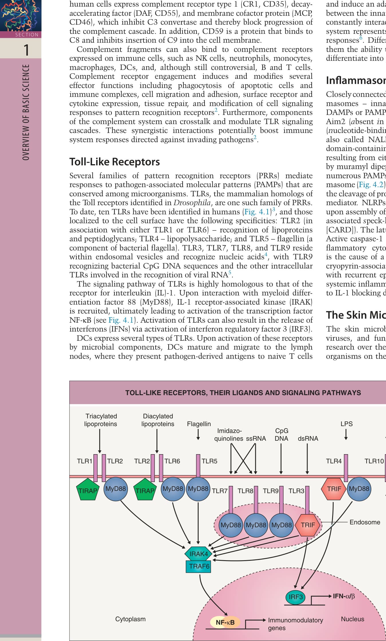
Fig. 4.1 Toll-like receptors, their ligands, and signaling pathways. Toll-like receptors (TLRs) recognize pathogen-associated molecular patterns and/or synthetic compounds in a specific fashion. A TLR2/TLR1 dimer recognizes triacylated lipoproteins, and a TLR2/TRL6 dimer interacts with diacylated lipoproteins. TLR5 recognizes flagellin and TLR4 recognizes lipopolysaccharide (LPS). These TLRs are located on the cell membrane and are internalized upon ligand interaction. TLR3, TLR7, TLR8, and TLR9 are located on intracellular membranes of endosomes and lysosomes. TLR3 recognizes viral double-stranded RNA (dsRNA); TLR7 and TLR8 viral single-stranded RNA (ssRNA); and TLR9 bacterial and viral hypomethylated DNA (CpG [5′-C-phosphate-G-3′] motifs). TLR7 and TLR8 also bind to synthetic compounds (imidazoquinolones). All TLRs except TLR3 utilize myeloid differentiation factor 88 (MyD88) for signaling, while TLR2 and TLR6 also require Toll-interleukin-1 receptor domaincontaining adaptor protein (TIRAP). MyD88 signaling via IL-1 receptor-associated kinase-4 (IRAK-4) and tumor necrosis factor receptor-activated factor-6 (TRAF-6) mostly results in activation of nuclear factor κB (NF-κB), ultimately inducing transcription of genes encoding immunomodulatory and proinflammatory molecules. TLR3 and TLR4 signal via TIR-domaincontaining adapter-inducing interferon-β (TRIF). The TRIF pathway induces the production of interferons (IFNs) via interferon regulatory factor-3 (IRF3). Both the ligand and signaling pathway of TLR10 are still unknown. Adapted from Miller LS. Toll-like receptors in skin. Adv Dermatol 2008;24:71–87 and from McInturff JE, Modlin RL, Kim J. The role of toll-like receptors in the pathogenesis and treatment of dermatological disease. J Invest Dermatol 2005;125:1–8.
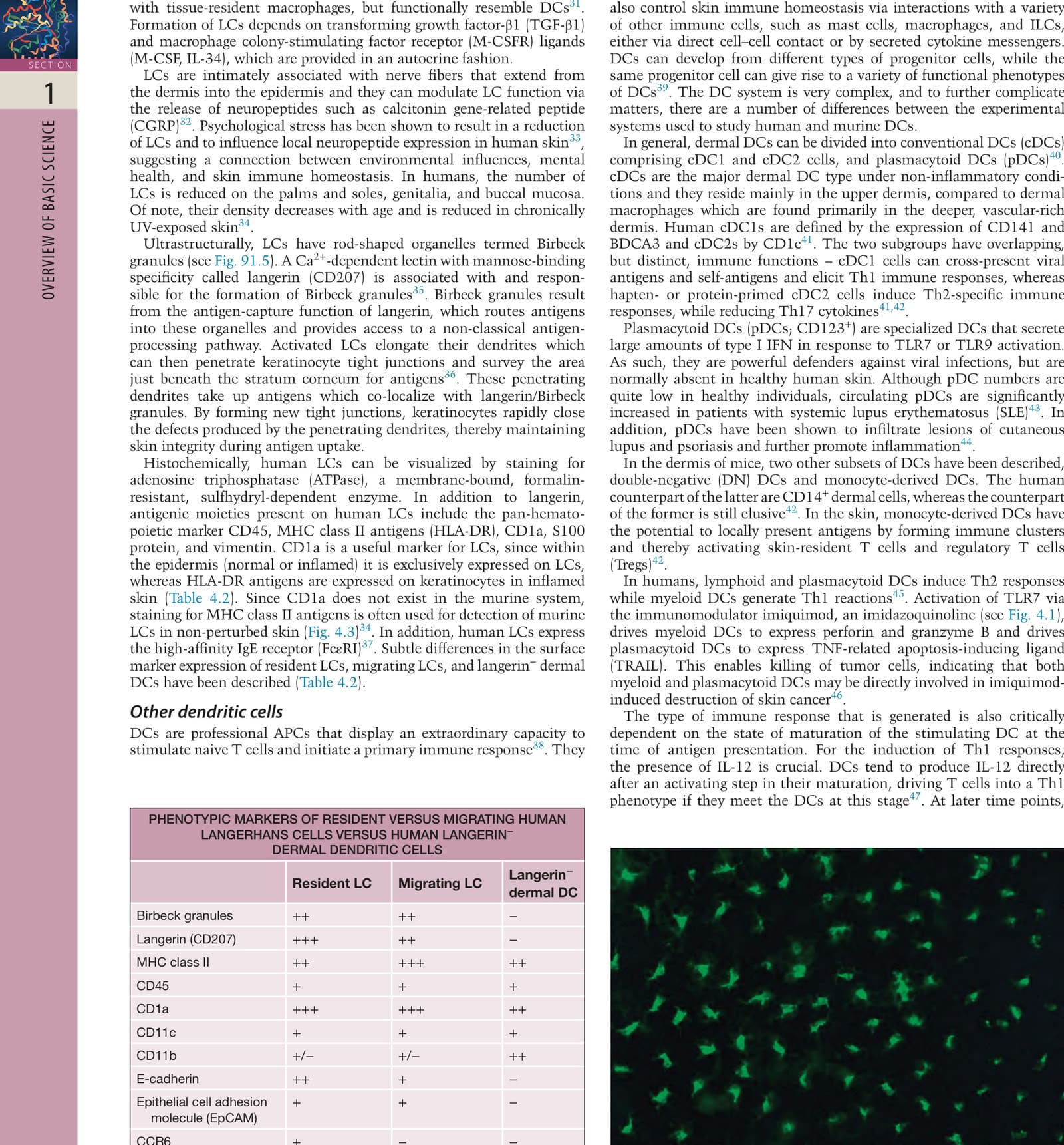
Fig. 4.3 Langerhans cells express MHC class II molecules. In a sheet preparation of murine epidermis, numerous Langerhans cells can be visualized by staining with an antibody against MHC class II molecules (Ia antigen). Note the dendritic shape of Langerhans cells. Courtesy N. Romani, Department of Dermatology, University of Innsbruck.
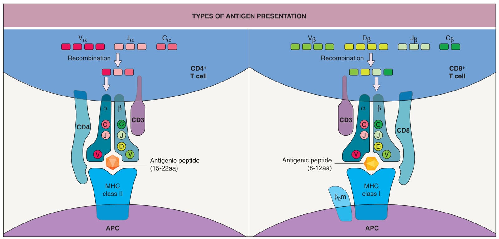
Fig. 4.4 Types of antigen presentation. Antigens are presented by antigen-presenting cells to the T cell receptor of CD4+ or CD8+ T cells in association with either MHC class II or MHC class I molecules. The diversity of the T cell receptors is generated by gene rearrangement. For reasons of clarity, the simplified gene rearrangement of the α-chain is shown only in the CD4+ T cell and that of the β-chain in the CD8+ T cell. aa, amino acids; APC, antigen-presenting cell; βm, β-microglobulin; V, variable; D, diversity; J, joining; C, constant. Adapted from Modlin RL. Lymphocytes. In: Freedberg IM, Eisen AZ, Wolf K, et al. (eds). Fitzpatrick’s Dermatology in General Medicine, vol. 1. New York: McGraw-Hill, 1999;32:400–5.
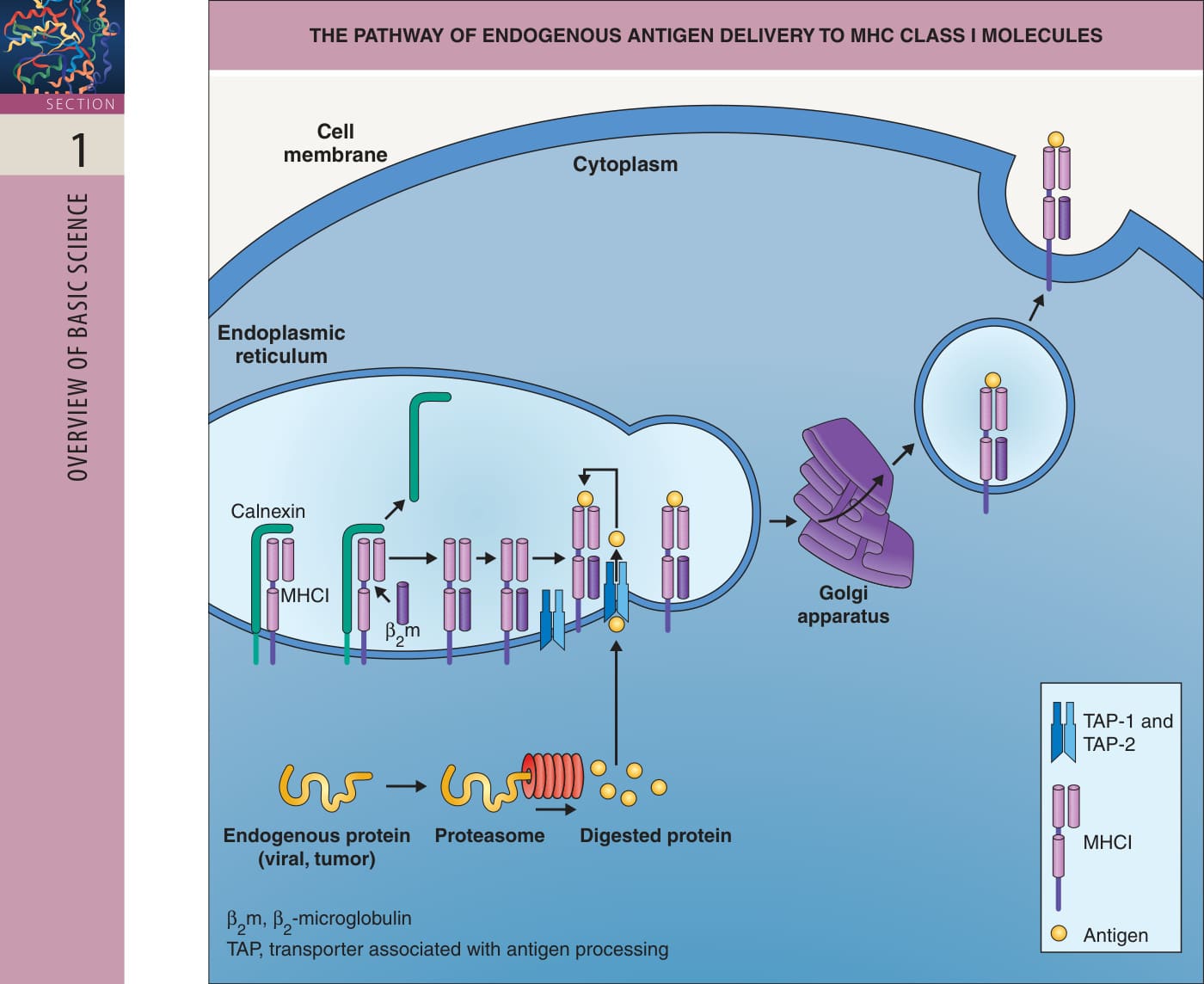
Fig. 4.5 The pathway of endogenous antigen delivery to MHC class I molecules. Newly synthesized MHC class I molecules are stabilized by calnexin. When β-microglobulin (βm) binds to the complex, calnexin dissociates. This complex associates with the TAP (transporter associated with antigen processing) protein waiting for a suitable peptide. Endogenous antigens are degraded by the proteasome and transported via TAP into the endoplasmic reticulum (ER), where they bind to the MHC class I/βm complex. Finally, the peptide/MHC complex is transported through the Golgi apparatus to the cell surface. Not pictured is a possible alternate pathway in which exogenous proteins are phagocytosed and the phagosome fuses to the ER. The proteins are then retransported out of the ER into the cytoplasm and degraded by proteasomes. The degraded peptides can now enter this pathway via the TAP protein. Adapted from Parkin J, Cohen B. An overview of the immune system. Lancet 2001;357:1777–89. With permission from Elsevier.
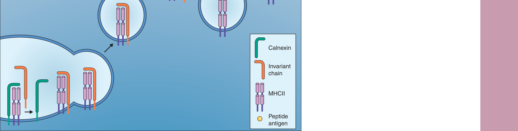
Fig. 4.6 The pathway of exogenous antigen delivery to MHC class II molecules. Exogenous antigens are taken up into the cell by endosomes. The protein is cleaved into peptides within the endosomes, which become increasingly acidic. In the endoplasmic reticulum (ER), newly synthesized MHC class II molecules bind to an invariant chain, which inhibits their disassociation while empty. During this assembly, the complex is stabilized by calnexin. The invariant chain transports the MHC class II molecule from the ER (via the Golgi apparatus) into the endosomal compartment, where the MHC class II complex meets the peptides. The invariant chain is cleaved, leaving a small fragment (CLIP) in the groove of the MHC class II molecule. Finally, CLIP is replaced by the antigenic peptide. The complex is transported to the cell membrane and expressed on the cell surface. CLIP, class II-associated invariant peptide. Adapted from Parkin J, Cohen B. An overview of the immune system. Lancet 2001;357:1777–89. With permission from Elsevier.
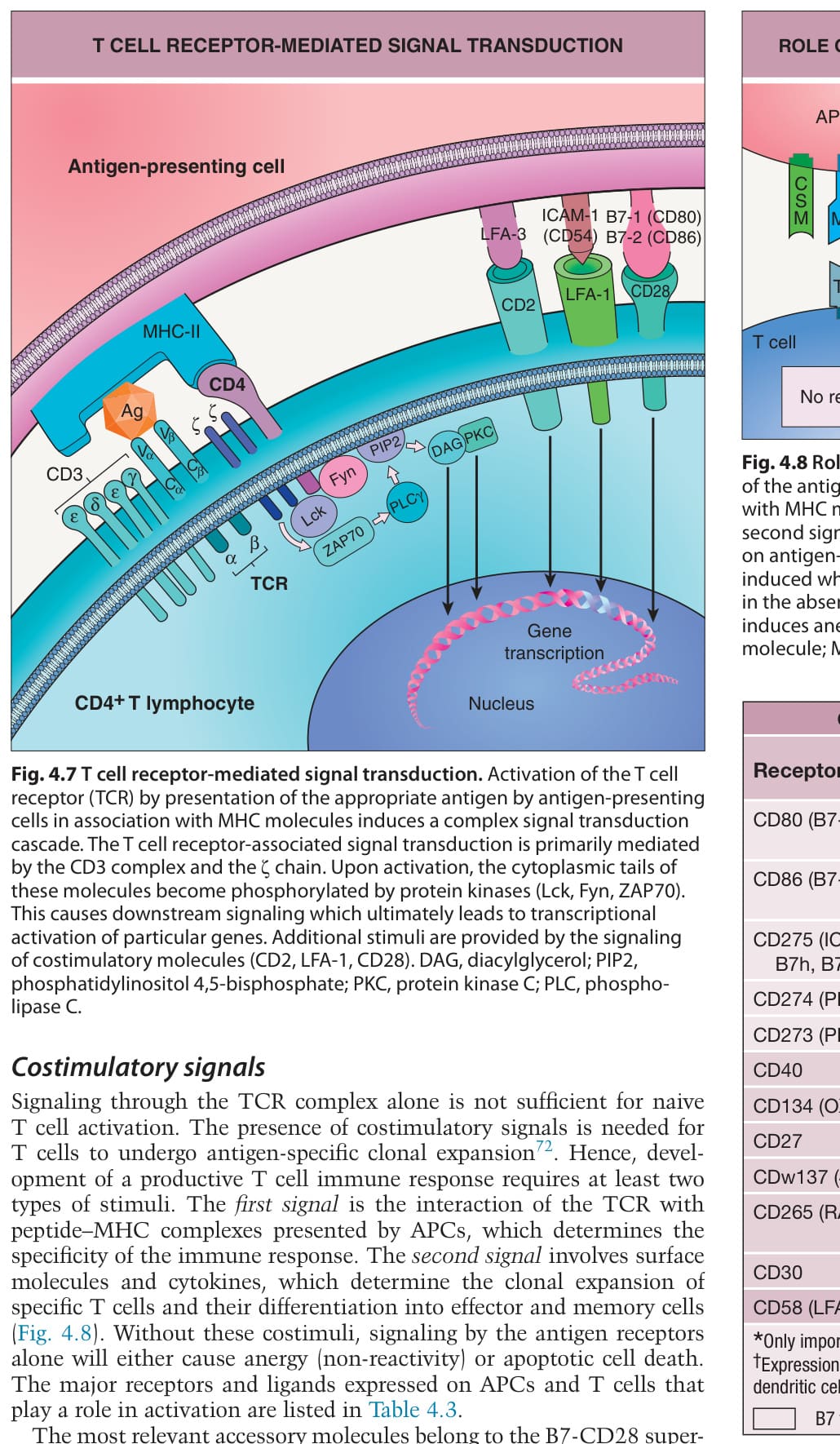
Fig. 4.7 T cell receptor-mediated signal transduction. Activation of the T cell receptor (TCR) by presentation of the appropriate antigen by antigen-presenting cells in association with MHC molecules induces a complex signal transduction cascade. The T cell receptor-associated signal transduction is primarily mediated by the CD3 complex and the ζ chain. Upon activation, the cytoplasmic tails of these molecules become phosphorylated by protein kinases (Lck, Fyn, ZAP70). This causes downstream signaling which ultimately leads to transcriptional activation of particular genes. Additional stimuli are provided by the signaling of costimulatory molecules (CD2, LFA-1, CD28). DAG, diacylglycerol; PIP2, phosphatidylinositol 4,5-bisphosphate; PKC, protein kinase C; PLC, phospholipase C.
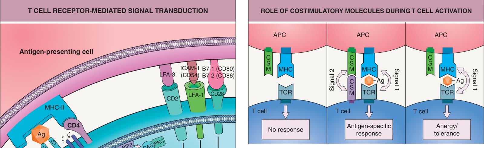
Fig. 4.8 Role of costimulatory molecules during T cell activation. Presentation of the antigen by antigen-presenting cells to the T cell receptor in association with MHC molecules delivers the first signal necessary for T cell activation. The second signal is provided by the interaction of costimulatory molecules present on antigen-presenting cells and T cells. An antigen-specific response is only induced when signal 1 and signal 2 are provided. Presentation of the antigen in the absence of signal 2 does not lead to an antigen-specific response but induces anergy and tolerance. APC, antigen-presenting cell; CSM, costimulatory molecule; MHC, major histocompatibility complex; TCR, T cell receptor.
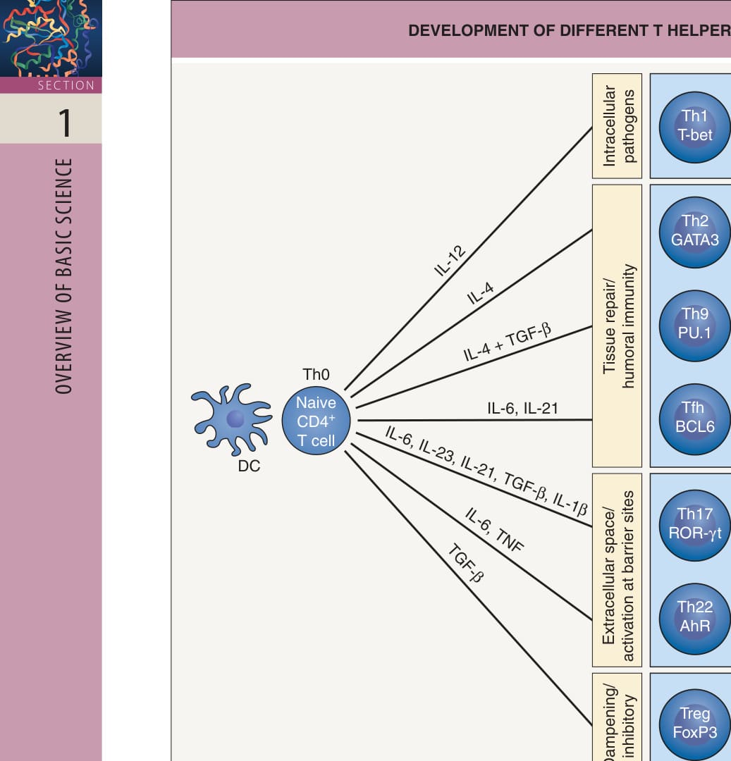
Fig. 4.9 Development of different T helper subsets. Key cytokines drive the differentiation of naive T helper (Th) cells into various Th cell subtypes. Th cell differentiation is non-linear and highly plastic, thus allowing trans-differentiation of already committed cells into other subtypes (plasticity model). Each CD4+ T cell subtype is depicted with its major driving cytokines (e.g. IL-12, IL-4, TGF-β) and fate-determining transcription factors (e.g. T-bet, GATA3, PU.1, BCL6, ROR-γt, AhR), which direct differentiation. There are additional transcription factors (e.g. STAT3) that play an important role. Differentiated Th cells secrete unique cytokines that influence other immune cells. Th1 cells are crucial for T cell-mediated immunity against intracellular pathogens whereas Th2, Th9, and Tfh cells support the development of humoral immunity and provide assistance in tissue repair. Th17 and Th22 cells are activated at barrier sites, such as the skin and gut mucosa, to protect the extracellular space and are involved in chronic inflammatory and autoimmune reactions. Tregs are crucial for maintaining immune homeostasis by dampening immune reactions. AhR, aryl hydrocarbon receptor; BCL6, B cell lymphoma 6; FoxP3, forkhead box P3; GATA3, transcription factor that binds to the DNA sequence “G-A-T-A”; IL, interleukin; IFN-γ, interferon-γ; PU.1, transcription factor that binds to purine-rich DNA sequence (PU-box); ROR-γt, retinoid acid receptor-related orphan receptor-γt; T-bet, T-box expressed in T cells; Tfh, T follicular helper cell; TGF-β, transforming growth factor-β; Th, T helper, Treg, T regulatory. Adapted from Tuzlak S, Dejean AS, Iannacone M, et al. Repositioning TH cell polarization from single cytokines to complex help. Nat Immunol 2021;22:1210–7 and O’Shea JJ, Paul WE. Mechanisms underlying lineage commitment and plasticity of helper CD4+ T cells. Science 2010;327:1098–102.
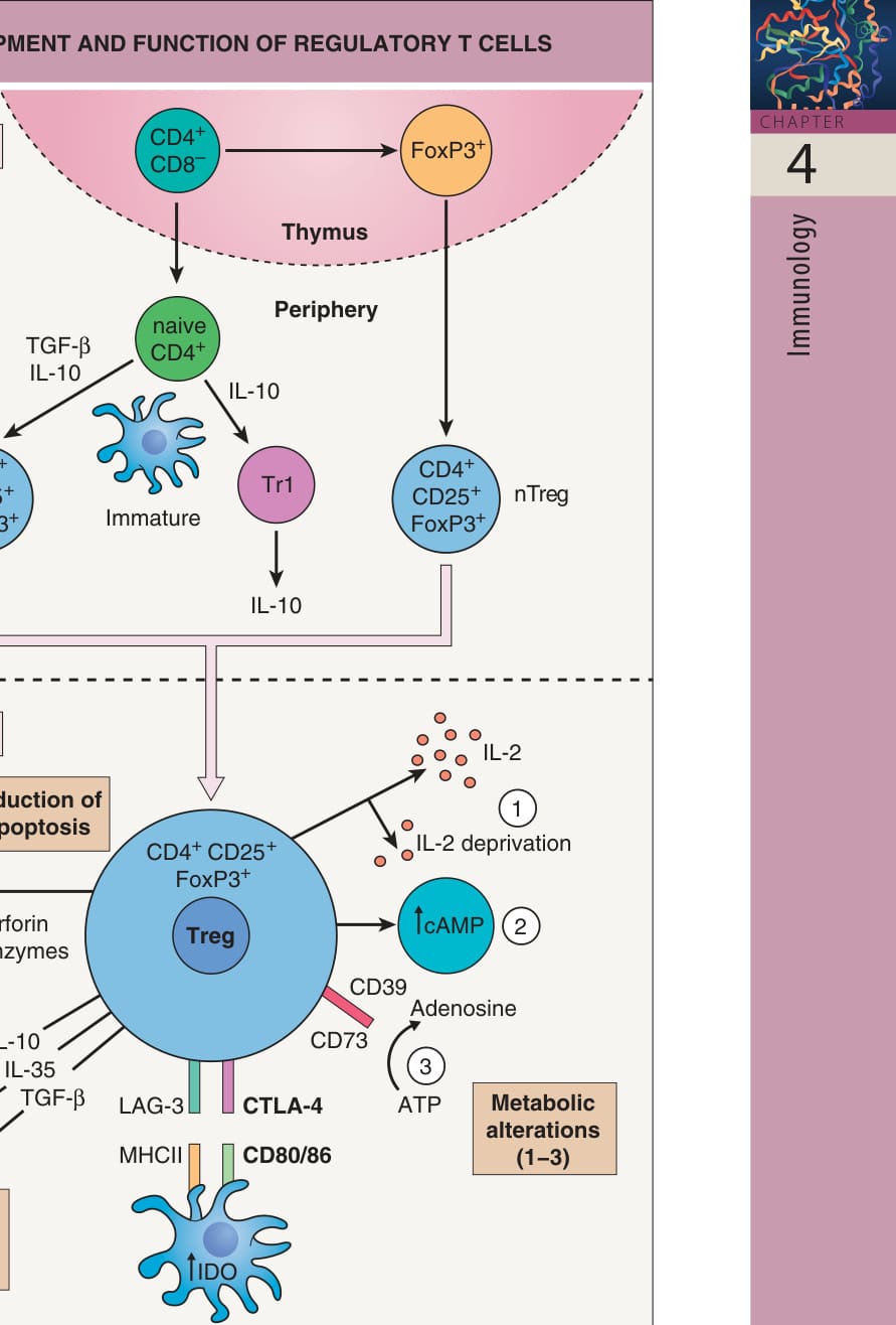
Fig. 4.10 Development and function of regulatory T cells. Natural regulatory T cells (nTregs), which express CD4, CD25 and FoxP3, develop from CD4+CD8− T cells in the thymus. In the periphery, naive conventional CD4+ T cells (in the presence of interleukin [IL]-10 and/or transforming growth factor-β [TGF-β]) can develop into FoxP3− regulatory type 1 cells (Tr1) or induced regulatory T cells (iTregs) that express CD24, CD25, and FoxP3. Tregs exert their inhibitory functions in several ways. They release inhibitory cytokines (e.g. IL-10, IL-35, TGF-β), induce apoptosis of their target effector T cells via release of granzymes and perforin, and interfere with metabolic functions of their target T cells. The latter can be accomplished by: (1) IL-2 deprivation, which triggers apoptosis; (2) transferring cAMP, which inhibits proliferation and IL-2 production, into target cells; and (3) increasing pericellular levels of cytotoxic adenosine via conversion of ATP to adenosine by CD39 and CD73, two ectonucleotidases expressed on the surface of Tregs. Tregs also modulate dendritic cell (DC) maturation and function via interaction of cytotoxic T lymphocyte-associated antigen-4 (CTLA-4) and lymphocyte-activation gene-3 (LAG-3) on Tregs with their ligands CD80/ CD86 and MHC class II, respectively, on DCs and induction of the tryptophandegrading enzyme indoleamine 2,3-dioxygenase [IDO] in DCs. Adapted from Workman CJ, Szymczak-Workman AL, Collison LW, et al. The development and function of regulatory T cells. Cell Mol Life Sci 2009;66:2603–22 and from Sakaguchi S, et al. Regulatory T cells and immune tolerance. Cell 2008;133:775–87.
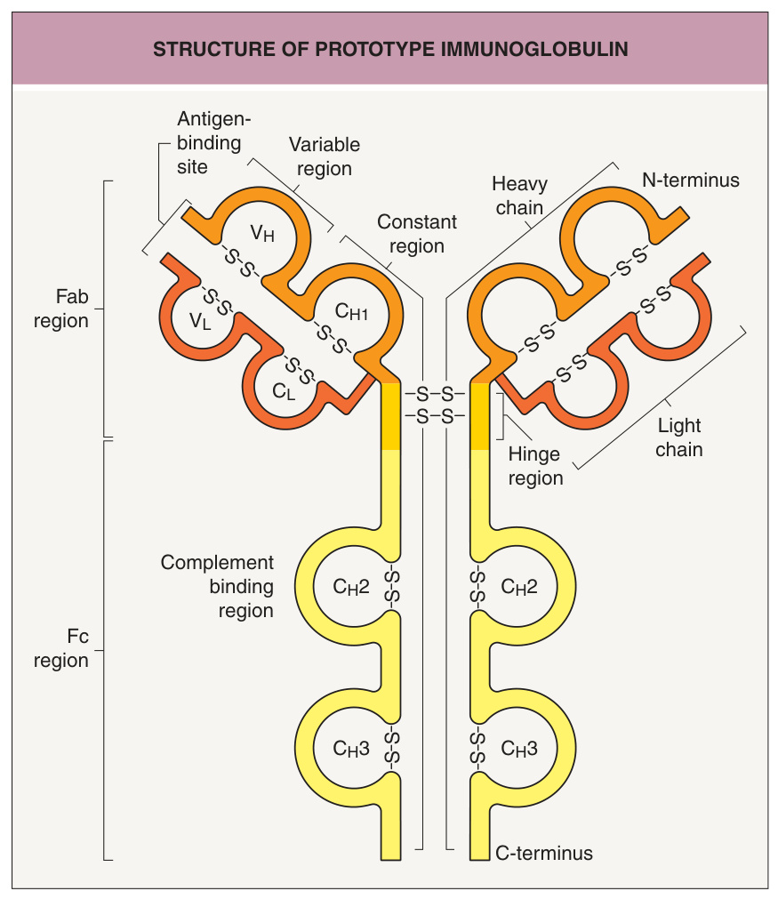
Fig. 4.11 Structure of a prototype immunoglobulin. The basic immunoglobulin structure consists of two identical light polypeptide chains and two identical heavy polypeptide chains, which are linked together by disulfide bonds. The antigen-binding site is at the N-terminal end.
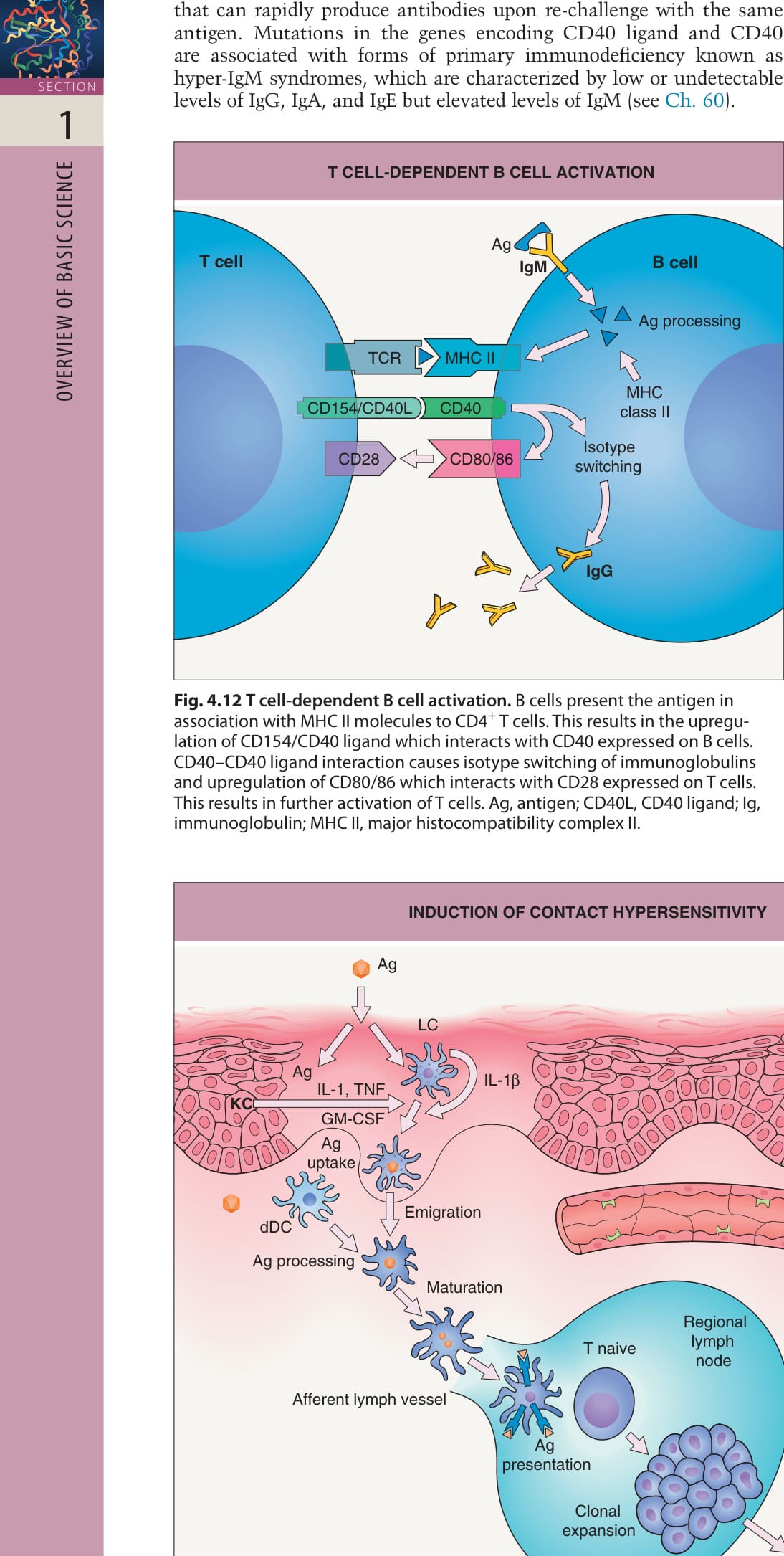
Fig. 4.12 T cell-dependent B cell activation. B cells present the antigen in association with MHC II molecules to CD4+ T cells. This results in the upregulation of CD154/CD40 ligand which interacts with CD40 expressed on B cells. CD40–CD40 ligand interaction causes isotype switching of immunoglobulins and upregulation of CD80/86 which interacts with CD28 expressed on T cells. This results in further activation of T cells. Ag, antigen; CD40L, CD40 ligand; Ig, immunoglobulin; MHC II, major histocompatibility complex II.
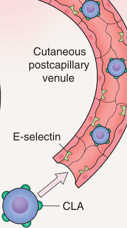
Fig. 4.13 Induction of contact hypersensitivity.
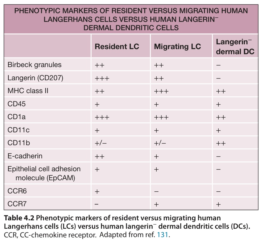
Table 4.2 Phenotypic markers of resident versus migrating human Langerhans cells (LCs) versus human langerin− dermal dendritic cells (DCs).
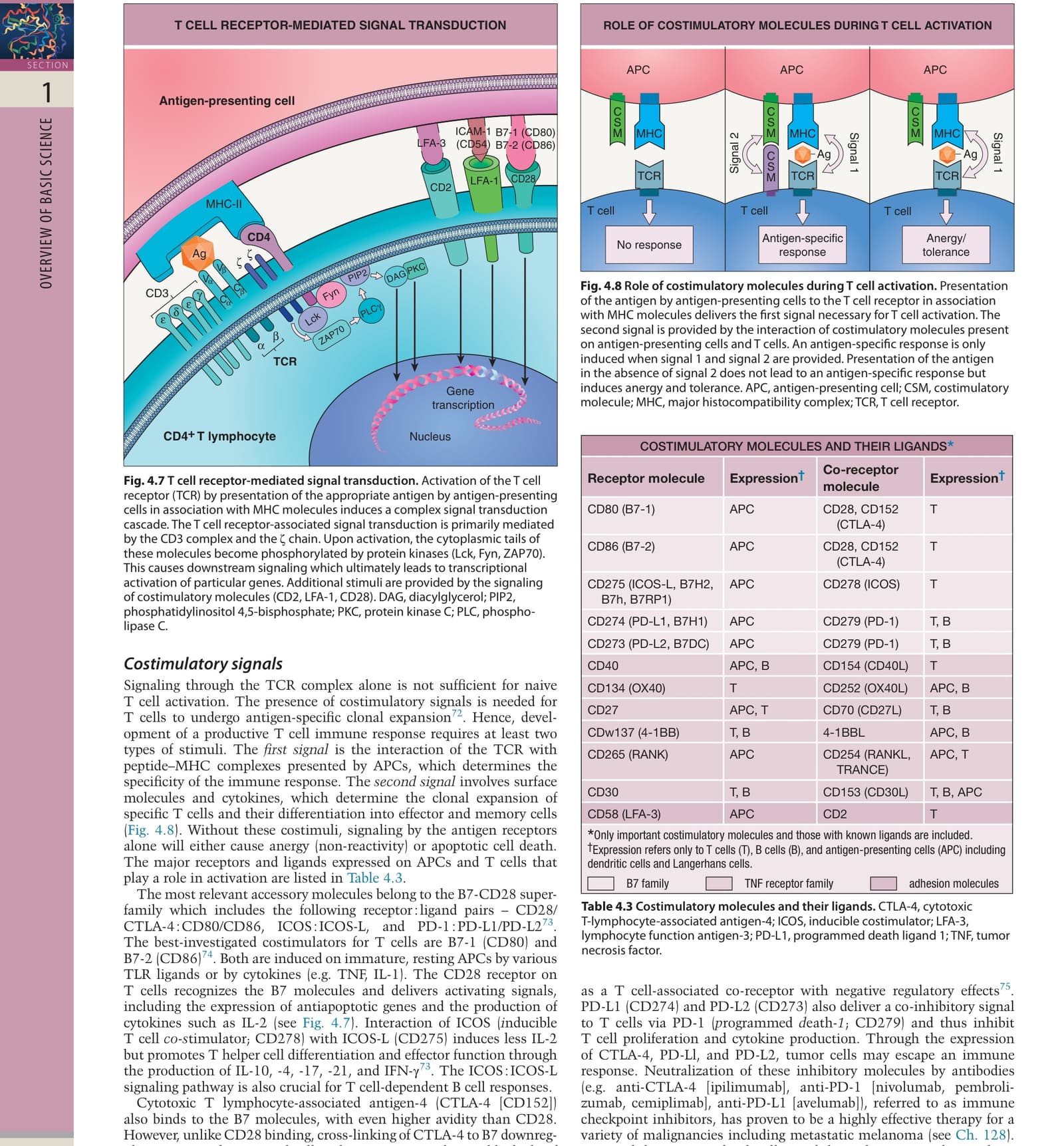
Table 4.3 Costimulatory molecules and their ligands. CTLA-4, cytotoxic T-lymphocyte-associated antigen-4; ICOS, inducible costimulator; LFA-3, lymphocyte function antigen-3; PD-L1, programmed death ligand 1; TNF, tumor necrosis factor.
Application of contact allergens (Ag) onto a sensitized individual causes the release of cytokines by keratinocytes (KCs) and Langerhans cells (LCs). These cytokines induce the expression of adhesion molecules and activation of endothelial cells, which ultimately attracts leukocytes to the site of antigen application. Among these cells, T effector cells are present, which are now activated upon antigen presentation either by LCs, dermal dendritic cells (dDCs), or infiltrating macrophages (Mphs). Antigen-specific T cell activation again induces the release of cytokines by T cells. This causes the attraction of other inflammatory cells, including granulocytes and macrophages, which ultimately cause the clinical manifestation of contact dermatitis. CLA, cutaneous lymphocyte antigen.
to enter the skin. These T cells become activated when they encounter their relevant hapten presented by APCs within the skin. However, in contrast to the sensitization phase, antigen presentation can now be effectively performed by other cells (including keratinocytes, dermal mast cells, and macrophages) that are capable of presenting antigen at least in a MHC class I-restricted fashion. Alternatively, inflammatory cells that infiltrate the site of hapten application very early during the response may function as APCs.
The earliest histopathologic findings during a CHS response are mast cell degranulation, vasodilation, and an influx of neutrophils, which is followed by infiltration of monocytes and T cells (Fig. 4.14). However, the pathophysiologic events that result in allergic contact dermatitis are clearly T cell-dependent, since T cell-deficient mice are unable to mount a CHS response. Furthermore, low doses of hapten that are sufficient to stimulate hapten-specific T cells are insufficient to elicit a CHS response. This indicates that the elicitation of a CHS response requires, in addition to hapten-specific recognition, a proinflammatory stimulus that can be provided in a dose-dependent manner by the hapten itself.
Additional figure available in our eBook (see inside front cover for access code).

Fig. 4.14 Elicitation of contact hypersensitivity.