PRURITUS PATHWAYS
The neurophysiology of itch involves specific itch mediators and nerve fibers that transmit itch both peripherally and centrally. While the concept of pruritus as a sensory modality separate from pain was not generally appreciated until the mid-twentieth century, studies have clearly identified individual histaminergic and non-histaminergic C fibers that uniquely transmit itch. C fibers that transmit itch have exceptionally slow conduction velocities (0.3–1.0 m/second) and innervate unusually wide territories.
Histamine-sensitive C fibers are sensitive to heat as well as pruritogenic stimuli but generally not to mechanical stimuli; in contrast, the vast majority of other C fibers, while sensitive to heat stimuli, have little or no response to histamine. The co-responsiveness of itch-transmitting C fibers to temperature explains aggravation of pruritus in a warm environment. However, the ineffectiveness of oral antihistamines for most types of pruritus suggests that other fibers play important roles in itch sensation.
Indeed, distinct parallel pathways of non-histaminergic C fibers that transmit itch have been identified in the peripheral nervous system of humans and the spinothalamic tract of other primates. Itch transmission can be elicited by spicules of the tropical legume cowhage (Mucuna pruriens), which produce an intense itch when rubbed, inserted, or injected into the skin, without producing a histaminergic axon reflex. Cowhage spicules induce itch via release of the protease mucunain, which activates proteinase-activated receptor (PAR)-2 and PAR-4. Cowhage-sensitive fibers transmit a burning sensation together with itch and are also sensitive to mechanical and other stimuli. PAR-2 receptors have been implicated in itch associated with atopic dermatitis (see Ch. 12). The non-histaminergic, polymodal C fibers stimulated by mucunain may also have clinical relevance in chronic itch. In addition to C fibers, A-delta fibers contribute to cowhage-evoked itch with a more rapid onset.
MAS-related G protein-coupled receptor D (MRGPRD) is a β-alanine receptor that is exclusively expressed by a subset of pruriceptive C fibers classified as type 1 non-peptidergic (NP1) neurons. Similarly, the pharmacologic pruritogen chloroquine directly activates NP2 (type 2) neurons, whereas the Th2 cytokine IL-31 elicits itch via NP3 (type 3) neurons. Collectively, recent studies suggest that a variety of sensory neurons respond to different itch stimuli via distinct pathways, explaining why there are so many types of itch.
Within the spinal cord, there is another layer of itch circuitry where signals from the skin are integrated and the perception of itch is then relayed to the brain. The first itch-specific receptor to be identified, gastrin-releasing peptide receptor (GRPR), is found in the spinal cord and represents a key downstream mediator of non-histaminergic itch responses. Overexpression of gastrin-releasing peptide (GRP) in cutaneous nerve fibers and GRPR in the spinal cord has been observed in mice and primates with chronic itch. B-type natriuretic peptide, an itch-selective neuropeptide found in dorsal root ganglia neurons, is believed to act upstream of the GRPR itch spinal circuit. Substance P is also expressed by a distinct population of murine dorsal horn neurons that respond to pruritic and noxious stimuli.
The perceived sensation of pruritus can vary greatly in quality. Patients may describe burning, pricking, “insects crawling” on the skin, or even a tickle, but the neurophysiologic and psychologic correlates of these qualitative differences have not yet been elucidated. However, information obtained from itch questionnaires has enabled a better understanding of the different characteristics of itch and its features in various skin diseases.
Central Pathways to Higher Nervous System Centers
In the spinal cord, pruriceptive C fibers synapse with secondary sensory neurons in the gray matter of the dorsal horn (Fig. 5.1). These neurons then cross over and ascend in the lateral spinothalamic tract to the
thalamus. In vivo studies using extracellular recordings from cats identified a subclass of lamina I spinothalamic tract neurons that are excited by iontophoretically administered histamine. Neurophysiologic experiments in primates found that cowhage-induced itch stimulates a distinct population of spinothalamic tract neurons that are not involved in the transmission of histamine-mediated itch (see above). Studies in humans utilizing functional MRI or positron emission tomography (PET) have shown that histamine and cowhage activate different brain areas, providing insight into the supraspinal processing of itch.
In healthy individuals, induction of itch by either histamine or cowhage can elicit activation of various regions, including the anterior and posterior cingulate cortex, precuneus, somatosensory areas I and II, supramarginal gyrus, inferior parietal lobe, and insula–claustrum complex. The activation of multiple brain areas argues against a single “itch center” and underscores the multidimensionality of the itch sensation. Of note, pain shows a similar pattern of brain activation across cortical regions (see Fig. 5.1). In mice, optogenetic studies, in which expression of light-induced proteins within neuronal cell membranes is measured, detected a spinoparabrachial pathway that sends itch signals to the parabrachial nucleus of the brain stem.
Seeing other people scratching can trigger itch and atopic dermatitis patients experience increased itch when exposed to visual stimuli. Studies utilizing functional MRI showed that “contagious itch” activates many of the neural regions linked to the physical perception of itch. In mice, the suprachiasmatic nucleus is a key visual organ that mediates contagious scratching behavior.
In addition to inhibiting activation of the cingulate cortex, scratching has been found to activate the prefrontal cortex and cerebellum. It is possible that the cerebellum plays a role in coordinating the itch–scratch cycle. Scratching inhibits histamine-evoked activity of spinothalamic neurons in primates, but not spontaneous or pain-triggered activity. Scratching also activates areas of the brain involved in reward processing, including the striatum and substantia nigra. While these responses correlate with the pleasure of scratching, activation of other areas is associated with itch relief.
Pruritus Receptor Units
Removal of the epidermis largely abolishes the perception of itch. Light microscopic and ultrastructural studies of human skin have shown the existence of intraepidermal nerve fibers with “free” non-specialized
nerve endings extending to the stratum granulosum. A subclass of C fibers in the epidermis that express MRGPRs, in particular MRGPRX1, are involved in chloroquine-induced pruritus (see Fig. 5.2F). Cathepsin S, an endogenous protease, can cleave and thereby activate MRGPRX2 (murine MrgprC11), which leads to scratching behavior, as well as PAR-2/4 receptors.
Keratinocytes express a variety of neural mediators and receptors involved in the sensation of itch (Table 5.2). These include opioids, proteases, substance P, NGF, and neurotrophin 4 in addition to their respective receptors, including μ- and κ-opioid receptors, PAR-2, neurotrophic tyrosine kinase receptor type 1 (NTRK1), and transient receptor potential vanilloid ion channels (particularly TRPV1 and TRPV3). Keratinocytes also have ATP-gated ion channels and adenosine receptor ligands similar to those observed in C fibers involved in pain transmission. These structural similarities to nerve fibers suggest that keratinocytes may be involved in the transduction and generation of itch.
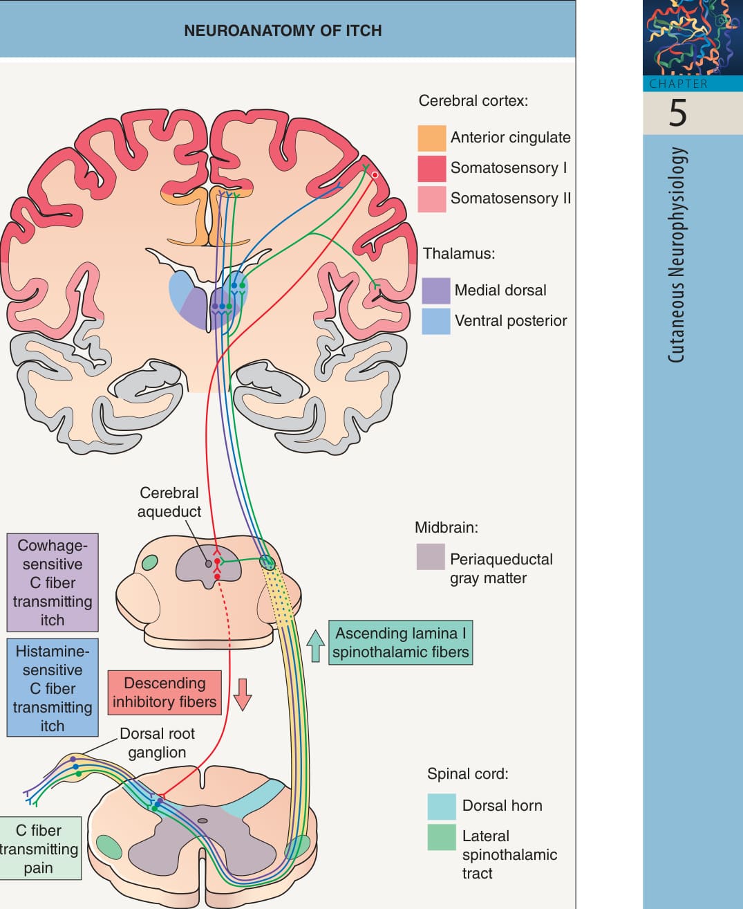
Fig. 5.1 Neuroanatomy of itch. Itch and pain transmission occur via unmyelinated C nerve fibers that excite lamina I neurons in the dorsal horn of the spinal cord. Two subsets of pruritoceptive C-fiber neurons (which respond to histamine and cowhage, respectively) are conducted through distinct lateral spinothalamic pathways, with projections to the thalamus. Processing of itch through either pathway activates several regions of the brain, which are similar to those involved in pain (see Table 5.4). Concomitant painful stimuli can reduce the sensation of itch, possibly by a descending inhibitory mechanism resulting from activation of the periaqueductal gray matter. In mice, gastrin-releasing peptide receptor-positive neurons transmit itch (but not pain) within the spinal cord; the role of such neurons in humans remains to be determined. Adapted from Yosipovitch G. Pruritus: an update. Curr Probl Dermatol 2003;15:137–64.
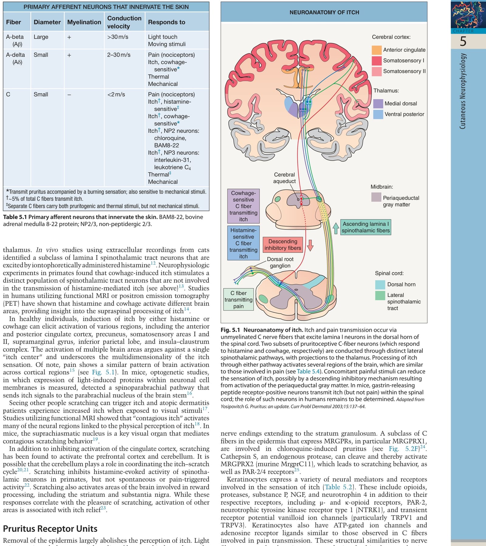
Table 5.1 Primary afferent neurons that innervate the skin. BAM8-22, bovine adrenal medulla 8-22 protein; NP2/3, non-peptidergic 2/3.
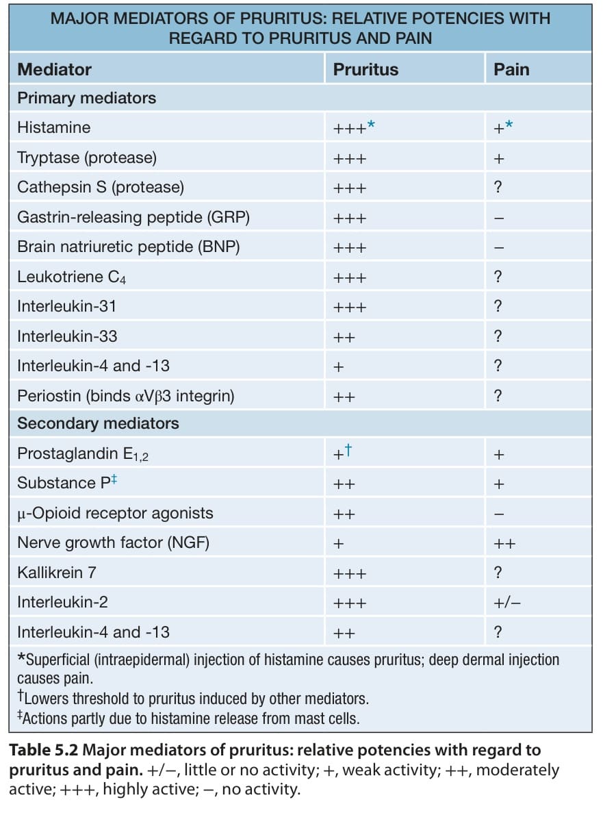
Table 5.2 Major mediators of pruritus: relative potencies with regard to pruritus and pain.+/−, little or no activity; +, weak activity; ++, moderately active; +++, highly active; −, no activity.