MEDIATORS OF PRURITUS
Various mediators that act centrally and/or peripherally are involved in pruritus, including histamine, proteases, substance P, opiates, NGF, and prostaglandins (Fig. 5.2A). In inflammatory skin diseases, proinflammatory mediators produce pruritus and other signs of inflammation, in particular erythema due to vasodilation and edema from increased vascular permeability. However, cytokines and other inflammatory mediators can also act directly on sensory neurons to influence itch sensation. The relative potencies of the major mediators are listed in Table 5.2.
Histamine
Histamine is the archetypal mediator of inflammation, including pruritus. In the skin, histamine is contained primarily within the granules of dermal mast cells. Histamine can be released from mast cells upon activation of a range of receptors, including the high-affinity IgE receptor (FcεRI), the KIT receptor, MRGPRX2, and receptors for various neuropeptides (e.g. NGF) and complement C5a. In IgE-mediated acute urticaria, histamine is released when a specific antigen/allergen cross-links adjacent receptor-bound specific IgE antibodies on mast cells. In autoimmune chronic urticaria, similar cross-linking occurs via functional circulating IgG that react with epitopes expressed on the α-chain of adjacent FcεRIs or less commonly anti-IgE autoantibodies (see Fig. 18.3). Histologically, dermal mast cells and unmyelinated neurons are juxtaposed (Fig. 5.2B,C,E), indicating a close (“synapselike”) functional relationship between the immune and nervous systems. However, evidence for histamine as the main mediator of pruritus is limited to a few skin diseases, including acute and chronic urticaria and mastocytosis.
Recognition of the histamine H4 receptor has expanded our under-standing of the physiologic actions of histamine beyond the traditional H1 receptor. The H4 receptor is expressed by neurons and immune cells such as eosinophils, mast cells, dendritic cells, monocytes, and CD8+ T cells. It mediates chemotaxis and plays a role in the inflammation and pruritus of atopic dermatitis. H4 antagonists are under development and have been shown to alleviate experimental pruritus. In animal models of itch, the effects of H4 antagonists are synergistic with those of centrally acting H1 antihistamines (e.g. diphenhydramine).
Gastrin-Releasing Peptide
As noted above, itch-specific GRPR-positive neurons have been identified in the dorsal horn of the spinal cord in mice. Although named for its role in regulating gastrointestinal functions, gastrin-releasing peptide (GRP) is expressed in many tissues. Whether peripheral sensory neurons express GRP is still debated.
B-Type Natriuretic Peptide
B-type natriuretic peptide (BNP; natriuretic polypeptide B) is a 32-amino acid peptide that is secreted by the cardiac ventricles to regulate blood pressure and fluid balance. BNP is also expressed by a subset of C fibers and may function in itch transmission in the spinal cord.
Proteases
Mast cells produce two proteases, tryptase and chymase. Tryptase, released by activated mast cells, cleaves PAR-2, a G protein-coupled receptor present on C-fiber terminals (Fig. 5.2B,D); this exposes a tethered ligand domain and thereby “self-activates” PAR-2, leading to itch transmission. Tryptase levels are increased four-fold in non-lesional forearm skin of atopic dermatitis patients, while expression of PAR-2 is significantly increased in the epidermis and cutaneous nerve fibers of eczematous lesions and to a lesser degree in non-lesional skin.
Kallikreins and cathepsins can also activate PAR-2 via their protease activity. In addition, kallikrein 7, which does not activate PAR-2, triggers itch in the setting of atopic dermatitis-like disease in mice and is enriched in lesional human atopic dermatitis skin. Cathepsin S can evoke itch by cleaving and thereby activating MRGPRX2 as well as PAR-2 (see above). Furthermore, proteases such as cathepsin B can be found in common allergens (e.g. grass pollen, house dust mites), and Staphylococcus aureus can induce secretion of proteases; both of these exogenous factors are known to aggravate atopic dermatitis and itch.
Transgenic mice overexpressing various serine proteases exhibit severe itch and scratching (Table 5.3). In Netherton syndrome, serine protease inhibitor deficiency leads to excess epidermal protease activity, resulting in pruritus and atopic manifestations (see Ch. 57). These observations demonstrate that interactions between proteases and receptors on C fibers play key roles in itch.
Opioid Peptides
Central pruritus, which involves pruritic mediators within the CNS, can occur in both cutaneous and systemic diseases. Endogenous opioids modify the perception of pruritus via central and peripheral opioid receptors. Generalized pruritus may be induced by an imbalance between the μ- and κ-opioid systems, activation of which stimulates and inhibits itch perception, respectively. κ-opioid receptor agonists can act within the skin, spinal cord, and brain to reduce itch and are utilized as therapies for chronic pruritus.
Morphine as well as other exogenous and endogenous μ-opioid receptor agonists may cause generalized pruritus. Morphine also produces local pruritus and erythema when injected intradermally; this response is only partially inhibited by the μ-opioid receptor antagonist naloxone but is substantially inhibited by topical pretreatment with the H1 antihistamine doxepin, indicating the ability of morphine to activate mast cells. Therefore, two possible mechanisms for opioidinduced itch are: (1) degranulation of cutaneous mast cells; and (2) activation of μ-opioid receptors with direct central and peripheral pruritogenic effects.
Substance P
Substance P, a neuropeptide with a widespread distribution in peripheral nerves and the CNS, is involved in itch perception. Substance P is
synthesized in the cell bodies of C neurons, transported towards the peripheral nerve terminals, and released by antidromic depolarization to cause vasodilation and increased vascular permeability. High concentrations of substance P can cause immediate mast cell degranulation via MRGPRX2 receptors, whereas low concentrations specifically activate neurokinin-1 (NK-1) receptors on mast cells, leading to sensitization of these cells and increased production of tumor necrosis factor (TNF; Fig. 5.2B).
Direct activation of mast cells by substance P triggers itch transmission. When injected intradermally, it provokes neurogenic inflammation, including erythema and the wheal-and-flare reaction, in addition to itch. Levels of substance P in the serum of patients with atopic dermatitis are elevated and correlate with disease severity.
Neurotrophins
Neurotrophins are factors that regulate the growth and function of neurons. Members of this family include the prototypic nerve growth factor (NGF) as well as brain-derived neurotrophic factor (BDNF) and neurotrophins 3, 4, and 5. Increased levels of epidermal NGF correlate with proliferation of terminal cutaneous nerves and upregulated expression of neuropeptides such as substance P. NGF can also induce sprouting of nerve fibers, sensitization of nerve endings, and axonal transport in dorsal root ganglia cells (Fig. 5.2C).
Keratinocytes express high levels of NGF, which is not only required for survival and regeneration of sensory neurons but also controls the responsiveness of neurons to external stimuli. NGF is thought to act as a signaling molecule between mast cells and keratinocytes in allergic skin diseases. Mast cell-derived histamine induces keratinocytes to increase NGF production, and the latter may promote infiltration of mast cells into inflamed skin. Patients with atopic dermatitis and psoriasis have increased expression of NGF in cutaneous mast cells, keratinocytes, and fibroblasts. Upregulation of other neurotrophins, such as neurotrophin 4, has also been observed in keratinocytes from atopic dermatitis patients.
Prostanoids and Leukotrienes
Prostaglandins are the products of the conversion of arachidonic acid by cyclooxygenase-1 (COX-1) or 2 (COX-2), and they enhance histamine-induced itch. When injected into the dermis, prostaglandin E (PGE) is not itself pruritogenic but enhances pruritus due to histamine subsequently injected into the same site. It appears that only itch-mediating neurons that display lasting activation following exposure to histamine are excited by PGE, and mechanosensitive fibers are unresponsive to both histamine and PGE. PGE has also been shown to have a direct, low-level pruritogenic effect in both atopic dermatitis patients and unaffected individuals without inducing protein extravasation. This suggests that prostanoids’ peripheral action is not solely via histamine and that prostanoids may potentiate pruritus via other effects on nerve fibers. Although oral administration of aspirin, a cyclooxygenase inhibitor, does not generally ameliorate pruritus, topical application of aspirin may reduce chronic localized itch.
The role of other eicosanoids, including leukotrienes and 12-hydroxy eicosatetraenoic acid (12-HETE), in the pathogenesis of pruritus is unclear. In mice, leukotriene B can provoke scratching and may be involved in skin disease-related itch. More recent studies have shown that basophil- and mast cell-derived leukotriene C directly activates NP3 neurons and acts as a pruritogen in acute itch flares of atopic dermatitis52,53,53a.
Mediators That Activate Transient Receptor Potential Receptors
Neuromediators that activate ion channels in the transient receptor potential (TRP) family are also involved in the sensation of itch. TRP vanilloid 1 (TRPV1) is located on C fibers (see Fig. 5.2E) and is activated by capsaicin, endogenous substances such as cannabinoids (e.g. anandamide), prostaglandins, neurotrophins, a lower pH, and temperatures >43°C (109°F). TRPV1-expressing C nerve fibers can mediate heat pain and they house many pruriceptors. In addition, experimental induction of histamine-mediated itch requires cooperation of TRPV1 ion channels. Stimulation of TRPV1-positive nerve fibers also leads to the release of pruritoceptive mediators such as neuropeptides.
TRPV3 is a thermosensor of warmth (>33°C, 91°F) that is expressed in keratinocytes and dorsal root ganglion neurons. Gain-of-function missense mutations in TRPV3 result in chronic itch, scratching, and an atopic-like dermatitis in mice and Olmsted syndrome in humans. The latter features pruritus as well as palmoplantar keratoderma and periorificial hyperkeratosis (see Ch. 58). PAR2 can induce itch via TRPV3 signaling in keratinocytes, and enhanced thermal sensitivity of TRPV3 in keratinocytes plays a role in heat-induced release of pruritogens such as thymic stromal lymphopoietin (TSLP), NGF, and PGE in patients with atopic dermatitis. Intradermal injection of citrusinine-II, a plant-based TRPV3 inhibitor, was recently shown to attenuate itch in mouse models. In contrast, TRP melastatin 8 (TRPM8) on C nerve fibers functions as a thermosensor of cool temperatures (<28°C, 82°F) and is activated by menthol and icilin, which provide a “cooling” sensation that may relieve itch.
TRP ankyrin 1 (TRPA1), a polymodal ion channel, functions as a downstream mediator of histamine-independent itch stimulated by MRGPRs in a subset of epidermal C fibers. TRPA1 can be activated directly by menthol as well as via pruriceptors such as the TSLP receptor subunit (see Fig. 5.2F).
Other Peripheral Mediators of Itch
Other neurotransmitters
Intradermal injection of acetylcholine, an important neurotransmitter in the autonomic nervous system, typically induces pain; however, in patients with chronic pruritus, it can induce itch. Activation of the muscarinic receptor 3 triggers itch in mice. Norepinephrine, a catecholamine neurotransmitter, exerts tonic inhibition of itch signaling in the spinal cord. Although serotonin can induce itch in mice, in humans it is a very mild pruritogen. Currently there are no data regarding the role of epinephrine or dopamine in itch transmission.
Other mediators with potential roles in itch
Nitric oxide may induce itch via neurogenic inflammation. The bovine adrenal medulla 8-22 (BAM8-22) peptide, a proteolytically cleaved product of proenkephalin A, is a potent activator of MRGPRs and can stimulate itch, usually accompanied by a stinging or burning sensation (see Fig. 5.2F). Chloroquine can also induce itch by activating MRGPRs, which are expressed by a subset of C fibers in the epidermis.
Immune Cells as Itch Mediators and Modulators
Interactions between the nervous and immune systems in the skin play important roles in itch induction. Neuropeptides such as substance P, CGRP, and vasoactive intestinal peptide, which are released from cutaneous sensory nerves, regulate the expression of adhesion molecules and proinflammatory cytokines, thereby modulating immune and inflammatory responses. These neuropeptides also influence cell proliferation and differentiation, tissue repair, and antigen presentation involving keratinocytes, mast cells, dermal microvascular endothelial cells, and Langerhans cells. These interactions are bidirectional, as cytokines and chemokines also regulate primary nerve afferents via receptor activation.
IL-31 is produced by Th2 cells and belongs to the IL-6 family. It induces pruritus by modulating the function of sensory neurons. The IL-31 receptor (IL-31R) is a heterodimer composed of the oncostatin M receptor (OSMR) β protein plus the IL-31 receptor A (IL-31RA). The IL-31R is found on TRPV1+/ TRPA1+ cutaneous C fibers and in dorsal root ganglia.
Signaling via either the IL-31R or OSMR, a heterodimer composed of the OSMR β protein and a gp130 subunit, can result in cutaneous inflammation as well as keratinocyte proliferation, differentiation, and apoptosis. Mutations in the gene encoding the OSMR β protein underlie familial primary localized cutaneous amyloidosis, an autosomal dominant disorder characterized by chronic localized itching and scratching that results in deposition of keratin-derived amyloid in the dermis. Of note, increased levels of IL-31 are found in the skin of patients with atopic dermatitis, prurigo nodularis, and cutaneous T cell lymphoma. In randomized controlled trials, nemolizumab, an anti-IL-31RA monoclonal antibody, reduced itch and skin lesions in patients with atopic dermatitis and prurigo nodularis. Vixarelimab, an anti-OSMRβ monoclonal antibody, is currently under investigation as a therapy for prurigo nodularis and other chronic pruritic skin conditions.
Thymic stromal lymphopoietin (TSLP) is produced by keratinocytes and promotes Th2 cell-associated responses. TSLP acts directly on TRPA1-expressing neurons to elicit itch in mice, and TSLP expression is increased within lesions of atopic dermatitis.
IL-4 and IL-13 produced by Th2 cells contribute to the itch of atopic dermatitis via activation of JAK (Janus kinase)/STAT (signal transducer and activator of transcription) signaling cascades in sensory neurons. In addition to reducing itch in patients with atopic dermatitis, both JAK inhibitors and monoclonal antibodies that target IL-4/-13 signaling (e.g. dupilumab, tralokinumab, lebrikizumab) lead to improvement of other chronic pruritic disorders such as prurigo nodularis.
IL-33 has been associated with chronic pruritus of unknown origin and mediates itch in the context of allergic contact dermatitis and xerotic skin.
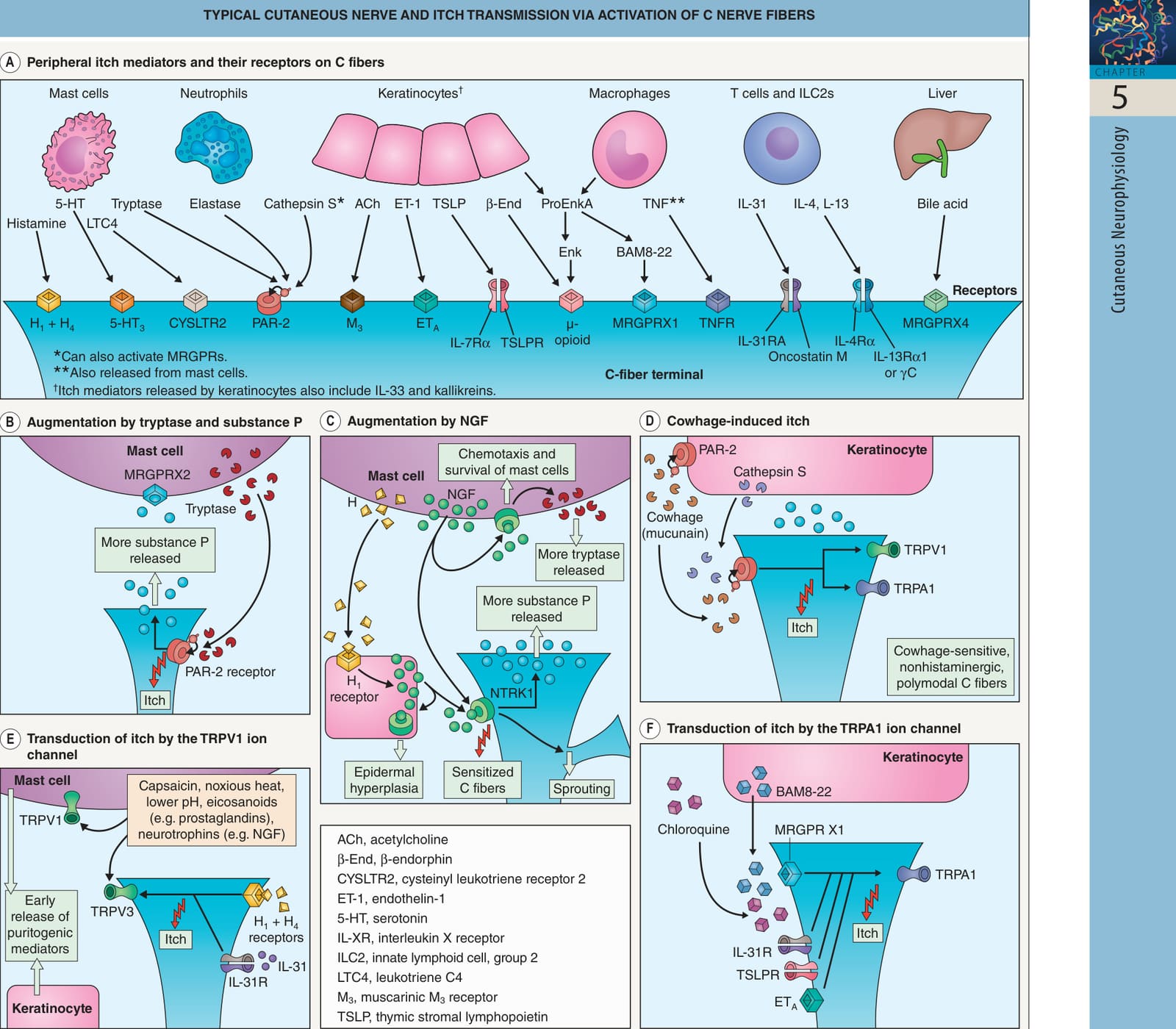
Fig. 5.2 Typical cutaneous nerve and itch transmission via activation of C nerve fibers. There are two categories of axons in a typical cutaneous nerve: (1) primary afferent Aβ, Aδ, and C fibers with cell bodies in dorsal root ganglia; and (2) sympathetic postganglionic fibers with cell bodies in sympathetic ganglia. Separate C fibers (~5% of the total) carry pruritogenic stimuli via two pathways: (1) histamine-induced itch that is transmitted by mechanically insensitive, capsaicin-sensitive fibers via the transient receptor potential vanilloid receptor 1 (TRPV1); and (2) cowhage (mucunain)-induced itch accompanied by a burning sensation that is transmitted by polymodal fibers sensitive to both capsaicin and mechanical stimuli. A Peripheral itch mediators released by immune cells, keratinocytes, and the liver activate pruriceptors on C-fiber terminals in the skin. For example, activated mast cells release histamine, which triggers itch via histamine H1 and H4 receptors on histaminesensitive C fibers. Proteases such as tryptase, elastase, and cathepsin S can cleave proteinase-activated receptor-2 (PAR-2), exposing a tethered ligand and thereby resulting in “self-activation”. Proenkephalin A (ProEnkA) may be cleaved to form enkephalin (ENK) or bovine adrenal medulla 8-22 peptide (BAM8-22). B Self-activation of PAR-2 following tryptase cleavage results in transmission of itch by the C fibers as well as release of additional substance P, which primes mast cells via neurokinin-1 receptors (NK1Rs). C Nerve growth factor (NGF) released by mast cells and keratinocytes (with NGF production stimulated by histamine [H]) activates neurotrophic tyrosine kinase receptor type 1 (NTRK1 [TRKA]) on C fibers, mast cells, and keratinocytes. This induces C fiber sprouting, sensitization to cowhage-induced itch, and increased substance P release; mast cell chemotaxis, survival, and increased tryptase release; and epidermal hyperplasia. D Cowhage-induced itch occurs through the release of mucunain, a protease that activates PAR-2 and PAR-4 receptors (the latter not yet identified in the skin); these receptors can also be activated by endogenous proteases such as cathepsin S as well as tryptase. Activation of PAR-2/4 sensitizes TRPV1 and TRP ankyrin 1 (TRPA1) channels, resulting in cross-talk and itch transmission. E Activation of H1/H4 receptors and the IL-31 receptor (IL-31R) heterodimer leads to membrane depolarization of C fibers via TRPV1, which is required for transduction of histamine-induced itch. The TRPV1 ion channel on C fibers, keratinocytes, and mast cells can also be activated by capsaicin, heat, low pH, eicosanoids, and neurotrophins; although this initially stimulates transmission of itch and release of pruritogenic mediators, it may eventually lead to desensitization, neuropeptide depletion, and attenuation of itch. F C fibers (including those responsive to histamine) in the epidermis express MAS-related G protein-coupled receptors (MRGPRs), which can be activated by chloroquine, BAM8-22, and cathepsin S. TRPA1 is involved in transduction of itch by MRGPRs as well as other pruriceptors, including the IL-31R, thymic stromal lymphopoietin receptor (TSLPR) heterodimer, and endothelin type A receptor (ETA).
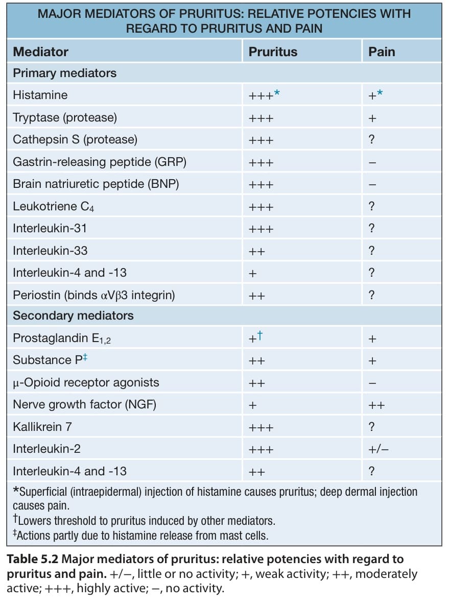
Table 5.2 Major mediators of pruritus: relative potencies with regard to pruritus and pain.+/−, little or no activity; +, weak activity; ++, moderately active; +++, highly active; −, no activity.
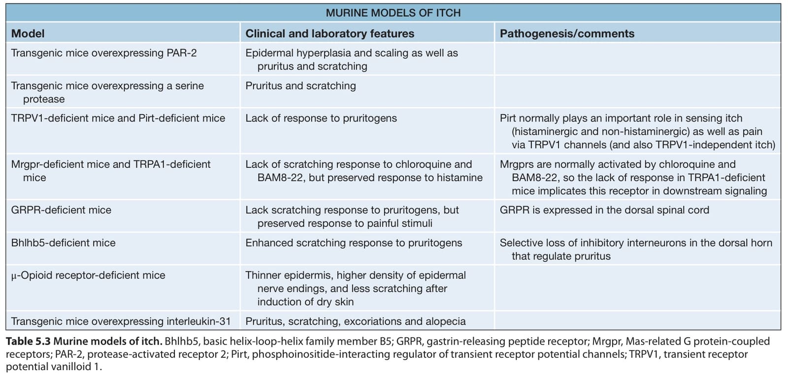
Table 5.3 Murine models of itch. Bhlhb5, basic helix-loop-helix family member B5; GRPR, gastrin-releasing peptide receptor; Mrgpr, Mas-related G protein-coupled receptors; PAR-2, protease-activated receptor 2; Pirt, phosphoinositide-interacting regulator of transient receptor potential channels; TRPV1, transient receptor potential vanilloid 1.