CHRONIC ITCH
Chronic itch may occur in the setting of inflammatory itch secondary to skin diseases, neuropathic itch due to pathology in the nervous system, and systemic or psychiatric disorders (see Ch. 6). It often has a significant effect on patients’ quality of life. Chronic itch and chronic pain share several features, with both potentially involving peripheral and central sensitization (Table 5.4; Fig. 5.3).
Peripheral Sensitization in Chronic Itch
Patients with chronic itch in the setting of atopic dermatitis have increased neurotrophin levels in involved skin, including NGF and neurotrophin 4. Chronic localized pain is also associated with elevated levels of the same neurotrophins, which are known to sensitize nociceptive neurons.
Central Sensitization in Chronic Itch
Chronic itch leads to sensitization of second-order neurons within the dorsal horn of the spinal cord (see Fig. 5.1). There are two forms of increased sensitivity to itch, alloknesis and hyperknesis. In alloknesis, stimuli that normally do not induce itch such as touch or gentle warming do so in the skin that surrounds a pruritic area. This phenomenon is analogous to allodynia, in which gentle mechanical stimuli give rise to a perception of pain. Like allodynia, alloknesis requires ongoing activity in primary afferent C fibers and is probably mediated by low-threshold myelinated mechanoreceptor Aβ fibers (see Fig. 5.2). Alloknesis is common and represents a prominent feature of atopic dermatitis, explaining pruritus associated with dressing and undressing. Expression of the channel protein Piezo2 on Merkel cells has been shown to limit alloknesis in mice.
Hyperknesis is characterized by more intense itch induced by a stimulus that usually produces slight itch and occurs within the skin surrounding an area of inflammation. It is similar to the phenomenon in chronic pain termed hyperalgesia. In mice, neurokinin-1
receptor-expressing spinal neurons play a major role in chronic itch, whereas gastrin-releasing peptide receptor-expressing spinal neurons contribute to hyperknesis, but not alloknesis or ongoing itch.
In patients with chronic itch, painful electrical and heat stimuli may be perceived as itch. An analogous phenomenon can occur in patients with chronic pain, in whom histamine iontophoresis may be perceived as painful. These findings indicate that pain-induced inhibition of pruritus may be compromised in patients with chronic itch. This may also explain why scratching aggravates itch in patients with chronic itch, thereby inducing a vicious itch–scratch cycle.
Itch Related to Impaired Skin Barrier Function
Itch is a common symptom of xerotic skin and is aggravated during the winter in cold climates when the relative humidity falls indoors. Damage to the stratum corneum and the impaired barrier function that results can induce itch even in the absence of inflammation. Environmental changes in pH, temperature, and humidity may activate C fibers to transmit the sensation of itch. Cross-talk between the stratum corneum and nerve fibers may explain the pruritus associated with impaired barrier function. Keratinocytes release neuromediators upon damage to the stratum corneum barrier, and nerve fibers sprout in the epidermis in response to this damage. Indeed, factors such as IL-33 have been shown to directly stimulate sensory neurons and evoke itch in the setting of xerotic skin.
Itch in Older Adults
Itch is particularly frequent in individuals over 65 years of age. Although dry skin is probably the most common trigger, elderly patients can have idiopathic itch without xerosis. Other possible explanations include age-related changes in nerve fibers and central disinhibition of itch due to loss of input from pain fibers. Additional changes that
may contribute to pruritus as well as xerosis in elderly patients include decreased skin surface lipids, diminished skin barrier repair, and immunosenescence (e.g. enhanced Th2 activity).
Cholestatic Pruritus
The enzyme autotaxin and its product lysophosphatidic acid (LPA), a neuronal activator, have a pathogenic role in cholestatic pruritus (see Ch. 6). Serum autotaxin levels correlate with itch intensity in patients with cholestasis. In addition, the G protein-coupled bile acid receptor-1 (Gpbar1; TGR5) has been found to have a role in cholestatic pruritus in mouse models. Murine MrgprA1 and human MRGPRX4 have been identified as itch receptors for bilirubin, suggesting another mechanism by which cholestatic itch is elicited.
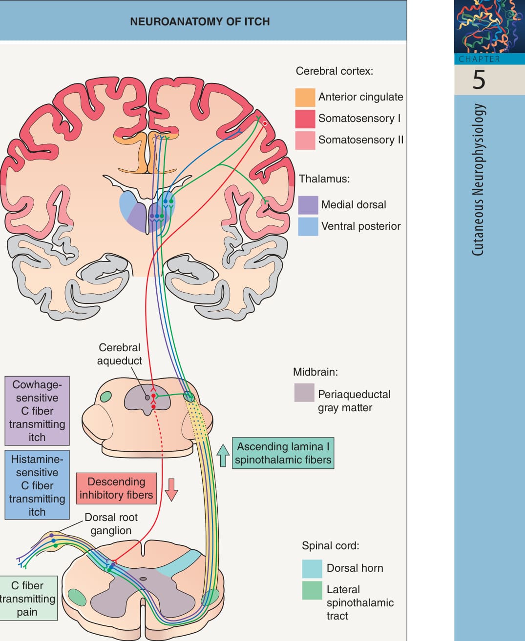
Fig. 5.1 Neuroanatomy of itch. Itch and pain transmission occur via unmyelinated C nerve fibers that excite lamina I neurons in the dorsal horn of the spinal cord. Two subsets of pruritoceptive C-fiber neurons (which respond to histamine and cowhage, respectively) are conducted through distinct lateral spinothalamic pathways, with projections to the thalamus. Processing of itch through either pathway activates several regions of the brain, which are similar to those involved in pain (see Table 5.4). Concomitant painful stimuli can reduce the sensation of itch, possibly by a descending inhibitory mechanism resulting from activation of the periaqueductal gray matter. In mice, gastrin-releasing peptide receptor-positive neurons transmit itch (but not pain) within the spinal cord; the role of such neurons in humans remains to be determined. Adapted from Yosipovitch G. Pruritus: an update. Curr Probl Dermatol 2003;15:137–64.
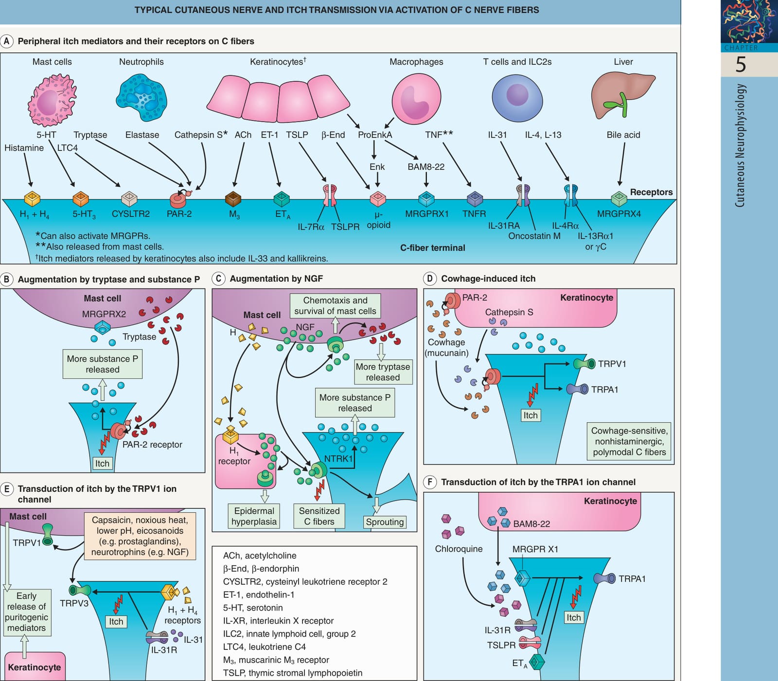
Fig. 5.2 Typical cutaneous nerve and itch transmission via activation of C nerve fibers. There are two categories of axons in a typical cutaneous nerve: (1) primary afferent Aβ, Aδ, and C fibers with cell bodies in dorsal root ganglia; and (2) sympathetic postganglionic fibers with cell bodies in sympathetic ganglia. Separate C fibers (~5% of the total) carry pruritogenic stimuli via two pathways: (1) histamine-induced itch that is transmitted by mechanically insensitive, capsaicin-sensitive fibers via the transient receptor potential vanilloid receptor 1 (TRPV1); and (2) cowhage (mucunain)-induced itch accompanied by a burning sensation that is transmitted by polymodal fibers sensitive to both capsaicin and mechanical stimuli. A Peripheral itch mediators released by immune cells, keratinocytes, and the liver activate pruriceptors on C-fiber terminals in the skin. For example, activated mast cells release histamine, which triggers itch via histamine H1 and H4 receptors on histaminesensitive C fibers. Proteases such as tryptase, elastase, and cathepsin S can cleave proteinase-activated receptor-2 (PAR-2), exposing a tethered ligand and thereby resulting in “self-activation”. Proenkephalin A (ProEnkA) may be cleaved to form enkephalin (ENK) or bovine adrenal medulla 8-22 peptide (BAM8-22). B Self-activation of PAR-2 following tryptase cleavage results in transmission of itch by the C fibers as well as release of additional substance P, which primes mast cells via neurokinin-1 receptors (NK1Rs). C Nerve growth factor (NGF) released by mast cells and keratinocytes (with NGF production stimulated by histamine [H]) activates neurotrophic tyrosine kinase receptor type 1 (NTRK1 [TRKA]) on C fibers, mast cells, and keratinocytes. This induces C fiber sprouting, sensitization to cowhage-induced itch, and increased substance P release; mast cell chemotaxis, survival, and increased tryptase release; and epidermal hyperplasia. D Cowhage-induced itch occurs through the release of mucunain, a protease that activates PAR-2 and PAR-4 receptors (the latter not yet identified in the skin); these receptors can also be activated by endogenous proteases such as cathepsin S as well as tryptase. Activation of PAR-2/4 sensitizes TRPV1 and TRP ankyrin 1 (TRPA1) channels, resulting in cross-talk and itch transmission. E Activation of H1/H4 receptors and the IL-31 receptor (IL-31R) heterodimer leads to membrane depolarization of C fibers via TRPV1, which is required for transduction of histamine-induced itch. The TRPV1 ion channel on C fibers, keratinocytes, and mast cells can also be activated by capsaicin, heat, low pH, eicosanoids, and neurotrophins; although this initially stimulates transmission of itch and release of pruritogenic mediators, it may eventually lead to desensitization, neuropeptide depletion, and attenuation of itch. F C fibers (including those responsive to histamine) in the epidermis express MAS-related G protein-coupled receptors (MRGPRs), which can be activated by chloroquine, BAM8-22, and cathepsin S. TRPA1 is involved in transduction of itch by MRGPRs as well as other pruriceptors, including the IL-31R, thymic stromal lymphopoietin receptor (TSLPR) heterodimer, and endothelin type A receptor (ETA).
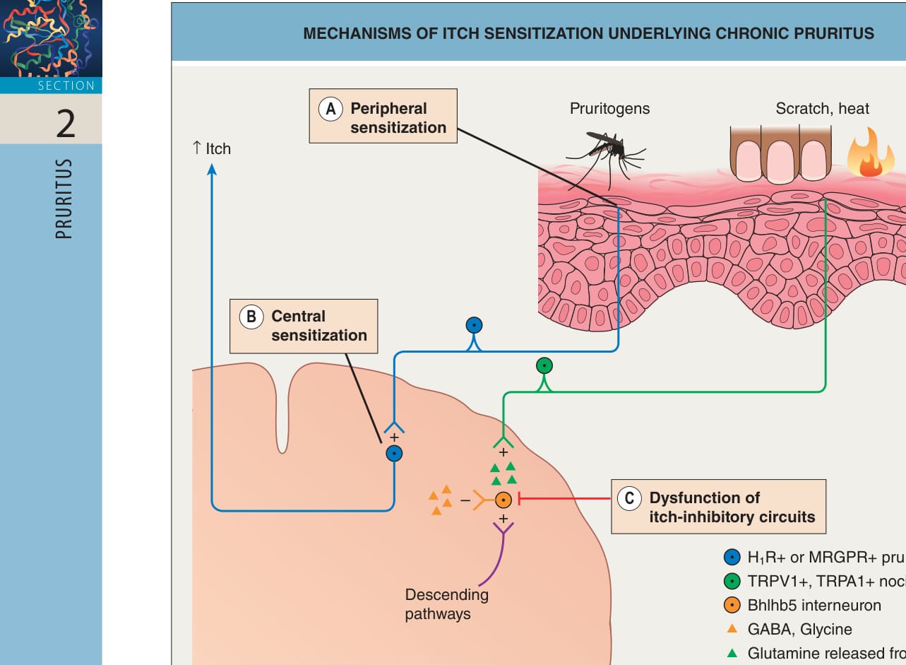
Fig. 5.3 Mechanisms of itch sensitization under- lying chronic pruritus. Three main mechanisms of itch sensitization have important roles in chronic pruritus. A Peripheral sensitization of C-fiber pruriceptors occurs in the setting of chronic pruritus, which may be associated with increased levels of neurotrophins such as nerve growth factor (NGF; see Fig. 5.2C). PAR-2 activation in C fibers can also play a role in itch sensitization by enhancing responses to MAS-related G protein-coupled receptor (MRGPR) agonists. B Spinal itch-transmitting neurons are sensitized in the setting of chronic pruritus. There may be increased synaptic efficacy and signaling via additional synapses. C Itch-inhibitory circuits are normally driven by vesicular glutamate transporter 2 (VGLUT2)- dependent glutamate release from nociceptors as well as by descending pathways; this results in activation of itch-inhibitory Bhlhb5 interneurons, which release GABA, glycine, and/or dynorphins. Decreased function of these interneurons can result in disinhibition of itch. HR, histamine H1 receptor; TRPA1, transient receptor potential ankyrin receptor 1; TRPV1, transient receptor potential vanilloid receptor 1.
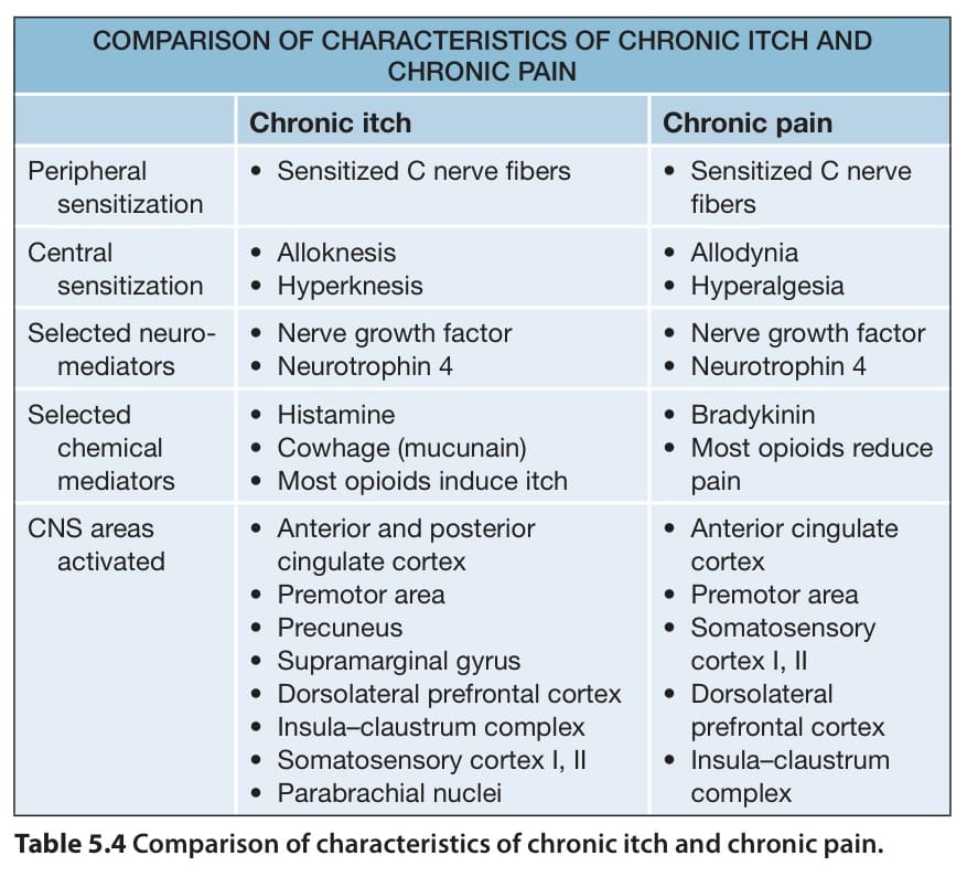
Table 5.4 Comparison of characteristics of chronic itch and chronic pain.