EVALUATION OF THE PATIENT
A structured approach to the evaluation of patients with a chief complaint of pruritus is presented in Fig. 6.2. Pruritus is a subjective sensation, and currently its presence or severity cannot be assessed objectively. Thus, a thorough history that includes the patient’s itch sensation plus a complete cutaneous examination for any evidence of skin lesions are important for correct diagnosis and allow assignment of the patient to one of the three clinical groups of pruritus (see above).
Patients with evidence of a particular skin condition should be treated accordingly, while those with no identifiable primary skin disease (i.e. an absence of or only secondary skin lesions) require further investigation to determine the etiology of their pruritus. Also, chronic pruritus can have multiple underlying factors, and a pruritic skin condition that has nonspecific clinical findings initially may over time develop diagnostic features. Therefore, longitudinal evaluation is essential.
History
A precise history may provide insight into the patient’s disease processes. In addition to the patient’s spontaneous description, specific questions regarding the onset, location, duration, and nature of the pruritus can help to determine its cause (Table 6.2). The use of standardized instruments to assess itch intensity (e.g. a numerical rating scale) and impairment of quality of life (e.g. ItchyQol) is recommended. A medical and medication history should also be obtained. Although chronic, progressive, generalized pruritus without primary skin lesions raises suspicion of an underlying systemic disease, no particular clinical characteristics reliably predict the likelihood of a systemic etiology.
Examination
Careful and complete examination of the skin, nails, scalp, hair, mucous membranes (e.g. oral, conjunctival), and anogenital area at the initial visit is recommended. The morphology and distribution of primary lesions and secondary changes (e.g. excoriations, crusts, lichenification) should be assessed, with particular attention to xerosis, the presence of dermographism (Fig. 6.3), and skin signs of systemic diseases (see Ch. 53). Lesions on the mid upper back suggest a primary skin disease, since this difficult-to-reach area is typically spared (the “butterfly sign”) in patients with skin lesions secondary to scratching (see Fig. 6.5A). However, this region can be accessed with “back-scratching” devices.
The examination should include palpation of major peripheral lymph node groups (e.g. cervical, supraclavicular, axillary, inguinal), especially
CLL, chronic lymphocytic leukemia; PCV, polycythemia vera.
in those with no obvious primary inflammatory skin disease. Together with a general physical examination performed by the patient’s primary care physician, this may disclose an undiagnosed extracutaneous disease (e.g. lymphoma) in individuals with pruritus of unknown etiology.
Laboratory Investigation and Radiographic Imaging
In the setting of pruritus of unknown etiology, a stepwise approach to laboratory tests and other investigations (e.g. radiographic imaging) is recommended (Table 6.3). Microscopic examination of skin scrapings for signs of scabies or a fungal infection as well as examination of stool samples for parasites can also be considered. Biopsies of representative skin lesions, even if nonspecific clinically, are occasionally informative, and direct immunofluorescence studies of perilesional skin or normal-appearing skin (in the vicinity of lesions if present) may point to a specific dermatologic disease such as bullous pemphigoid or dermatitis herpetiformis, respectively. Radiographic examinations may be performed to exclude an underlying malignancy or other systemic condition as well as peripheral nerve impingement when neuropathic itch is suspected.
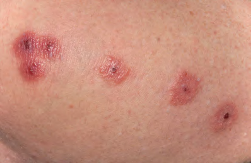
Fig. 6.1 Insect bites. Linear pruritic papules with central crusts demonstrating the “breakfast, lunch, and dinner” sign. Courtesy Antonio Torrelo, MD.
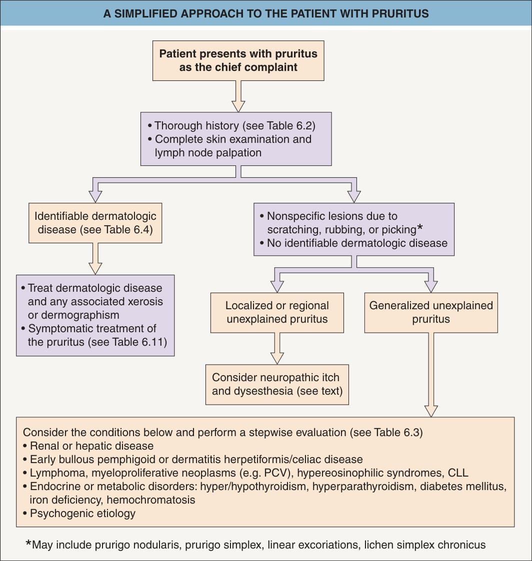
Fig. 6.2 A simplified approach to the patient with pruritus.
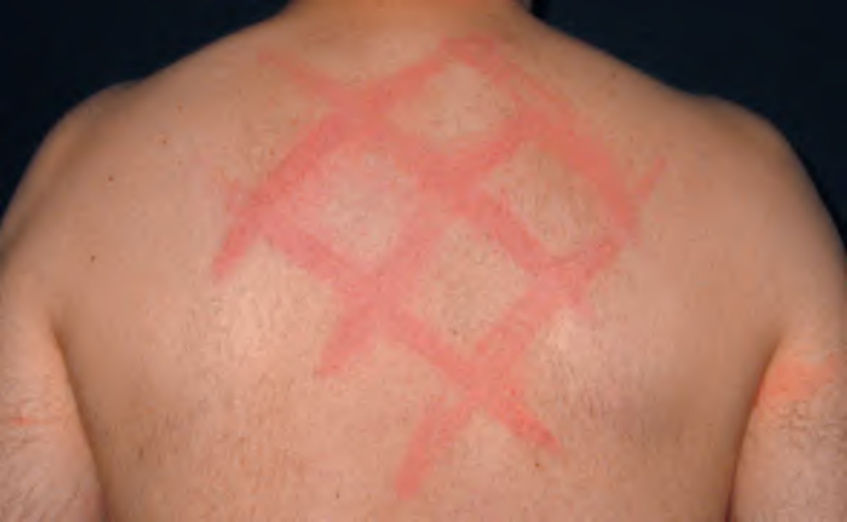
Fig. 6.3 Dermographism. Linear streaks of urticaria induced by scratching the skin. Assessment for dermographism should be performed in all patients with pruritus.
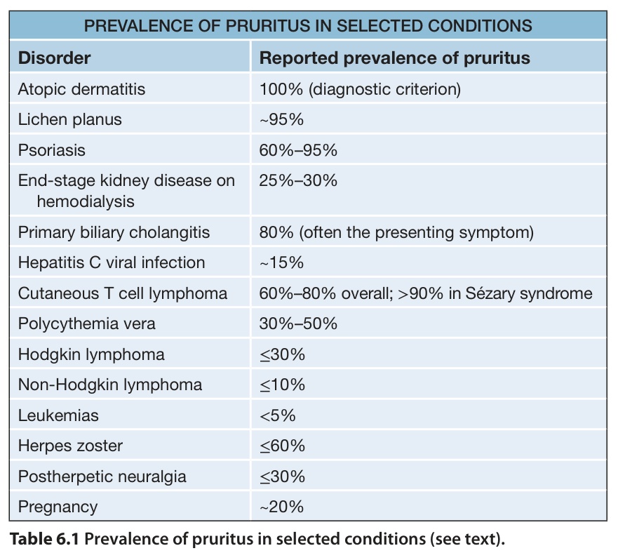
Table 6.1 Prevalence of pruritus in selected conditions (see text).
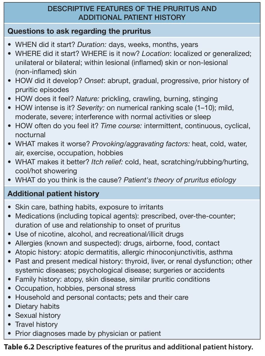
Table 6.2 Descriptive features of the pruritus and additional patient history.
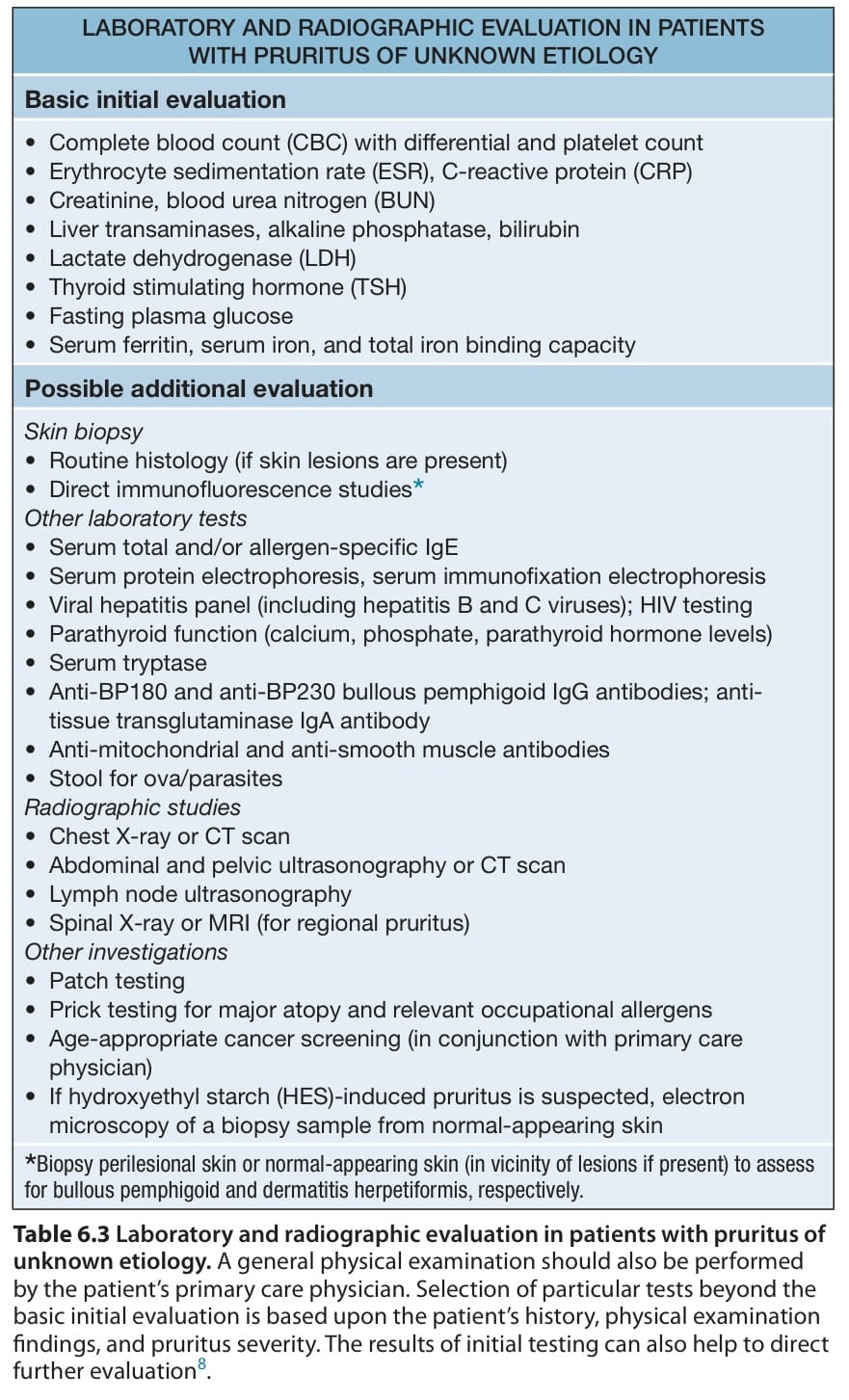
Table 6.3 Laboratory and radiographic evaluation in patients with pruritus of unknown etiology. A general physical examination should also be performed by the patient’s primary care physician. Selection of particular tests beyond the basic initial evaluation is based upon the patient’s history, physical examination findings, and pruritus severity. The results of initial testing can also help to direct further evaluation. life. Often, pruritus is the first indication of a disease flare. Moreover, scratching as a response to itch leads to excoriations and lichenification, which represent additional clinical features of atopic dermatitis. Pruritus can be provoked by exposure to aeroallergens or ingestion of foods to which the patient is sensitized as well as non-immunologic triggers, including emotional stress, overheating, perspiration, and contact with rough fabrics or even air (atmokinesis) (Fig. 6.4). Although most affected children experience worsening of symptoms during the winter, others have exacerbations primarily during the summer. Of note, pruritus may persist after resolution of inflammatory skin lesions.