PRURITUS IN DERMATOLOGIC DISEASE
This section focuses on the sensation of pruritus in selected skin diseases where this symptom is a prominent feature. A more comprehensive discussion of the diseases mentioned and the many additional dermatoses in which pruritus is a characteristic feature (Table 6.4) may be found in other chapters.
Inflammatory Dermatoses
Urticaria
Urticaria is often intensely pruritic and can also produce stinging or prickling sensations. Early lesions and small superficial wheals are
particularly symptomatic. Histamine plays a primary role in the pruritus of urticaria, and H1-receptor antagonists usually decrease or eliminate this sensation as well as the wheals themselves. Several other mediators are involved in the pathophysiology of urticaria, and interleukin (IL)-31 levels correlate with itch intensity. When refractory to higher doses of antihistamines, omalizumab, and cyclosporine represent alternative treatments for urticaria (see Ch. 18).
Atopic dermatitis
Pruritus is such an important aspect of atopic dermatitis that “the diagnosis of active atopic dermatitis cannot be made if there is no history of pruritus” (see Ch. 12). The itch sensation typically comes in “attacks”, which may be severe and significantly impact quality of
The minimal effectiveness of antihistamines in alleviating the itch of atopic dermatitis indicates that histamine is probably not the predominant mediator of the pruritic sensation. Rather, the benefit of antihistamines in atopic dermatitis is most likely due to their sedative effects. Type 2 cytokines such as IL-4, IL-13, IL-31, and thymic stromal lymphopoietin play a key role in the inflammation of atopic dermatitis. As a result, therapies that target Th2 signaling via IL-4/-13 (e.g. dupilumab, tralokinumab, lebrikizumab), IL-31 (nemolizumab), or Janus kinase (JAK) 1/2 have anti-pruritic effects.
Other mediators and receptors with possible roles in the pruritus of atopic dermatitis include neuropeptides (e.g. substance P, calcitonin gene-related peptide [CGRP]), neurotrophic factors (e.g. nerve growth factor [NGF], artemin [causes warmth-provoked itching]), epidermal opioid receptors (downregulated), and endothelin-1. In addition, itch transmission results from kallikrein-related peptidase and mast
Psoriasis
Although not traditionally regarded as a pruritic disease, studies have shown that up to 60%–95% of psoriasis patients suffer from pruritus, with xerosis, heat, sweating, and emotional stress serving as exacerbating factors. While lesions on the scalp, back, and legs are most often pruritic, generalized pruritus can occur in plaque-type as well as erythrodermic and pustular psoriasis. Patients frequently describe the pruritus as having tickling, crawling, and burning components, and antihistamines rarely provide relief. Although the pathophysiology of psoriatic itch is still largely unknown, it is thought to be multifactorial and a variety of itch mediators (e.g. substance P, NGF, protein gene product 9.5, CGRP, neuropeptide Y, IL-2, opioid receptors) have been
implicated. Coadministration of β-blockers, angiotensin-converting enzyme (ACE) inhibitors, or antacids may be associated with the development of psoriatic itch. Successful treatment of psoriasis can reduce associated pruritus (see Ch. 8).
Infestations
Scabies
In patients with scabies, pruritus can be localized or generalized, and it may have a burning component. It is usually more intense at night and typically spares the head. The pruritus usually begins 3–6 weeks after a first-time infestation, and within a few days in subsequent infestations; multiple family members are often affected (see Ch. 84). The itch reflects various components of the immune response against mites, eggs, and scybala. Bacterial superinfection and eczematization of skin lesions may also contribute to itch in patients with scabies.
Pediculosis (lice)
Pruritus at the primary sites of infestation (e.g. scalp, groin) is a clue to the diagnosis of head and pubic lice (see Ch. 84). Generalized pruritus is characteristic of body lice but can also occur with other forms of lice infestation.
Cutaneous T Cell Lymphoma (CTCL)
Pruritus affects >60% of patients with CTCL, with an increased frequency and intensity in those with later-stage disease, especially Sézary syndrome (>90% of patients) and folliculotropic mycosis fungoides (MF). A variant of Sézary syndrome presenting with generalized pruritus and minimal or no clinically evident skin disease has also been described. Pruritus significantly reduces the quality of life in CTCL patients, and it has been associated with an increased risk of disease progression and death.
IL-31 is a putative mediator of itch in CTCL, especially in late-stage disease, and lower serum IL-31 levels have been observed in association with reduced pruritus following treatment with histone deacetylase inhibitors and mogamulizumab (anti-CC-chemokine receptor type-4 antibody). Other possible mediators of CTCL-related itch include IL-4, IL-5, IL-10, IL-13, substance P, NGF, endogenous opioids, and visfatin.
Treatment strategies for severe pruritus not responsive to diseasedirected therapy in CTCL patients include gabapentin (900–2400 mg daily in divided doses) and/or mirtazapine (7.5–15 mg nightly) as well as opioid antagonists (e.g. naltrexone 50–150 mg daily). Aprepitant has also been reported to relieve intractable pruritus in patients with Sézary syndrome and other forms of CTCL, suggesting a role for neurokinin-1 (NK1) receptors and substance P in CTCL-related itch. However, in a recent randomized controlled trial (RCT), aprepitant failed to demonstrate an antipruritic effect compared to placebo in patients with Sézary syndrome.
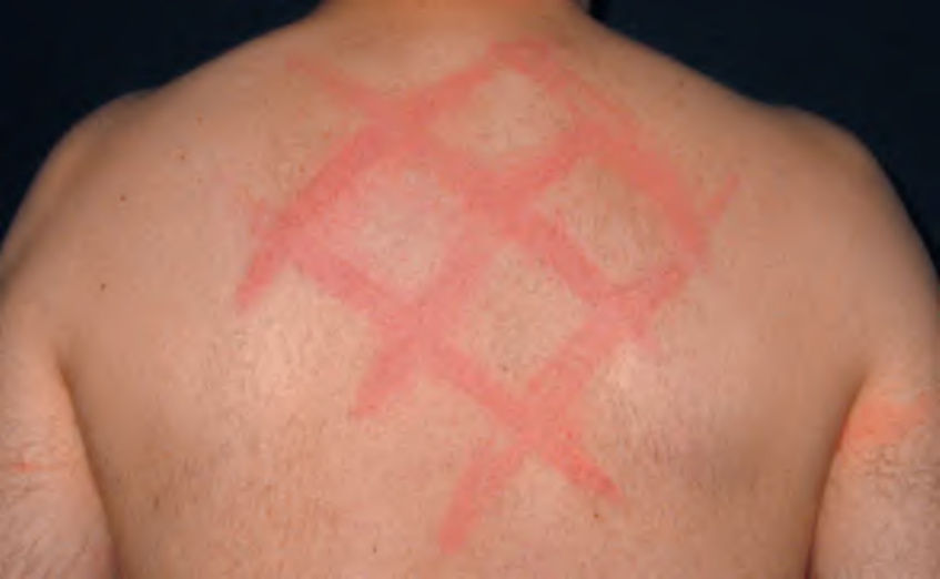
Fig. 6.3 Dermographism. Linear streaks of urticaria induced by scratching the skin. Assessment for dermographism should be performed in all patients with pruritus.
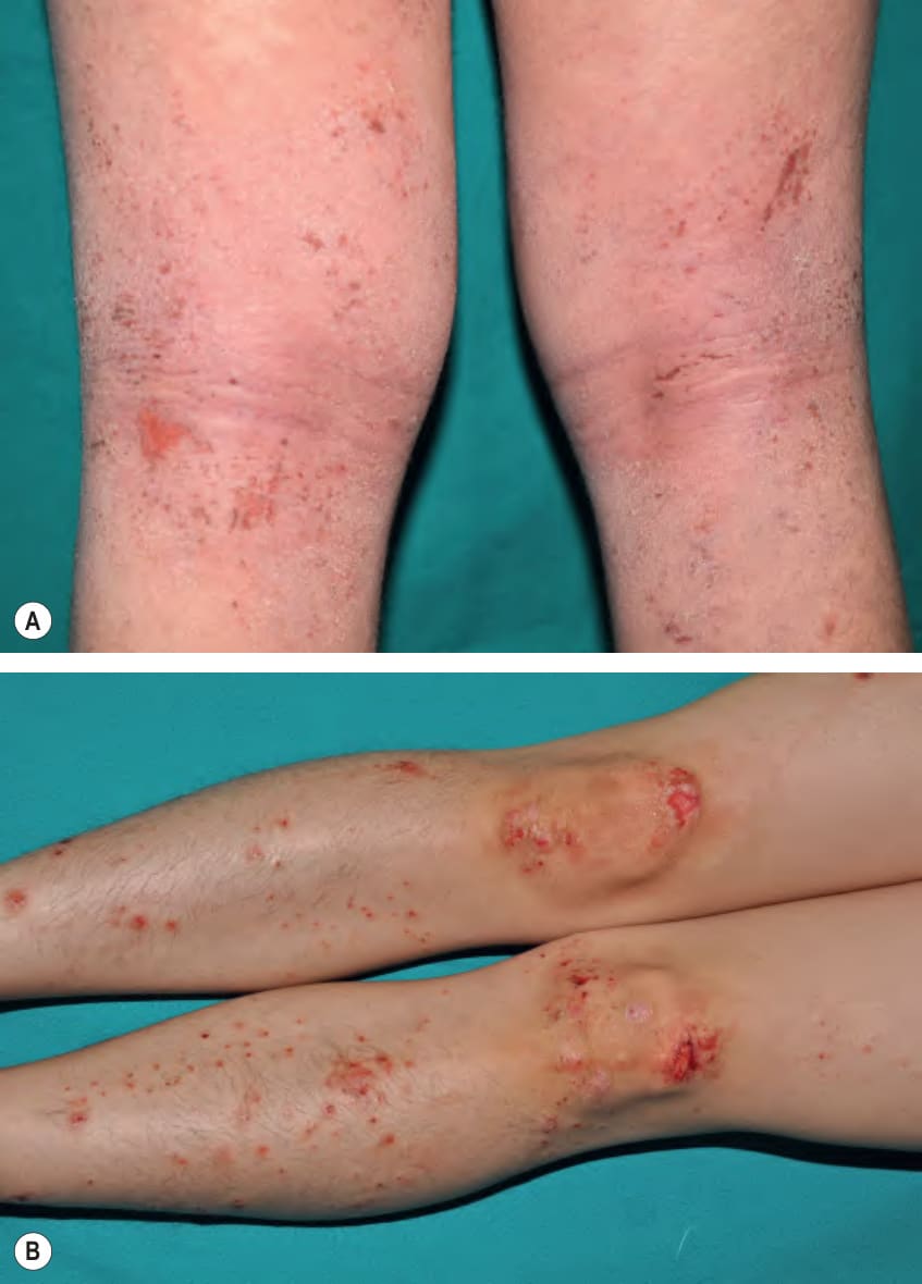
Fig. 6.4 Atopic dermatitis.A Excoriated eczematous plaques in the popliteal fossae, a classic flexural location for atopic dermatitis. B Prurigo simplex, prurigo nodularis-like lesions, and angulated ulcerations; the latter two are primarily on the knees. Courtesy Antonio Torrelo, MD.
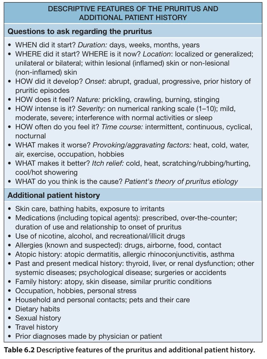
Table 6.2 Descriptive features of the pruritus and additional patient history.
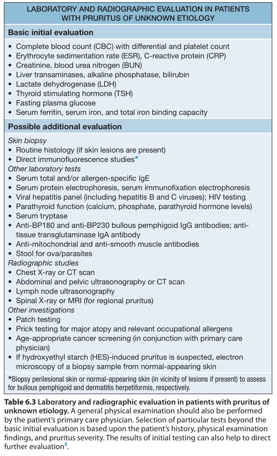
Table 6.3 Laboratory and radiographic evaluation in patients with pruritus of unknown etiology. A general physical examination should also be performed by the patient’s primary care physician. Selection of particular tests beyond the basic initial evaluation is based upon the patient’s history, physical examination findings, and pruritus severity. The results of initial testing can also help to direct further evaluation. life. Often, pruritus is the first indication of a disease flare. Moreover, scratching as a response to itch leads to excoriations and lichenification, which represent additional clinical features of atopic dermatitis. Pruritus can be provoked by exposure to aeroallergens or ingestion of foods to which the patient is sensitized as well as non-immunologic triggers, including emotional stress, overheating, perspiration, and contact with rough fabrics or even air (atmokinesis) (Fig. 6.4). Although most affected children experience worsening of symptoms during the winter, others have exacerbations primarily during the summer. Of note, pruritus may persist after resolution of inflammatory skin lesions.
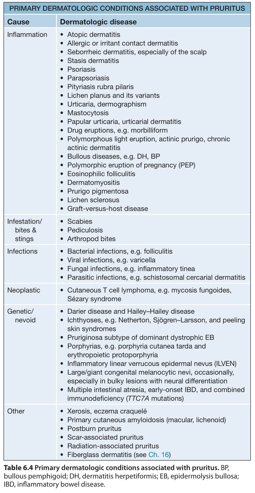
Table 6.4 Primary dermatologic conditions associated with pruritus. BP, bullous pemphigoid; DH, dermatitis herpetiformis; EB, epidermolysis bullosa; IBD, inflammatory bowel disease. cell tryptase binding to protease-activated receptors (PAR-2; see Ch. 5). In atopic patients, morphologic alterations of cutaneous nerves and neuronal sensitization may also contribute to an increased itch sensation and even the perception of painful stimuli as itch.