Dermatitis Artefacta
Synonyms: Factitial dermatitis Factitious disorder
Key features
More common in women than men
Motive is usually subconscious and patients deny their role in causation
Self-inflicted cutaneous lesions that are often induced by foreign objects
Typically located in areas that are easily reached by the hands
Introduction
Dermatitis artefacta represents a type of factitious disorder within the broad DSM-5™ category of somatic symptom and related disorders. Patients inflict cutaneous lesions upon themselves to satisfy a psychologic need of which they are usually not consciously aware. If asked, the patients deny having any role in creating the skin lesions. While some of these individuals create skin lesions as a maladaptive response to an acute psychosocial stress, many suffer from borderline personality disorder. Because patients may dissociate in the process of creating these lesions, they are often unable to give an accurate history, making this disorder challenging to diagnose and treat. “Factitious disorder imposed on another” (previously known as Munchausen syndrome by proxy) is a related condition in which an individual surreptitiously inflicts lesions on another person in order to satisfy a subconscious psychologic need.
Epidemiology
Dermatitis artefacta is uncommon and is reported to have a female : male ratio of up to 20 : 1. Although the onset is most frequently during adolescence and young adulthood, it can occur at any age. Many patients either work in or have a close family member who works in the healthcare field.
Clinical features
Dermatitis artefacta can mimic a wide variety of dermatoses. The lesions may be single or multiple and unilateral or bilateral. Although usually within easy reach of the hands, the lesions can be caused by methods such as carving with sharp instruments, applying chemicals, injecting foreign substances, thermal burns, and the “salt and ice challenge” publicized on the Internet. The morphology ranges from vesicles and bullae to purpura to subcutaneous emphysema to erosions and ulcerations. A clue to the diagnosis is unusual shapes, particularly with angulated edges, that suggest an external method of induction (Fig. 7.8).
Pathology
Histopathologic changes seen with dermatitis artefacta vary considerably, depending upon the manner in which the lesions were produced. The findings are generally not diagnostic and can include erosion, ulceration, epidermal necrosis with multinucleated keratinocytes, hyperkeratosis, irregular acanthosis, vascular proliferation, hypertrophied nerves (as observed in prurigo nodularis), and fibroplasia. Polarizing exogenous material with a variable foreign body response is occasionally evident.
Differential diagnosis
In addition to excluding a primary skin disorder (e.g. ecthyma), dermatitis artefacta must be distinguished from delusional infestation, excoriation disorder, and malingering; the latter is characterized by skin lesions that are self-inflicted for a conscious gain. In nonsuicidal self-injury (see below), patients acknowledge that they inflicted the lesions on themselves.
Treatment
Initially, wound care to help with healing is often indicated and the possibility of a primary dermatologic disorder needs to be excluded. There is controversy as to whether or not to confront the patient upon recognition of dermatitis artefacta, as this might acutely exacerbate their self-harm behaviors. Therefore, it is often more fruitful to provide a supportive environment early in the doctor–patient relationship and then advance to the psychological aspects of the disease at subsequent visits. Antidepressant, antianxiety, or antipsychotic medications may be indicated when affected individuals have an underlying psychiatric disorder (see Table 7.1). Children or adults in whom the lesions represent a response to a transient stress have the most favorable prognosis. Dermatitis artefacta usually has a chronic course, with waxing and waning depending upon circumstances in the affected individual’s life.
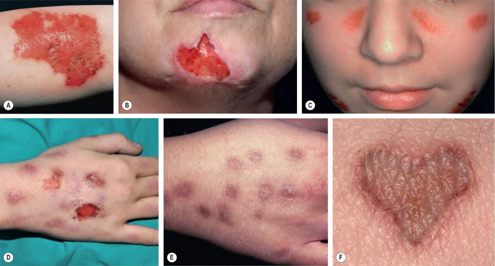
Fig. 7.8 Dermatitis artefacta. Erosion (A) and ulcer (B) with “bizarre” shapes and angulated borders, possibly created with sharp instruments. C Symmetric erosions on the cheeks of an adolescent girl. D Erosions in different stages of healing. E Scars from cigarette burns. F The patient carved this heart-shaped lesion into her skin. These patients all denied knowing how the lesions had developed or having any role in the process. B, Courtesy Kalman Watsky, MD; D, Courtesy Antonio Torrelo, MD; E Courtesy Ronald P. Rapini, MD; F, Courtesy Department of Dermatology, Medical University of Graz.
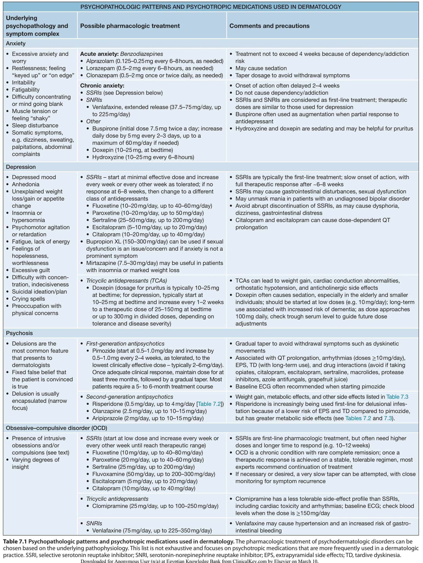
Table 7.1 Psychopathologic patterns and psychotropic medications used in dermatology. The pharmacologic treatment of psychodermatologic disorders can be chosen based on the underlying pathophysiology. This list is not exhaustive and focuses on psychotropic medications that are more frequently used in a dermatologic practice. SSRI, selective serotonin reuptake inhibitor; SNRI, serotonin-norepinephrine reuptake inhibitor; EPS, extrapyramidal side effects; TD, tardive dyskinesia.