PATHOGENESIS
Because it primarily affects the interfollicular epidermis, psoriasis was long regarded as an epidermal disease in which the fundamental biochemical or cellular defect resided within the keratinocyte. Accordingly, prior to the early 1980s24, a number of biochemical mediators, enzymes, and pathways involved in epidermal function were incriminated as being abnormal in psoriasis, including cyclic AMP, eicosanoids, protein kinase C, phospholipase C, polyamines, and transforming growth factor (TGF)-α. Although associated immunologic abnormalities were reported in the late 1970s25, a major paradigm shift occurred when T cell suppressive agents such as cyclosporine were found to result in dramatic improvement of psoriasis. Over the past three decades, psoriasis has been regarded as a T cell-driven disease. The role of lymphocyte subsets as well as cytokines involved in chemotaxis, homing, and activation of inflammatory cells has been extensively investigated, culminating in the development of novel therapeutic approaches. In sum, psoriasis is a chronic, inflammatory, autoimmune skin disease elicited by environmental factors or autoantigens in genetically predisposed individuals and it involves complex interactions amongst the innate and adaptive immune systems as well as keratinocytes and other cells residing within the skin.
Immunopathogenesis
Role of T cells and dendritic cells
A pathogenic role for antigen-presenting cells and T cells is strongly suggested by the association of psoriasis with particular MHC alleles, e.g. HLA-Cw6, as well as (in individuals who carry such alleles) variants in ERAP1, which encodes an aminopeptidase involved in antigen processing. The presence of specific T cell subsets within the epidermis and dermis of lesional skin is well documented (Fig. 8.1A). Also, a number of compounds that affect T cell function (e.g. by targeting the IL-2 receptor, CD2, CD11a, or CD4) were found to result in clinical improvement of psoriasis. Another argument supporting involvement of the adaptive immune system is the disappearance or development of psoriasis following hematopoietic stem cell transplantation. In addition, analysis of lesional T cells has shown oligoclonality, indicating potential antigen-specific expansion of T cell subpopulations, possibly triggered by exogenous microbial or viral antigens or cross-reacting autoantigens, e.g. keratins. Other implicated autoantigens include the antimicrobial peptide LL37 (cathelicidin) and the melanocyte antigen ADAMTSL5.
Animal models for psoriasis have also demonstrated the importance of T cells. In xenograft models in which uninvolved psoriatic skin was transplanted onto immunodeficient mice, donor immune cells (in particular resident T cells) were capable of expanding and inducing the complete lesional phenotype. Induction of psoriatic lesions in these models was also shown to be dependent on TNF and on IFN-α derived from plasmacytoid DCs (pDCs). These experiments demonstrated that T cells can trigger psoriasis in a suitable patient-derived environment.
In humans, several cell types have been implicated in the initiation and maintenance of psoriatic lesions (see Fig. 8.1A). Most of the epidermal T cells are CD8+, whereas the dermal infiltrate is a mixture of CD4+ and CD8+ cells. The majority of the cells in both locations are memory T cells that express cutaneous lymphocyte antigen (CLA; the skin homing receptor) and chemokine receptors such as CCR4. Expression of αβ integrin (VLA-1) on psoriatic T cells, which allows
their interaction with basement membrane collagen IV, is key for the entrance of these cells into psoriatic epidermis and establishment of the psoriatic epithelial phenotype. Unconventional T cells also contribute to the pathogenesis of psoriasis via secretion of proinflammatory cytokines and chemokines. These include natural killer (NK) T cells, gamma delta (γδ) T cells, and innate lymphoid cells (ILCs).
DCs are present in both uninvolved and lesional psoriatic skin, and because of their potent immunostimulatory capacity, they are likely to be involved in its pathogenesis. There is an increased number of dermal
DCs in psoriatic skin, and they have an enhanced ability to activate T cells when compared to DCs from normal skin. The role of DCs in psoriasis has been validated by the presence of a prominent genomic DC signature and the decrease of DCs during effective targeted therapy.
Based upon studies in humans and a xenograft model, a subtype of DC, the pDC, was shown to initiate psoriasis via the production of IFN-α. Complexes of self DNA or RNA (from keratinocytes) plus antimicrobial peptide LL37 trigger IFN-α release by pDCs via a Toll-like receptor 9 (TLR9)-dependent mechanism (see Fig. 8.1A). This leads to a breaking of tolerance to self nucleic acids and potentially explains the start of the inflammatory cascade in psoriasis.
The presence of neutrophils in the epidermis, either in spongiform pustules of Kogoj or in microabscesses of Munro, is a typical histopathologic feature of psoriasis, especially acute or pustular forms.
Neutrophils are typically prominent in active lesions and in the marginal zone of expanding plaques, but, in contrast to T cells, they are not a consistent feature of lesional skin. Although activated neutrophils could contribute to its pathogenesis, they are not considered to be the primary cause of psoriasis.
Prominent angiogenesis is observed within plaques of psoriasis. There is increased expression of vascular endothelial growth factor (VEGF), and anti-VEGF therapy leads to improvement in mouse models of psoriatic inflammation.
Cytokines, chemokines, and transcription factors
Psoriasis is considered to be a disease with prominent involvement of T helper cell subsets and their secreted cytokines. This is supported by the significant clinical responses observed following administration of monoclonal antibodies that target specific cytokines (Fig. 8.1B). Increased amounts of Th1 (IFN-γ and IL-2), Th17 (IL-17A and -17F), and Th22 cytokines (IL-22) are observed, whereas levels of the anti-inflammatory cytokine IL-10 are reduced. The concerted action of these cytokines leads to proliferation of keratinocytes and dermal inflammation (see Fig. 8.1A).
Following activation of pDCs, large amounts of type I IFN are produced, leading to maturation and activation of dDCs. The latter cells then secrete IL-23, which together with other cytokines (e.g. IL-6, TGF-β), drives naive T cells in a Th17 direction. Within the skin-draining lymph nodes, a naive T cell can differentiate into a Th1, Th17, or Th22 cell. After migrating into the skin, these cells release their signature cytokines; release of IL-17A and IL-17F leads, often in synergy with TNF, to an activation of the MAPK and NF-κB signaling pathways in keratinocytes and other skin resident cells. This creates a “feed-forward” inflammatory response that can be self-amplifying and result in activation and recruitment of Th1 and Th22 cells into the psoriatic lesion.
Any model of pathogenesis has to also account for the dramatically increased proliferation rate of keratinocytes. Within psoriatic plaques, keratinocytes express the transcription factor STAT3, and in a transgenic animal model, epidermal expression of STAT3 (in cooperation with T cells) was found to induce psoriasis-like lesions. STAT3 induces upregulation of a number of psoriasis-relevant genes (e.g. ICAM1, TGFA), with TGF-α having been shown to stimulate proliferation of keratinocytes in psoriasis via an autocrine loop. Furthermore, STAT3 activation occurs downstream of several cytokines involved in the pathogenesis of psoriasis such as IL-6, IL-22, and IL-23. In addition,
STAT3 activation induces differentiations of Th17 cells and it directly activates the promoters of IL17A and IL17F.
IFN-γ is released by activated T cells and NKT cells within the epidermis, and it activates members of the STAT transcription factor family, which drive the expression of a large number of immune-related genes that have roles in psoriasis pathogenesis (see above). The IFN-γ- activated pathway is a key feature of psoriasis and explains several phenotypic alterations such as vasodilation (by the induction of nitric oxide synthase [iNOS]) and accumulation of T cells (via the expression of various chemokines). The innate immune cytokines IL-1, IL-6, and TNF are also upregulated in psoriatic skin. TNF is a particularly relevant cytokine and its importance is underscored by the therapeutic efficacy of TNF inhibitors (see Fig. 8.1B).
Chemokines are important mediators in the trafficking of leukocytes, and the increase in several chemokines and their cognate receptors within psoriatic lesions has been extensively documented. CXCL8 is thought to mediate the often-prominent infiltration by neutrophils. CCL17, CCL20, CCL27, and CXCL9–11 are implicated in attracting T cells to the psoriatic plaque. A pDC-attracting chemokine, chemerin, is increased in psoriatic skin and might contribute to the early recruitment of pDCs into psoriatic lesions.
Lastly, there is significant overlap between the cytokines, transcription factors, and subsequent signaling pathways discussed in this section and the psoriasis susceptibility genes outlined in Table 8.1.
Innate immunity and role of keratinocytes
In the skin, various cell types are involved in innate (non-adaptive) immune response pathways. These include DCs (myeloid DCs and pDCs), NKT cells, γδT cells, ILCs, and neutrophils, as well as epidermal keratinocytes. For example, keratinocytes constitutively express antimicrobial proteins such as β-defensin-1 (hBD1) and secretory leukocyte protease inhibitor (SLPI), which have direct antimicrobial activity against a broad spectrum of pathogens. In addition, keratinocytes can be stimulated to express a wide variety of other inducible antimicrobials such as hBD2, the cathelicidin LL37, and SKALP/elafin. In addition to these effector molecules, keratinocytes express TLRs and secrete signaling molecules such as IL-1, IL-6, IL-8, and TNF (see Fig. 8.1A). Interestingly, the antimicrobial effector protein hBD2 was also shown to have chemotactic activity via CCR6 and to bind to TLR4. Since most of these proteins are highly expressed in lesional psoriatic skin, it is likely that they are involved in the initiation or control of the inflammatory process; however, their precise roles remain to be determined.
Disease memory
It is well known that psoriasis often relapses at previously affected body sites after treatment cessation. The origin of this disease memory is unknown, but both tissue-resident memory T cells and epithelial stem cells have been proposed as candidate cells responsible for this phenomenon. The hypothesis that disease memory develops when lesions become chronic has led to the speculation that early and intensive interventions could avoid this phenomenon.
Triggering Factors
Triggering factors, both external (directly interacting with the skin) and systemic, can elicit psoriasis in genetically predisposed individuals.
External triggering factors
The Koebner or isomorphic phenomenon, i.e. the elicitation of psoriatic lesions by injury to the skin, is observed in ~25% of patients with psoriasis. A particular patient may be “Koebner-negative” at one point in time and later become “Koebner-positive”. The Koebner phenomenon suggests that psoriasis is a generalized skin disease that can be triggered locally. Psoriatic lesions can also be induced by other forms of cutaneous injury, e.g. sunburn, morbilliform drug eruption, viral exanthem. The lag time between the trauma and the appearance of skin lesions is usually 2–6 weeks.
Systemic triggering factors
Infections, particularly bacterial infections, may induce or aggravate psoriasis. Provoking infections have been observed in up to 45% of psoriatic patients. Streptococcal infections, especially pharyngitis, are the most common offenders. In the palatine tonsils of psoriasis patients, immune responses have been shown to be dysregulated, with elevated expression of CLA and the IL-23 receptor. Streptococci can also be isolated from other sites of infection, e.g. dental abscesses, perianal cellulitis, impetigo. Recently, group A streptococcal infections were shown to induce clonal expansion of CD1a-restricted T cells that can drive autoreactivity in psoriasis51a.
HIV infection has also been shown to aggravate psoriasis (see Ch. 78).
Hypocalcemia has been reported to be a triggering factor for generalized pustular psoriasis. Although active vitamin D analogues improve psoriasis, abnormal vitamin D levels have not been shown to induce psoriasis. Pregnancy may alter disease activity, e.g. 50% of the patients in one series reported improvement during pregnancy. However, pregnant women may develop pustular psoriasis, also referred to as impetigo herpetiformis, and sometimes in association with hypocalcemia. As in pustular psoriasis in non-pregnant individuals, the pustules in impetigo herpetiformis are sterile.
Psychogenic stress is a well-established systemic triggering factor in psoriasis, with a heightened cortisol response to stress having been demonstrated in affected patients. Stress is associated with initial presentations of the disease as well as flares of pre-existing psoriasis. In a prospective study, cognitive and behavioral patterns of worrying and scratching were both independently related to an increase in disease severity and pruritus 4 weeks later.
Several drugs have been incriminated as inducers of psoriasis, in particular lithium, IFNs, anti-PD-1 antibodies, β-blockers, and antimalarials. The association with immune checkpoint inhibitors may be related to increased levels of IL-6. A rapid taper of systemic corticosteroids can induce pustular psoriasis as well as flares of plaque psoriasis. Patients receiving TNF inhibitors, e.g. for Crohn disease or rheumatoid arthritis, may experience new-onset or worsening of psoriasis, including pustulosis of the palms and soles (see Fig. 21.24). In addition, patients with atopic dermatitis who receive dupilumab can develop psoriasiform lesions, presumably due to a phenotypic shift from predominantly Th2-mediated inflammation to a Th1 immune response.
Obesity, increased alcohol consumption, and smoking have all been associated with psoriasis. In one analysis, smoking appeared to have a role in the onset of psoriasis, while obesity appeared to be a consequence of psoriasis. However, other studies have suggested that weight gain often precedes the development of psoriasis. Some studies have found that the prevalence of psoriasis in a population of individuals who stop smoking or who lose weight eventually reverts to background levels.
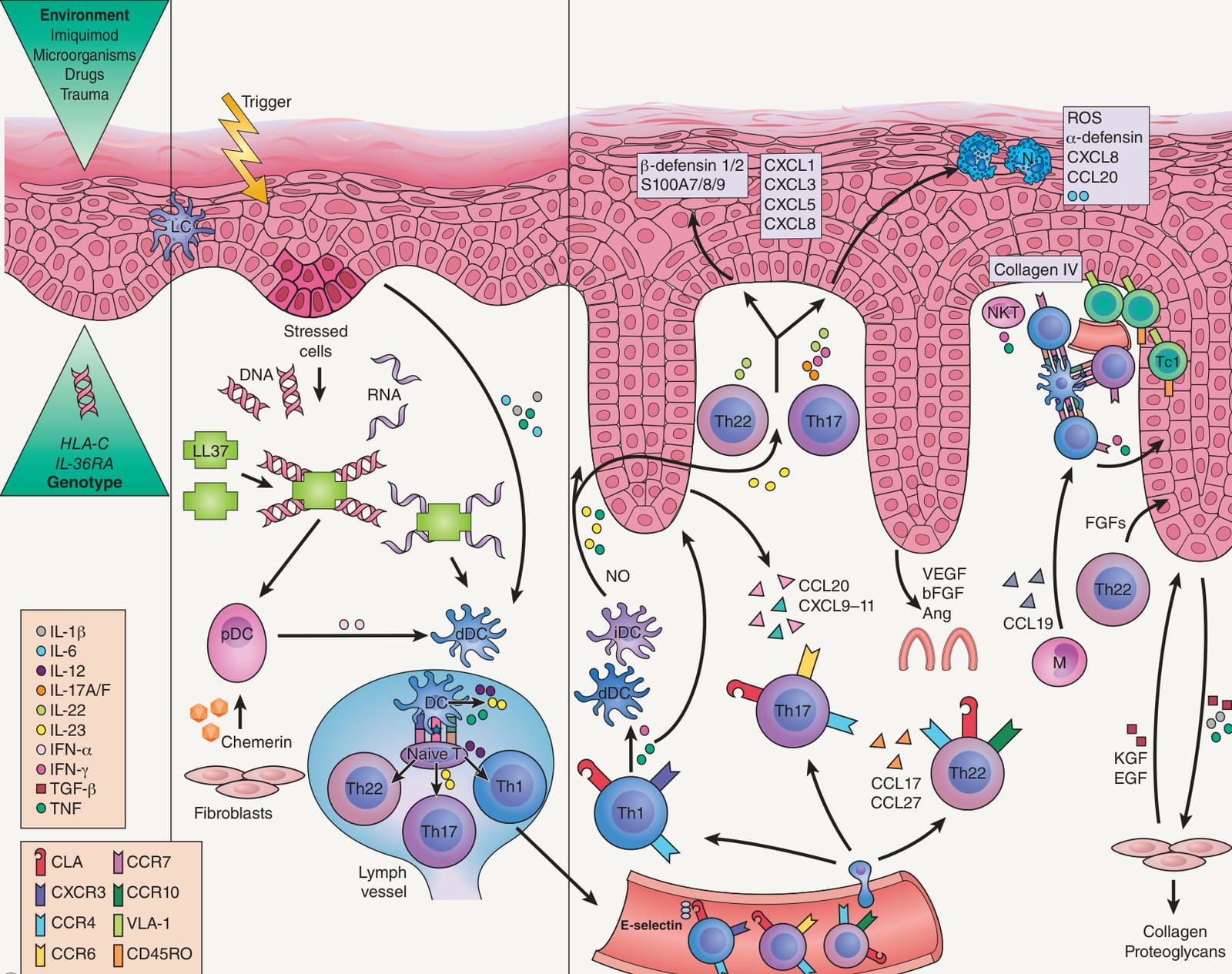
Fig. 8.1A Immunopathogenesis of psoriasis. The occurrence of triggering environmental factors in genetically predisposed individuals, carrying susceptibility alleles of psoriasis-associated genes, results in disease development. During the initiation phase, stressed keratinocytes can release self DNA and RNA, which form complexes with the cathelicidin LL37 that then induce interferon-α (IFN-α) production by plasmacytoid dendritic cells (pDCs; recruited into the skin via fibroblastreleased chemerin), thereby activating dermal DCs (dDCs). Keratinocyte-derived interleukin-1β (IL-1β), IL-6, and tumor necrosis factor (TNF) also contribute to the activation of dDCs. Activated dDCs then migrate to the skin-draining lymph nodes to present an as-yet-unknown antigen (either of self or of microbial origin) to naive T cells and (via secretion of different types of cytokines such as IL-12 and IL-23 by DCs) promote their differentiation into T helper 1 (Th1), Th17, and Th22 cells. Th1 cells (expressing cutaneous lymphocyte antigen [CLA], CXC-chemokine receptor 3 [CXCR3], and CC-chemokine receptor 4 [CCR4]), Th17 cells (expressing CLA, CCR4, and CCR6), and Th22 cells (expressing CLA, CCR4, and CCR10) migrate via lymphatic and blood vessels into psoriatic dermis, attracted by the keratinocyte-derived chemokines CCL20, CXCL9–11, CCL17, and CCL27; this ultimately leads to the formation of a psoriatic plaque. Th1 cells release IFN-γ and TNF, which amplify the inflammatory cascade, acting on keratinocytes and dDCs. Th17 cells secrete IL-17A and IL-17F (and also IFN-γ and IL-22), which stimulate keratinocyte proliferation and the release of β-defensin 1/2, S100A7/8/9, and the neutrophil-recruiting chemokines CXCL1, CXCL3, CXCL5, and CXCL8. Neutrophils (N) infiltrate the stratum corneum and produce reactive oxygen species (ROS) and α-defensin with antimicrobial activity, as well as CXCL8, IL-6, and CCL20. Th22 cells secrete IL-22, which induces further release of keratinocyte-derived T cell-recruiting chemokines. Moreover, inflammatory DCs (iDCs) produce IL-23, nitric oxide (NO) radicals, and TNF, while natural killer T (NKT) cells release TNF and IFN-γ. Keratinocytes also release vascular endothelial growth factor (VEGF), basic fibroblast growth factor (bFGF), and angiopoietin (Ang), thereby promoting neoangiogenesis. Macrophage (M)-derived chemokine CCL19 promotes clustering of Th cells expressing chemokine receptor CCR7 with DCs in the proximity of blood vessels, with further T cell activation. At the dermal–epidermal junction, memory CD8+ cytotoxic T cells (Tc1) expressing very-late antigen-1 (VLA-1) bind to collagen IV, allowing entry into the epidermis and contributing to disease pathogenesis by releasing both Th1 and Th17 cytokines. Cross-talk between keratinocytes, producing TNF, IL-1β and transforming growth factor-β (TGF-β), and fibroblasts, which in turn release keratinocyte growth factor (KGF), epidermal growth factor (EGF) and TGF-β, and possibly Th22 cells releasing FGFs, contribute to tissue reorganization and deposition of extracellular matrix (e.g. collagen, proteoglycans).
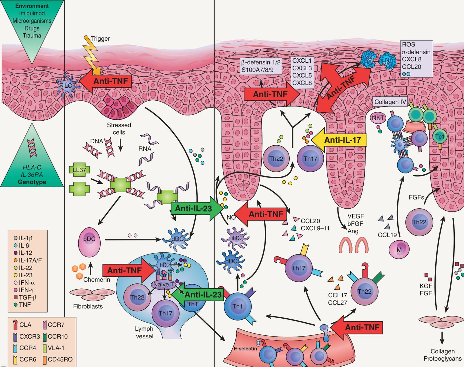
Fig. 8.1B Sites of action of systemic immunomodulators (“biologics”) used to treat psoriasis. Note the multiple sites for TNF inhibitors while anti-IL-17 and -IL-23 antibodies are more selective. In the epidermis of psoriatic lesions, IL-17A, IL-17F, and IL-22 induce host defense proteins and several proinflammatory cytokines, including IL-36; there is greater expression of IL-36 in patients with generalized pustular psoriasis and in those with a loss-of-function mutation in the gene that encodes the IL-36 receptor antagonist. Recently, the first anti-IL-36 receptor antibody was approved by the FDA to treat patients with flares of generalized pustular psoriasis. LC, Langerhans cell. Courtesy Dr Paola DiMeglio.
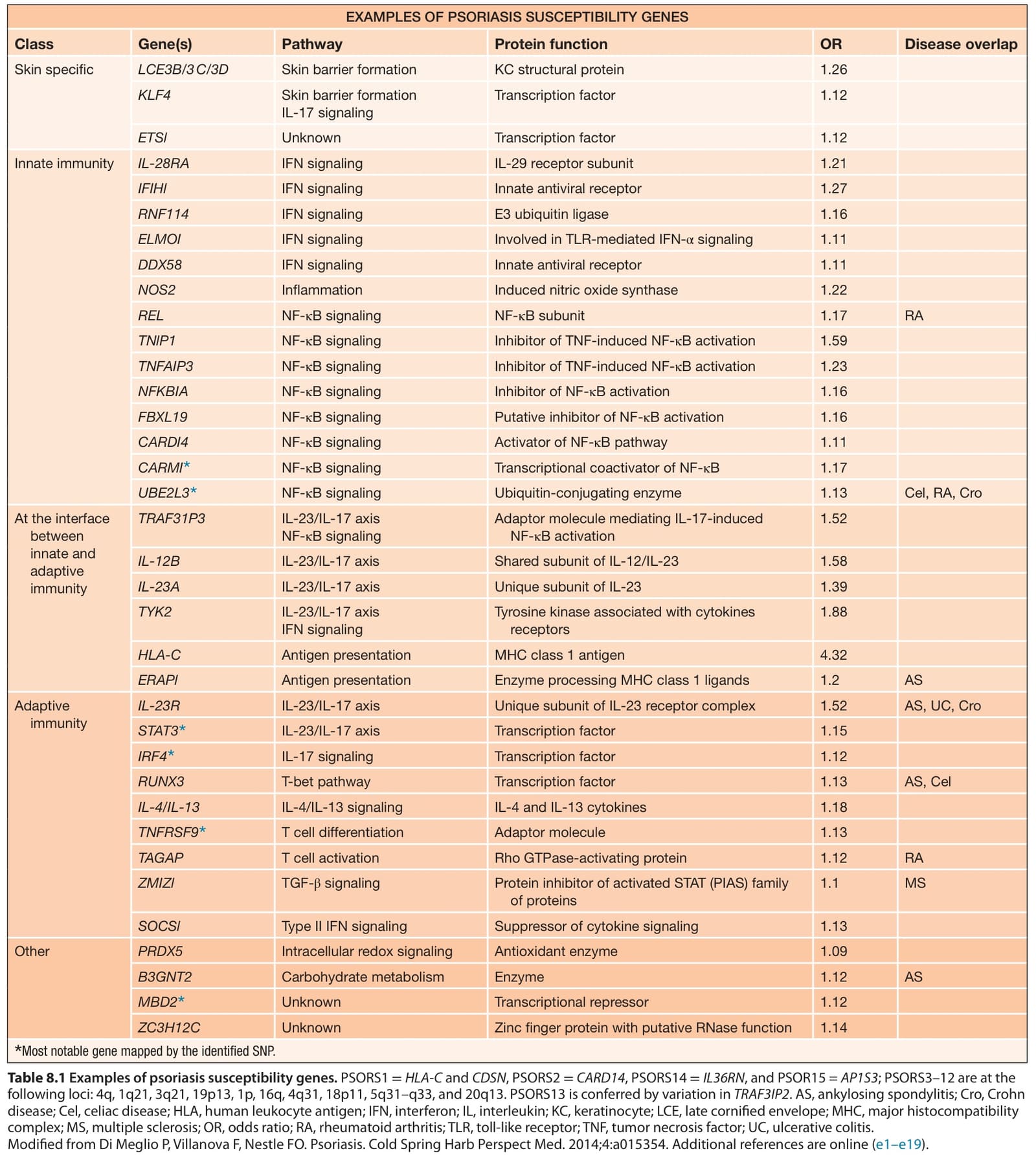
Table 8.1 Examples of psoriasis susceptibility genes. PSORS1 =HLA-C and CDSN, PSORS2 =CARD14, PSORS14 =IL36RN, and PSOR15 = AP1S3; PSORS3–12 are at the following loci: 4q, 1q21, 3q21, 19p13, 1p, 16q, 4q31, 18p11, 5q31–q33, and 20q13. PSORS13 is conferred by variation in TRAF3IP2. AS, ankylosing spondylitis; Cro, Crohn disease; Cel, celiac disease; HLA, human leukocyte antigen; IFN, interferon; IL, interleukin; KC, keratinocyte; LCE, late cornified envelope; MHC, major histocompatibility complex; MS, multiple sclerosis; OR, odds ratio; RA, rheumatoid arthritis; TLR, toll-like receptor; TNF, tumor necrosis factor; UC, ulcerative colitis. Modified from Di Meglio P, Villanova F, Nestle FO. Psoriasis. Cold Spring Harb Perspect Med. 2014;4:a015354. Additional references are online (e1–e19).