CLINICAL FEATURES
The extent to which psoriasis represents a spectrum of different manifestations of a single disease versus several separate disease entities remains a matter of debate. As knowledge regarding genotypes expands, will entities be defined by the associated genetic findings (e.g. in CARD14 or IL36RN) or will there continue to be classifications based upon different phenotypes? Either way, recognition of characteristic skin findings remains essential.
Chronic plaque psoriasis, the most common variant of psoriasis vulgaris, presents as sharply demarcated and erythematous papulosquamous lesions. Less often, nearly all of the body surface is involved (erythrodermic psoriasis) or numerous, small, widely disseminated papules and plaques are seen (guttate psoriasis).
Given the spectrum of cutaneous manifestations, at any one point in time, different variants may coexist in a single individual. However, the skin lesions all share the same important hallmarks: erythema, thickening, and scale. As previously noted, there is also significant interindividual variability. For example, in patients with chronic plaque psoriasis, those with type I disease (HLA-Cw6+) have an earlier onset,
more widespread disease and frequent recurrences, compared to those with type II psoriasis.
Although the size of a lesion may vary from a pinpoint papule to over 20 cm in diameter, the outline of the lesion is usually circular, oval, or polycyclic (the latter indicating that the lesion is derived from several smaller units). The configuration of psoriatic lesions due to the Koebner phenomenon reflects the etiology of the trauma. In addition to their highly characteristic sharp demarcation, psoriatic lesions are sometimes surrounded by a pale blanching ring, which is referred to as Woronoff’s ring.
The classic findings of erythema, thickening, and scale are reflections of the histologic findings of elongated dilated capillaries that are close to the skin surface, epidermal acanthosis plus cellular infiltrates, and abnormal keratinization, respectively. If the superficial silvery white (micaceous) scales are removed, then a wet surface is seen with characteristic pinpoint bleeding. This finding, called Auspitz sign, is the clinical reflection of elongated vessels in the dermal papillae together with thinning of the suprapapillary epidermis.
During exacerbations, psoriatic lesions often itch. Pinpoint papules surrounding existing psoriatic plaques indicate that the patient is in an unstable phase of the disease. In addition, expanding psoriatic lesions are characterized by an active edge with a more intense erythema. Inflamed lesions may even be slightly tender. The involution of a lesion usually starts in its center, resulting in annular psoriatic lesions (Fig. 8.2).
Chronic Plaque Psoriasis
In chronic plaque psoriasis, there is a relatively symmetric distribution of sharply defined, erythematous, scaly plaques (Figs. 8.3 & 8.4). The degree of body surface area involvement can vary, from limited to extensive. The scalp, elbows, knees and lumbosacral area are sites of predilection, as are the hands and feet (Figs. 8.5 & 8.6). The genitalia are involved in up to 45% of patients (Fig. 8.7; see Ch. 73).
Plaques can persist for months to years at the same locations. In contrast, periods of complete remission do occur and remissions lasting for at least 5 years have been reported in ~15% of patients. When the disease is flaring, including in response to a triggering factor, pinpoint papules and deep erythematous rims may develop. The latter serve as clues the psoriasis has entered an unstable phase and occasionally sterile pustules may be observed. Differentiation of unstable psoriasis with pustule formation from generalized pustular psoriasis can be difficult.
Because the percentage of body surface area involved does not reflect the severity of the individual lesions with respect to erythema, induration, and scaling, the Psoriasis Area and Severity Index (PASI) was formulated (Table 8.2). This is a single calculated score that is based on the body surface area involved (in each of four anatomic areas – head, upper extremities, trunk, and lower extremities) and clinical grading of lesional erythema, induration, and scaling. The PASI is a cumbersome
calculation and is more commonly utilized for clinical trials than for the routine management of patients with psoriasis. Other outcome measures of disease severity include the Palmoplantar Psoriasis Physician Global Assessment, Psoriasis Scalp Severity Index, Nail Psoriasis Severity Index, and Static Physician Global Assessment of Genitalia.
Guttate Psoriasis
Guttate psoriasis is more commonly seen in children and adolescents and is frequently preceded by an upper respiratory tract infection (Fig. 8.8). In over half of the patients, an elevated antistreptolysin O,
anti-DNase B, or streptozyme titer is found, indicating a recent streptococcal infection (see previous section).
Erythrodermic Psoriasis
This variant of psoriasis is characterized by generalized erythema and scaling, and its onset can be gradual or acute. Although there are many causes of erythroderma (see Ch. 10), clues to the diagnosis of psoriatic erythroderma include previous plaques in classic locations, characteristic nail changes, and central facial sparing.
CARD14-associated papulosquamous eruption
In patients with CARD14-associated papulosquamous eruption that is due to CARD14 mutations, there are features of both psoriasis and pityriasis rubra pilaris (PRP). Features include an early age of onset, prominent involvement of the cheeks, chin, and ears, a family history of psoriasis or PRP, minimal response to conventional topical and systemic psoriasis therapies, and improvement with ustekinumab (Fig. 8.9). NF-κB is upregulated in the skin of those with this disorder and CARD14 mutations, resulting in an increase in IL-8 and CCL20 which recruit and activate inflammatory cells. The latter leads to production of IL-23 by dendritic cells and IL-17 and IL-22 by T cells. This provides an explanation for the therapeutic response to ustekinumab.
Pustular Disease
Generalized pustular psoriasis
In generalized pustular psoriasis, infiltration of neutrophils dominates the histologic picture, explaining the bright erythema and sterile pustules (Fig. 8.10). It is an unusual manifestation of psoriasis, and triggering factors include pregnancy, rapid tapering of corticosteroids (or
Diffuse involvement with erythema and scaling of the palmar (A) and plantar surfaces (B).
other systemic therapies), hypocalcemia, infections, and, in the case of the localized pattern, topical irritants. Generalized pustular psoriasis during pregnancy is also referred to as impetigo herpetiformis.
Of note, some patients with the phenotype of pustular psoriasis have an autoinflammatory disorder with a distinct genotype (see Tables 45.6 & 45.7). This group includes DIRA (deficiency of IL-1 receptor antagonist; see Figs. 45.11A & 45.12A) and DITRA (deficiency of IL-36 receptor antagonist; see Figs. 45.11B & 45.12B). Future pathogenetic discoveries may lead to additional discrete entities.
Pustular psoriasis has four distinct phenotypic patterns:
●von Zumbusch pattern. This is a generalized eruption starting abruptly with erythema and pustulation (see Fig. 8.10). The skin is painful during this phase, and the patient has a fever and feels ill. After several days, the pustules usually resolve and extensive scaling is observed. Sometimes, chronic plaques of psoriasis, if present, can resolve. In the original case report by von Zumbusch (1910), nine episodes of pustulation occurred over a period of 10 years.
●Annular pattern. The eruption is characterized by annular lesions, consisting of erythema and scaling with pustulation at the advancing edge (Fig. 8.11). The lesions enlarge by centrifugal expansion over a period of hours to days, while healing occurs centrally.
A Well-demarcated erythematous plaques with scale on the glans and shaft of the penis in an adult. B Infantile psoriasis with a well-demarcated erythematous plaque of the diaper area, along with involvement of the penis and scrotum. This is in contrast to atopic dermatitis where there is often sparing of the diaper area. A, Courtesy Lorenzo Cerroni, MD; B, Courtesy Julie V. Schaffer, MD.
●Exanthematic type. This is an acute eruption of small pustules, abruptly appearing and disappearing over a few days. It usually follows an infection or may occur as a result of administration of specific medications, e.g. lithium. Systemic symptoms usually do not occur. There is overlap between this form of pustular psoriasis and pustular drug eruptions, also referred to as acute generalized exanthematous pustulosis (AGEP; see Ch. 21).
●“Localized” pattern. Sometimes pustules appear within or at the edge of existing psoriatic plaques. This can be seen during the unstable phase of chronic plaque psoriasis and following the application of irritants, e.g. tars, anthralin.
Pustulosis of the palms and soles
Pustulosis of the palms and soles is characterized by “sterile” pustules of the palmoplantar surfaces admixed with yellow–brown macules (Fig. 8.12); scaly erythematous plaques may also be seen. A minority of patients have chronic plaque psoriasis elsewhere. In contrast to the natural history of generalized pustular psoriasis, the pustules remain localized to the palmoplantar surfaces and the course of this disease is chronic. Focal infections and stress have been reported as triggering factors and smoking may aggravate the condition. Pustulosis of the palms and soles is one of the entities most commonly associated with sterile inflammatory bone lesions within the context of SAPHO
syndrome, which consists of synovitis, acne, pustulosis, hyperostosis and osteitis. Several neutrophilic dermatoses are associated with SAPHO (see Table 25.18). As noted previously, pustulosis of the palms and soles can occur as a paradoxical reaction in patients receiving TNF inhibitors for disorders such rheumatoid arthritis or inflammatory bowel disease (see Fig. 21.24).
Acrodermatitis continua of Hallopeau
This is a rare manifestation of psoriasis. Clinically, pustules are seen on the distal portions of the fingers (Fig. 8.13) and occasionally the toes. Pustulation is often followed by scaling and crust formation. Pustules may also form in the nail bed (beneath the nail plate), and there may be shedding of nail plates. Transition into other forms of psoriasis can
occur and acrodermatitis continua may be accompanied by annulus migrans of the tongue (see below).
Special Locations
Scalp psoriasis
The scalp is one of the most common sites for psoriasis. Unless there is complete confluence, the individual lesions are discrete, in contrast to the less well-defined areas of involvement in seborrheic dermatitis. At times, however, it is not possible to distinguish seborrheic dermatitis from psoriasis, and the two disorders may coexist. The lesions of psoriasis often advance onto the periphery of the face, the retroauricular areas and the posterior upper neck (Fig. 8.14). The scales sometimes have an asbestoslike appearance and can adhere to hair shafts in clumps (pityriasis amiantacea). Although pityriasis amiantacea can also be seen in patients with seborrheic dermatitis, secondarily infected atopic dermatitis, and tinea capitis, psoriasis is the most common cause. Alopecia occasionally develops within involved areas, including in the setting of TNF inhibitorinduced psoriasis (see Ch. 69). In addition, patients with dermatomyositis involving the scalp may have lesions that resemble psoriasis.
Flexural psoriasis
Flexural lesions are characterized by shiny, pink to red, sharply demarcated thin plaques (Fig. 8.15). There is much less scale than in untreated chronic plaque psoriasis. Often a central fissure is seen. The most common sites of involvement are the retroauricular fold, intergluteal cleft, inguinal crease, axilla, and inframammary region. When flexural areas are the only sites of involvement, the term “inverse” psoriasis is sometimes used. Localized dermatophyte, candidal, or bacterial infections can be a trigger for flexural psoriasis.
Oral mucosa
Migratory annular erythematous lesions with hydrated white scale (annulus migrans) have been observed in patients with acrodermatitis continua of Hallopeau and generalized pustular psoriasis. The most common location is the tongue, and the clinical (and histologic) appearance is similar to geographic tongue (see Fig. 72.3). Occasionally, lesions are observed on the buccal mucosa.
Nail psoriasis
Nail involvement has been reported in 10%–80% of psoriatic patients, depending upon the series. The fingernails are more often affected than the toenails (Fig. 8.16). In a survey from the Netherlands, 79% of patients reported involvement of their nails, with 52% experiencing associated pain and 14% major restrictions in daily life due to the nail changes. Patients with nail involvement appear to have an increased incidence of psoriatic arthritis.
Psoriasis affects the nail matrix, nail bed, and hyponychium. Small parakeratotic foci in the proximal portion of the nail matrix lead to pits in the nails (see Fig. 71.2). Leukonychia and loss of transparency (less common findings) are due to involvement of the midportion of the matrix. If the entire nail matrix is involved, a whitish, crumbly, poorly adherent “nail” is seen. Psoriatic changes of the nail bed result in the “oil drop” or “salmon patch” phenomenon, which reflects exocytosis of leukocytes beneath the nail plate (see Fig. 71.13). Splinter hemorrhages are the result of increased capillary fragility, and subungual hyperkeratosis and distal onycholysis are due to parakeratosis of the distal nail bed. Vigorous removal of distal subungual debris may be an exacerbating factor.
Psoriatic Arthritis
Psoriatic arthritis occurs in 5%–30% of patients with cutaneous psoriasis, with some clinical investigators suggesting that classic studies of psoriatic arthritis may have underestimated its prevalence. In a minority of patients (10%–15%), the symptoms of psoriatic arthritis appear before involvement of the skin. Currently, there are no specific serologic tests for establishing the diagnosis of psoriatic arthritis, but an important hallmark is erosive change radiographically, which may occur years after the presenting peri-articular inflammation. Psoriatic arthritis is more prevalent among patients with relatively severe psoriasis. Risk factors for a more severe course of the arthritis include: initial presentation at an early age, female sex, polyarticular involvement, genetic predisposition, and radiographic signs of the disease early on. The five major forms of psoriatic arthritis are outlined in Table 8.3.
In one study involving 1511 patients with chronic plaque psoriasis, ~20% of the affected individuals had psoriatic arthritis. DIP
involvement was observed in ~40% of the patients with arthritis, and 5% suffered from arthritis mutilans. Patients with psoriatic arthritis can have involvement of juxta-articular tendons (tendonitis) and the sites where they insert into bone (enthesitis) as well as swelling of the fingers (dactylitis). Enthesitis and dactylitis have been reported in ~20% and 15%–30%, respectively, of patients with “probable” or definite psoriatic arthritis. In patients with psoriasis, nail involvement is a strong predictor of concomitant psoriatic arthritis.
Early diagnosis of psoriatic arthritis is important, as disease progression often results in loss of function and irreversible joint destruction.
Disorders Related to Psoriasis
A few disorders share important clinical and histologic features with psoriasis, but are distinct disease entities based upon genetic, epidemiologic, or clinical features.
Inflammatory linear verrucous epidermal nevus (ILVEN)
ILVEN is characterized by linear psoriasiform lesions (i.e. scaling and erythematous plaques) that follow the lines of Blaschko (see Ch. 62). Based upon its chronicity and resistance to therapy, ILVEN is thought to be an entity separate from linear psoriasis. However, a subset of patients were found to have mosaicism for heterozygous missense variants in CARD14.
Reactive arthritis
This syndrome features urethritis, arthritis, ocular findings, and oral ulcers, in addition to psoriasiform skin lesions. The disorder is uncommon in children and occurs more frequently in men than in women. The urethritis may be mild or severe and accompanied by complications such as cystitis, cervicitis, and salpingitis. Chlamydia trachomatis is a major cause of urethritis and may trigger the entire syndrome, as may other infections such as shigellosis. Conjunctivitis is a common eye finding in affected patients, although iritis, uveitis with glaucoma, and keratitis may also occur. Polyarthritis and sacroiliitis are the most frequent joint manifestations. Cutaneous lesions occur in ~5% of reactive arthritis patients, with a predilection for the soles, extensor surfaces of the legs, penis, dorsal aspects of the hands, fingers, nails, and scalp (Fig. 8.19). The lesions on the plantar surface usually have thick yellow scale and are often pustular (keratoderma blennorrhagicum). Psoriatic plaques on the penis are referred to as balanitis circinata.
Reactive arthritis has a strong association with HLA-B27. Although the course is often self-limited, lasting weeks to months, some patients have disease that is chronic and disabling. Of note, HIV-infected patients can also develop this disorder and it may be severe.
Sneddon–Wilkinson disease (subcorneal pustular
This disorder is characterized by annular or polycyclic lesions, usually commencing in the flexures (Fig. 8.20A). Very superficial (subcorneal) sterile pustules are the hallmark of Sneddon–Wilkinson disease, hence its second name (Fig. 8.20B). There may be a gravity-induced demarcation in some vesiculopustules, with clear fluid superiorly and pus inferiorly. This disease has a cyclic course, i.e. as the pustules resolve they are replaced by superficial scaling and then new pustules form again. Some patients with Sneddon–Wilkinson disease have an associated IgA paraproteinemia. Its response to dapsone, combined with subcorneal pustules (in the absence of spongiform pustules), provide support for this condition being a disease entity distinct from pustular psoriasis, although some authors have questioned the existence of Sneddon– Wilkinson disease as a distinct entity. Of note, immunofluorescence studies are required to distinguish the subcorneal pustular dermatosis type of IgA pemphigus from Sneddon–Wilkinson disease.
Associations Between Psoriasis and Other Diseases
Associations with skin diseases
There is a pronounced under-representation of allergic skin diseases in psoriatic patients as compared to age-matched controls without
Annular and polycyclic plaques in the axilla with small pustules, erosions, and scale in the border. B Numerous, fragile, subcorneal pustules arising within a background of erythema. There is dependent pooling of the pustular contents in some of the larger lesions. There is significant overlap with pustular psoriasis. B, Courtesy Department of Dermatology, Medical University of Graz.
psoriasis. The frequencies of atopic dermatitis, asthma, urticaria, and allergic contact dermatitis have been found to be lower in psoriatic patients. For example, in one study atopic dermatitis was seen ~50 times less often in psoriatic compared with non-psoriatic patients. An obvious explanation is the immunologic difference between these two conditions, with a predominantly Th1 and Th17 response in psoriasis and a predominantly Th2 response in atopic dermatitis. However, in patients with psoriasis in atypical locations and/or treatment-resistant lesions, the possibility of allergic or irritant contact dermatitis as a triggering factor needs to be considered.
A bi-directional relationship exists between lichen simplex chronicus (LSC) and psoriasis. If a psoriatic lesion is pruritic, superimposed
LSC may develop and the surface becomes shiny with increased skin markings. Because the LSC itself is pruritic, the resultant rubbing may worsen the psoriasis (Koebner phenomenon). The patient then enters a vicious cycle. Therapeutic regimens must address both disorders.
Seborrheic dermatitis is characterized by pink to red patches with yellowish, sometimes greasy, scales (see Ch. 13). The sites of predilection are the scalp, central face, ears, presternal area, and inter-triginous zones. As the clinical features of psoriasis and seborrheic dermatitis can be seen in the same patient, some authors use the term “sebopsoriasis”, especially when diagnostic lesions of psoriasis are not present elsewhere.
Infections
In contrast to atopic dermatitis, psoriatic lesions are seldomly impetiginized, i.e. secondarily infected by bacteria. One explanation for this resistance to secondary bacterial infections is increased production of skin-derived antimicrobial peptides, e.g. defensins, SKALP/elafin (see Pathogenesis).
The prevalence of onychomycosis, due to Candida species or dermatophytes, is increased in psoriatic patients (average, ~18%), compared to control groups. In flexural psoriasis, concomitant Candida infections are frequently observed and have been incriminated as a local triggering factor.
Cancer
Patients with psoriasis may have a slightly higher incidence rate of all malignancies compared to the general population. With the exception of a significantly higher rate of non-melanoma skin cancer in those who had received phototherapy (type not specified), differences in rates were minimal when various systemic therapies were compared. It has been shown that psoriatic patients who have had >200 PUVA treatments are at increased risk for the development of skin cancers, especially squamous cell carcinomas (SCCs). Of note, the use of cyclosporine in patients previously treated with PUVA significantly increases the number and rate of appearance of SCCs. Lastly, there is controversy as to whether PUVA-treated patients have an increased risk of cutaneous melanoma.
Association with internal diseases (including comorbidities)
Cardiovascular diseases, e.g. myocardial infarctions, peripheral arterial disease, cerebrovascular accidents, are more common in patients with severe psoriasis. The latter is associated with a threefold increased risk for myocardial infarction and a 3.5- to 4.5-year reduction in life expectancy. This is largely due to an increased risk for having metabolic syndrome (see Table 53.5). In Mendelian randomization studies, it has been shown that the presence of cardiovascular disease can have a significant causal effect on psoriasis, but not vice versa, in that systemic inflammation (which causes cardiovascular disease) can increase the risk of psoriasis67a. In patients with psoriatic arthritis, serum levels of C-reactive protein (CRP) have been reported to be elevated (as compared to healthy controls), and elevated CRP levels represent a risk factor for the development of cardiovascular disease. It has also been shown that TNF and IL-6 can target adipocytes and induce dyslipidemia. Several studies have found that treatment with methotrexate and/or targeted immunomodulators (“biologics”) may reduce the risk of atherosclerotic cardiovascular disease. Patients with psoriasis may also be at increased risk for the development of venous thromboembolism.
Non-alcoholic steatohepatitis, characterized by fatty infiltration, periportal inflammation, and focal necrosis, is more commonly observed in patients with psoriasis. In a study of 142 adults with psoriasis, non-alcoholic fatty liver disease was detected in 59%, and it correlated with the presence of obesity, hyperlipidemia, the metabolic syndrome, an AST : ALT ratio >1, and psoriatic arthritis. Chronic administration of methotrexate is associated with a significant risk for hepatic damage in patients with psoriasis, whereas similar methotrexate dosages in patients with rheumatoid arthritis do not pose such hepatotoxic potential. Although the reason for this difference is not known, several possible explanations have been put forward, including a genetic predisposition, increased alcohol consumption by psoriasis patients, and a higher incidence of non-alcoholic fatty liver disease.
Crohn disease, ulcerative colitis, and psoriasis share an association with sacroiliitis and HLA-B27 positivity. In genome-wide association studies of Crohn disease and psoriasis, shared susceptibility loci were identified. Additional disorders that share susceptibility genes with psoriasis are listed in Table 8.1. Of note, patients with psoriasis have also been shown to be at increased risk of depression, anxiety, and suicidal ideation. Lastly, in a population-based cohort study, an association between psoriasis and kidney disease was suggested.
Patients with both psoriasis and lupus erythematosus, Sjögren syndrome, or dermatomyositis have been reported in the literature. It is not clear whether this is true-true related or true-true unrelated. However, the presence of a coexisting autoimmune connective tissue disease does have therapeutic implications in an individual patient.
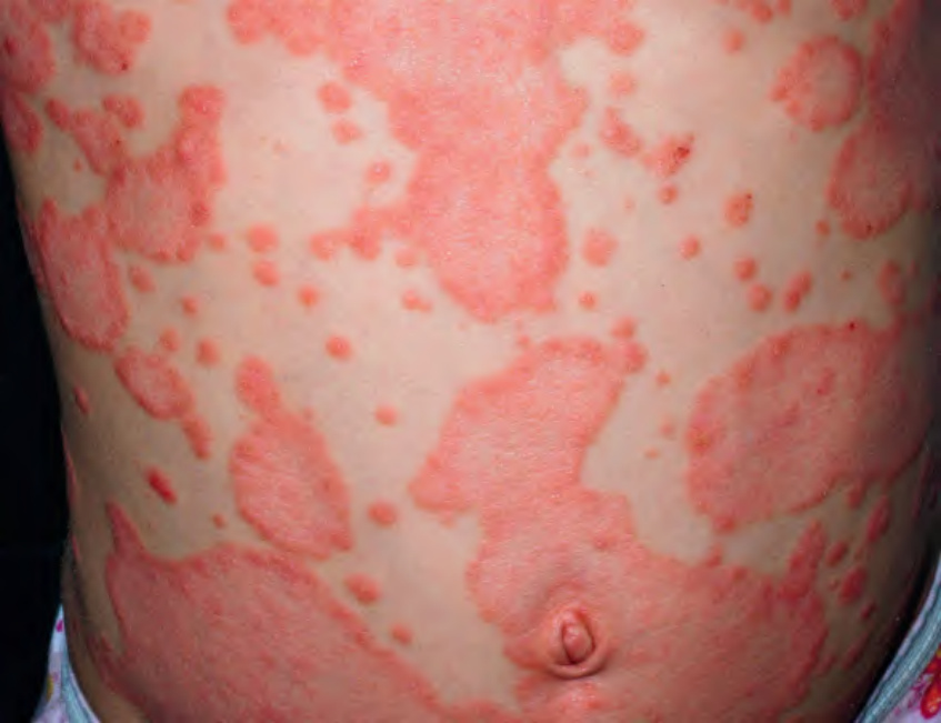
Fig. 8.2 Annular plaques of psoriasis due to central clearing.Courtesy Julie V. Schaffer, MD.
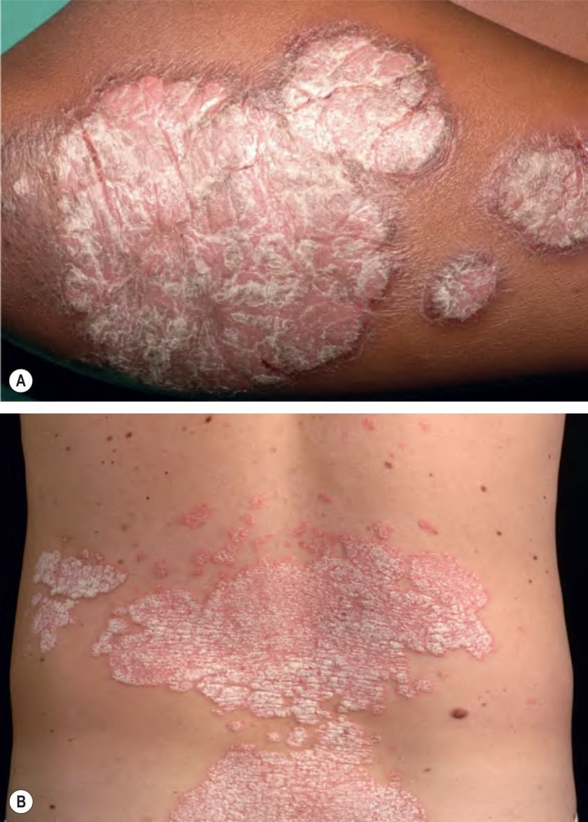
Fig. 8.3 Psoriatic plaques.A, B Note the sharp demarcation and silvery scale. The plaques can vary in thickness and amount of scale. B, Courtesy Lorenzo Cerroni, MD.
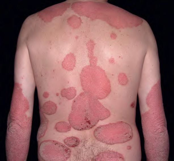
Fig. 8.4 Multiple large plaques of psoriasis. There is obvious symmetry of the plaques on the upper extremities. Some patients have associated pruritus and hemorrhagic crusts due to scratching. Courtesy Luis Requena, MD.
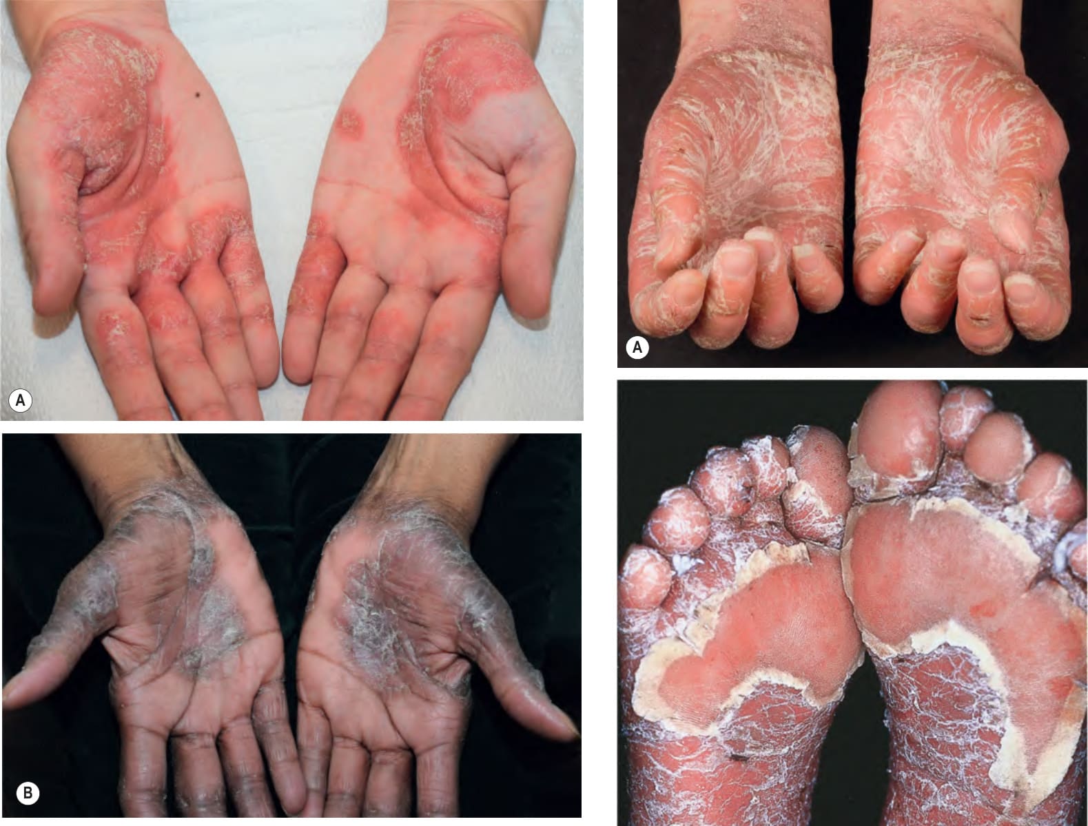
Fig. 8.5 Palmar psoriasis. Well demarcated scaly plaques of the volar wrist and palms. Comparison of the color of the plaques in a lightly pigmented (A) versus darkly pigmented (B) patient. Courtesy Kalman Watsky, MD.
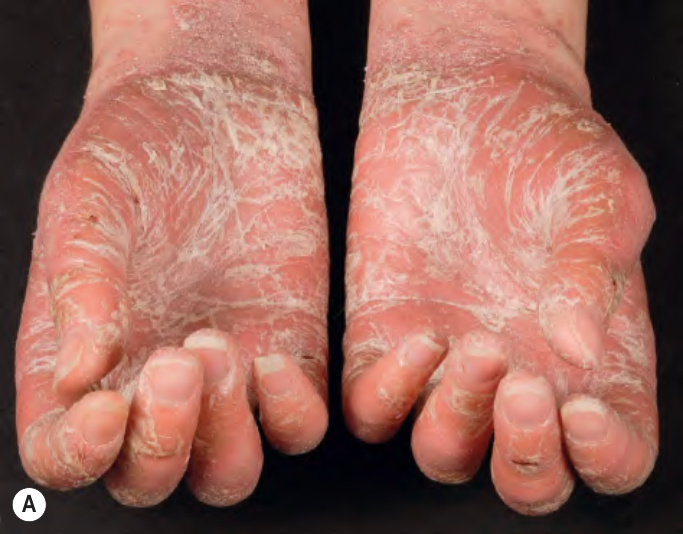
Fig. 8.6 Palmoplantar psoriasis.
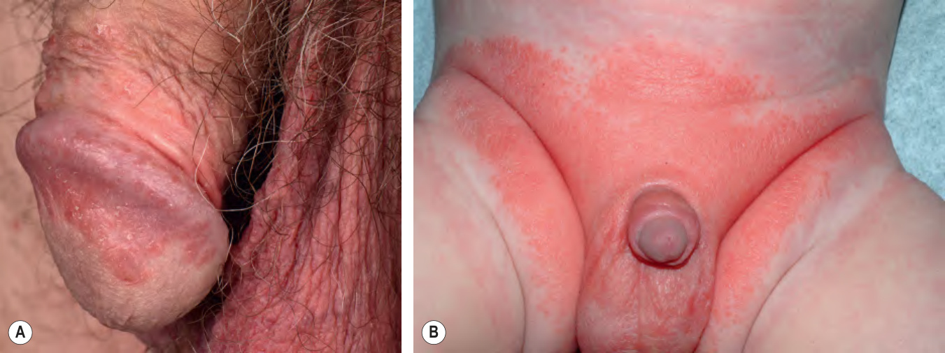
Fig. 8.7 Psoriasis of the genitalia.
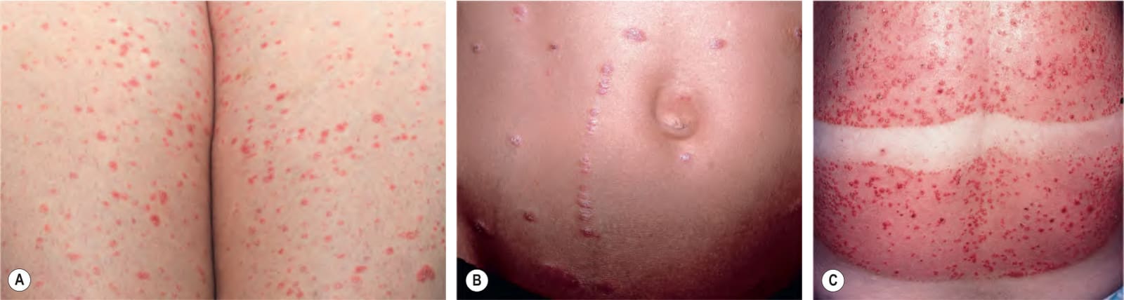
Fig. 8.8 Guttate psoriasis.A Numerous, small, discrete papules with scale on the thighs. B This adolescent has evidence of a linear Koebner phenomenon in addition to more widely scattered guttate lesions. C Numerous papules due to a sunburn-related Koebner phenomenon. A, Courtesy Kalman Watsky, MD; C, Courtesy Ronald P. Rapini, MD.
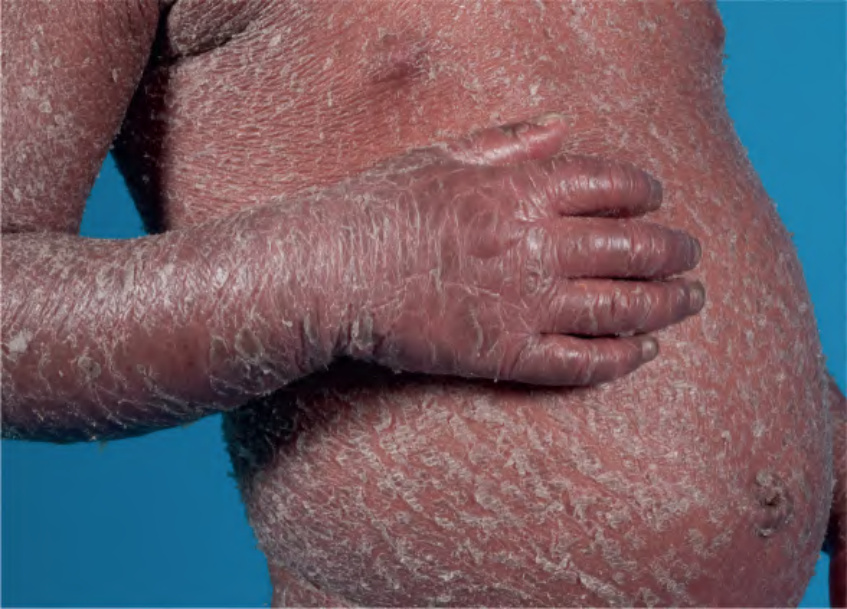
Fig. 8.9 CARD14-associated erythroderma. Extensive erythema and scale resembling erythrodermic psoriasis. Courtesy Edward Cowen, MD.
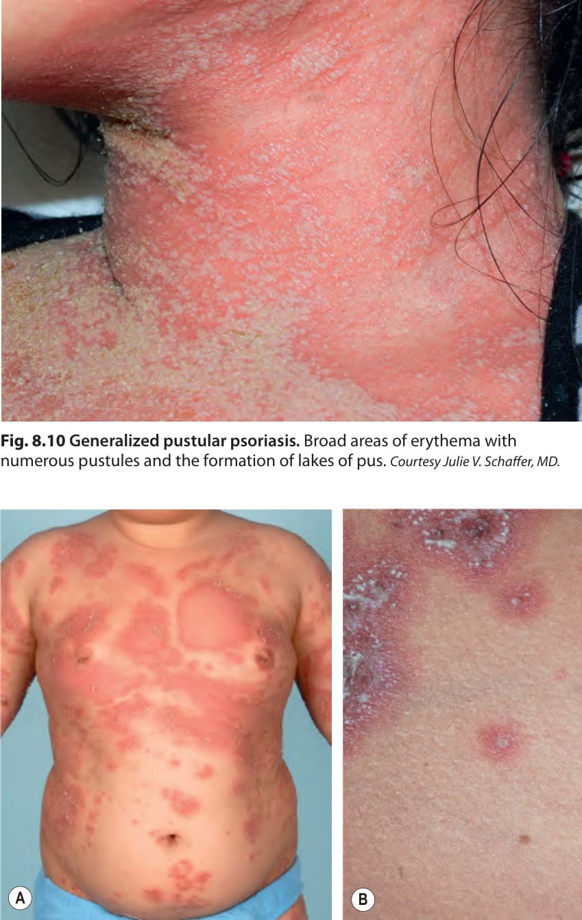
Fig. 8.10 Generalized pustular psoriasis. Broad areas of erythema with numerous pustules and the formation of lakes of pus. Courtesy Julie V. Schaffer, MD.
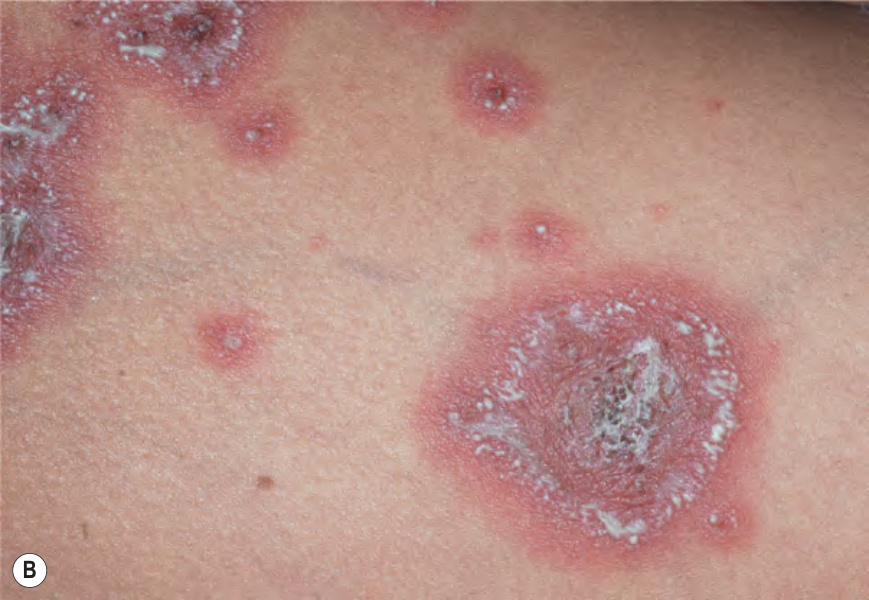
Fig. 8.11 Annular pustular psoriasis.A, B Multiple annular inflammatory plaques whose active borders are studded with pustules followed by desquamation. As these lesions enlarge, there is central clearing. The intensity of erythema can vary and the color is influenced by the skin phototype. A, Courtesy Marieke M. B. Seyger, MD, PhD; B, Courtesy Julie V. Schaffer, MD.
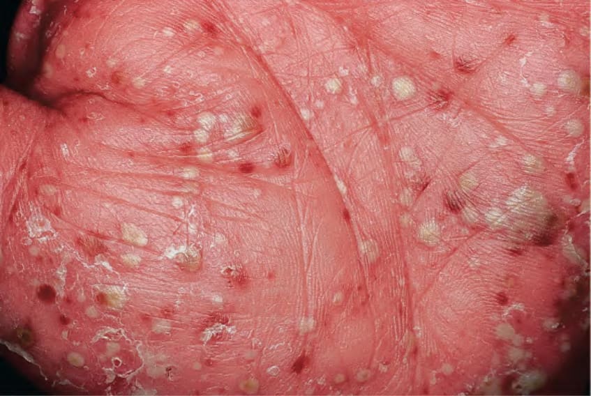
Fig. 8.12 Pustulosis of the palms and soles. Multiple sterile papules are admixed with yellow–brown macules on the palm.
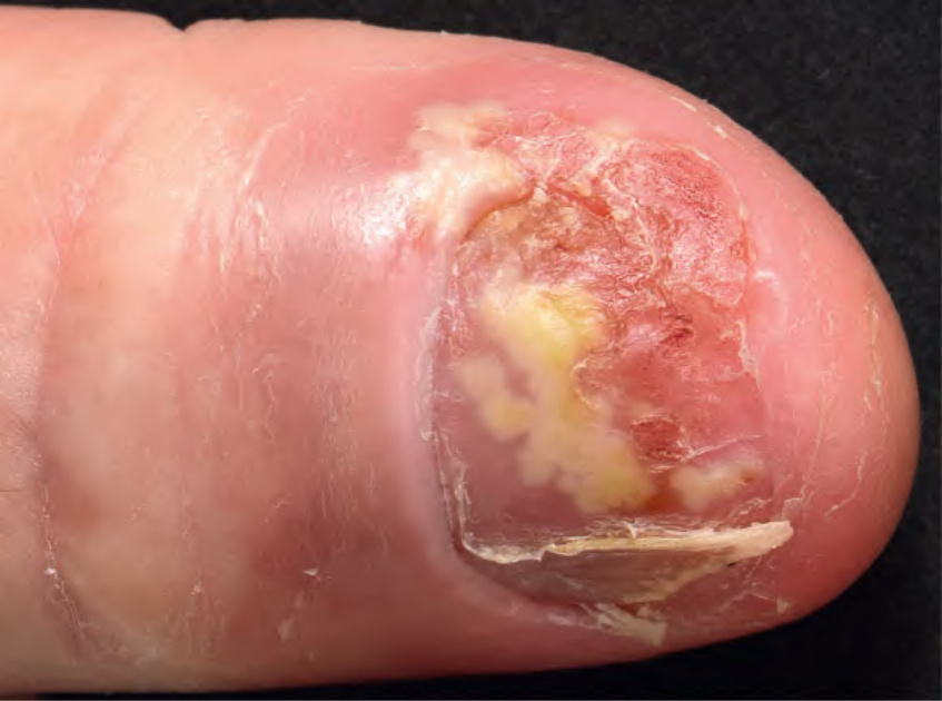
Fig. 8.13 Acrodermatitis continua of Hallopeau. Erythema and slight scale of the distal digit, pustules within the nail bed, and partial shedding of the nail plate.
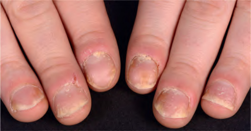
Fig. 8.14 Scalp psoriasis with extension onto the neck. Note the involvement of the external auditory canal.
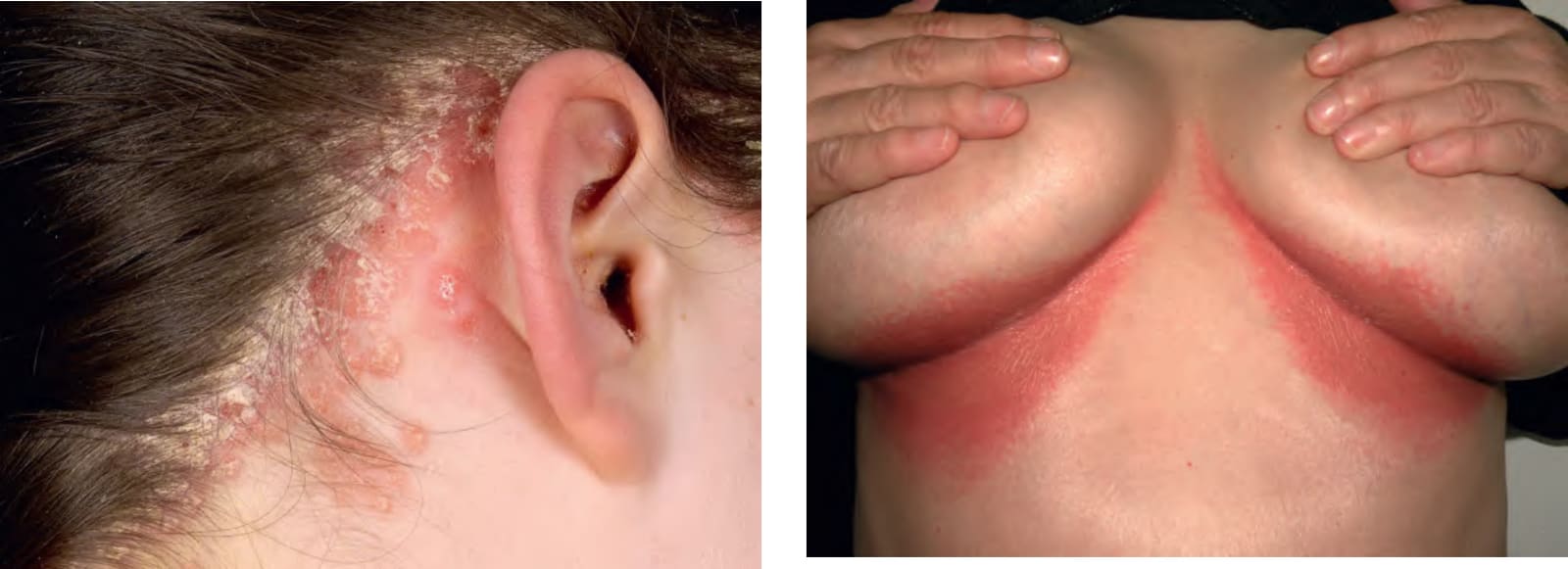
Fig. 8.15 Inverse psoriasis. Shiny erythematous plaques of the inframammary folds that lack scale. Courtesy Luis Requena, MD.
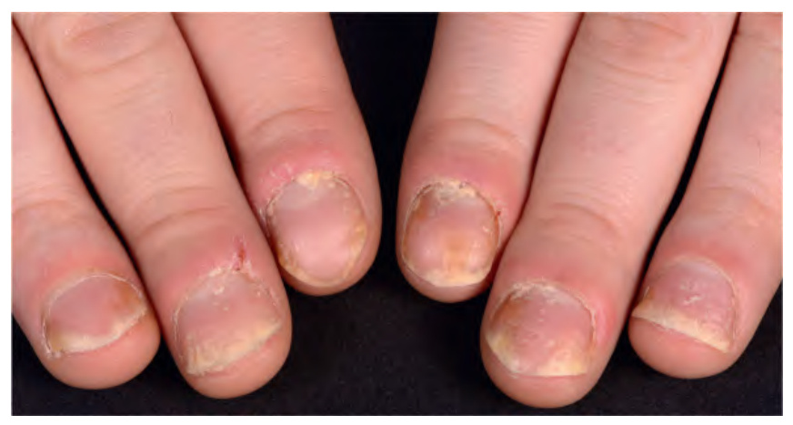
Fig. 8.16 Nail psoriasis. Nail plate pitting, distal onycholysis, oil drop changes, and subungual and proximal hyperkeratosis are seen. There is also proximal nail-fold inflammation with loss of the cuticle, especially of the forefingers. Courtesy Marcel C. Pasch, MD.
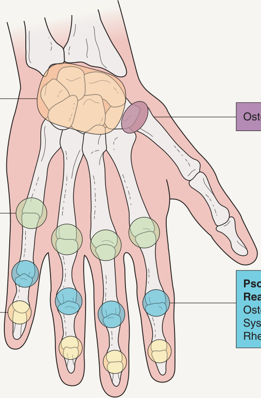
Fig. 8.17 Sites of psoriatic arthritis and reactive arthritis (formerly Reiter disease).Adapted from Cush JJ. Evaluation of musculoskeletal complaints. In: Rheumatology: Diagnosis and Therapeutics, 2nd ed. Lippincott, 2003.
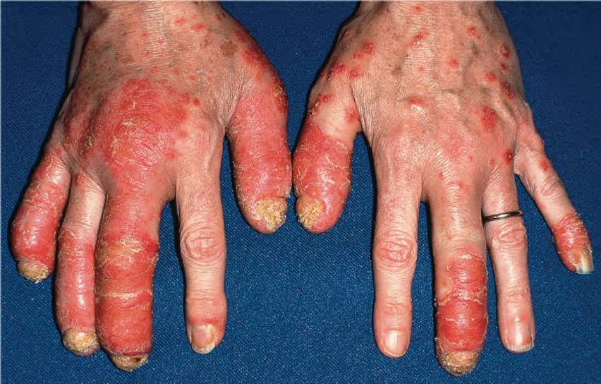
Fig. 8.18 Psoriatic arthritis. Asymmetric involvement of the distal interphalangeal (DIP) and proximal interphalangeal (PIP) joints. A “sausage” digit (third digit bilaterally) results from involvement of both the DIP and PIP joints. Note the yellowing, thickening, and crumbling of several fingernails.
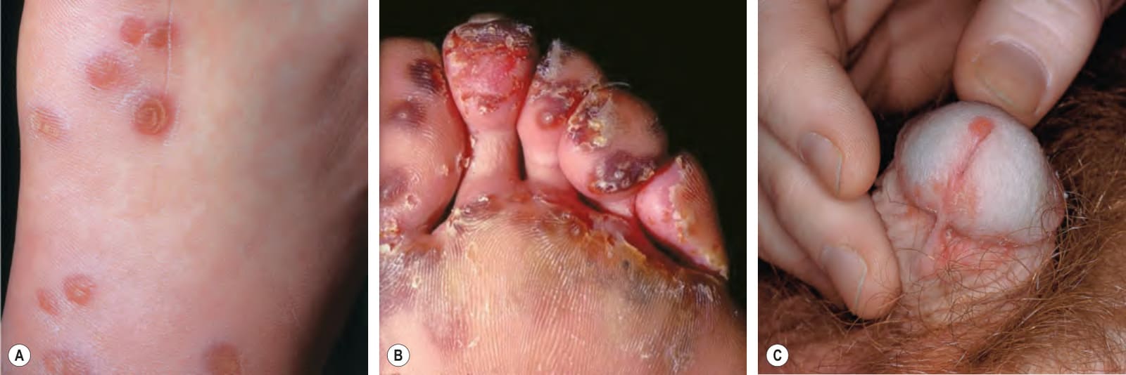
Fig. 8.19 Reactive arthritis (formerly Reiter disease). A, B Plantar lesions of keratoderma blennorrhagicum. The amount of scale can vary and when sterile pustules form, they are often admixed with yellow–brown macules. C Papulosquamous lesions on the glans and shaft of the penis. B, Courtesy Eugene Mirrer, MD.
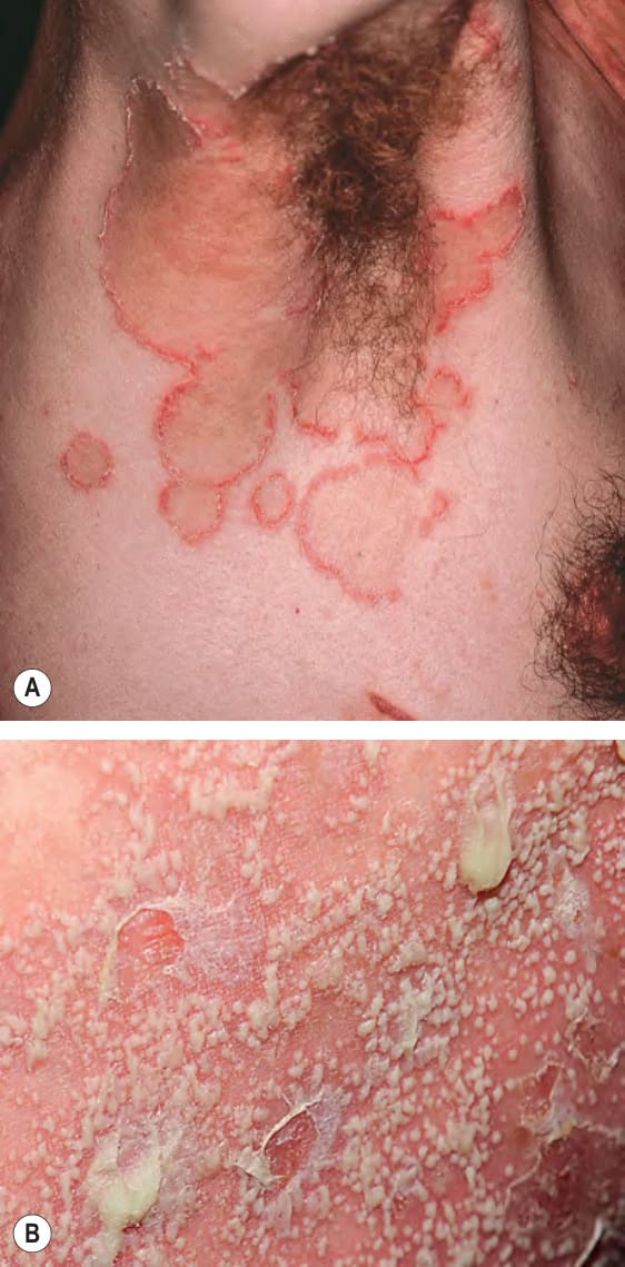
Fig. 8.20 Sneddon– Wilkinson disease. A
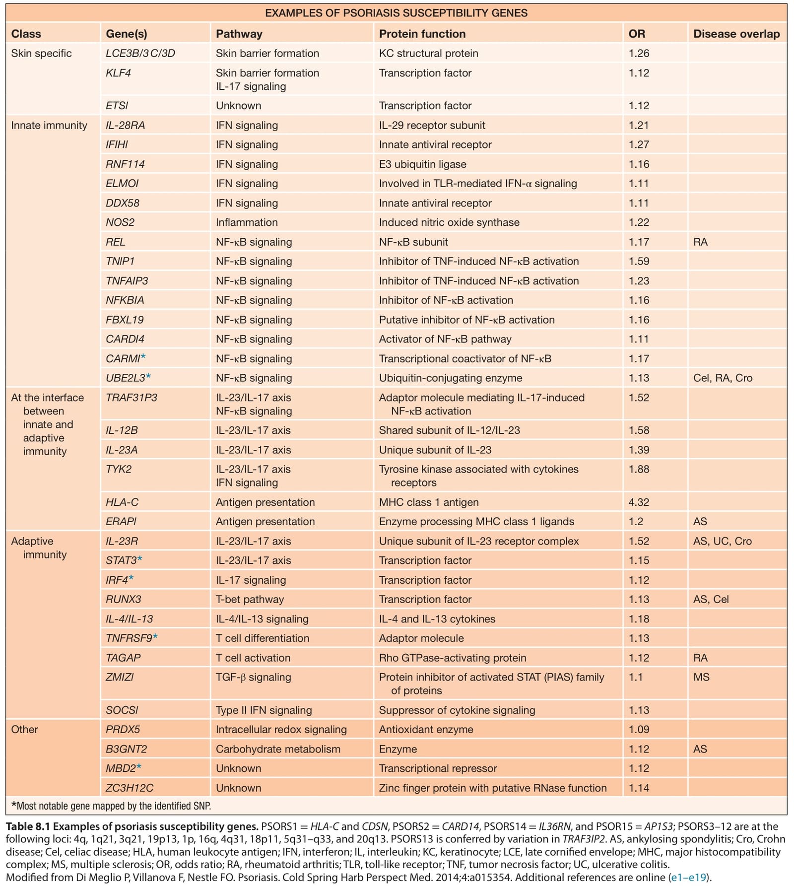
Table 8.1 Examples of psoriasis susceptibility genes. PSORS1 =HLA-C and CDSN, PSORS2 =CARD14, PSORS14 =IL36RN, and PSOR15 = AP1S3; PSORS3–12 are at the following loci: 4q, 1q21, 3q21, 19p13, 1p, 16q, 4q31, 18p11, 5q31–q33, and 20q13. PSORS13 is conferred by variation in TRAF3IP2. AS, ankylosing spondylitis; Cro, Crohn disease; Cel, celiac disease; HLA, human leukocyte antigen; IFN, interferon; IL, interleukin; KC, keratinocyte; LCE, late cornified envelope; MHC, major histocompatibility complex; MS, multiple sclerosis; OR, odds ratio; RA, rheumatoid arthritis; TLR, toll-like receptor; TNF, tumor necrosis factor; UC, ulcerative colitis. Modified from Di Meglio P, Villanova F, Nestle FO. Psoriasis. Cold Spring Harb Perspect Med. 2014;4:a015354. Additional references are online (e1–e19).
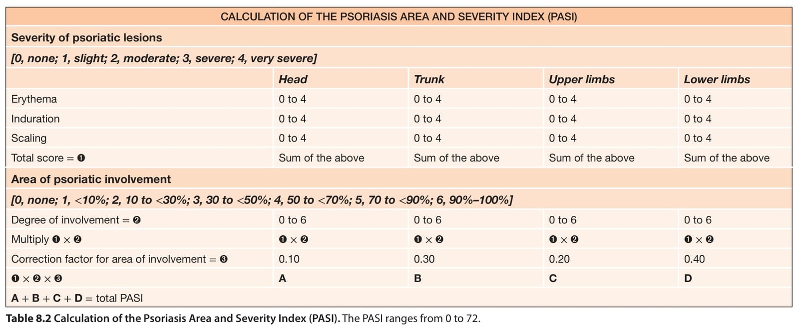
Table 8.2 Calculation of the Psoriasis Area and Severity Index (PASI). The PASI ranges from 0 to 72.
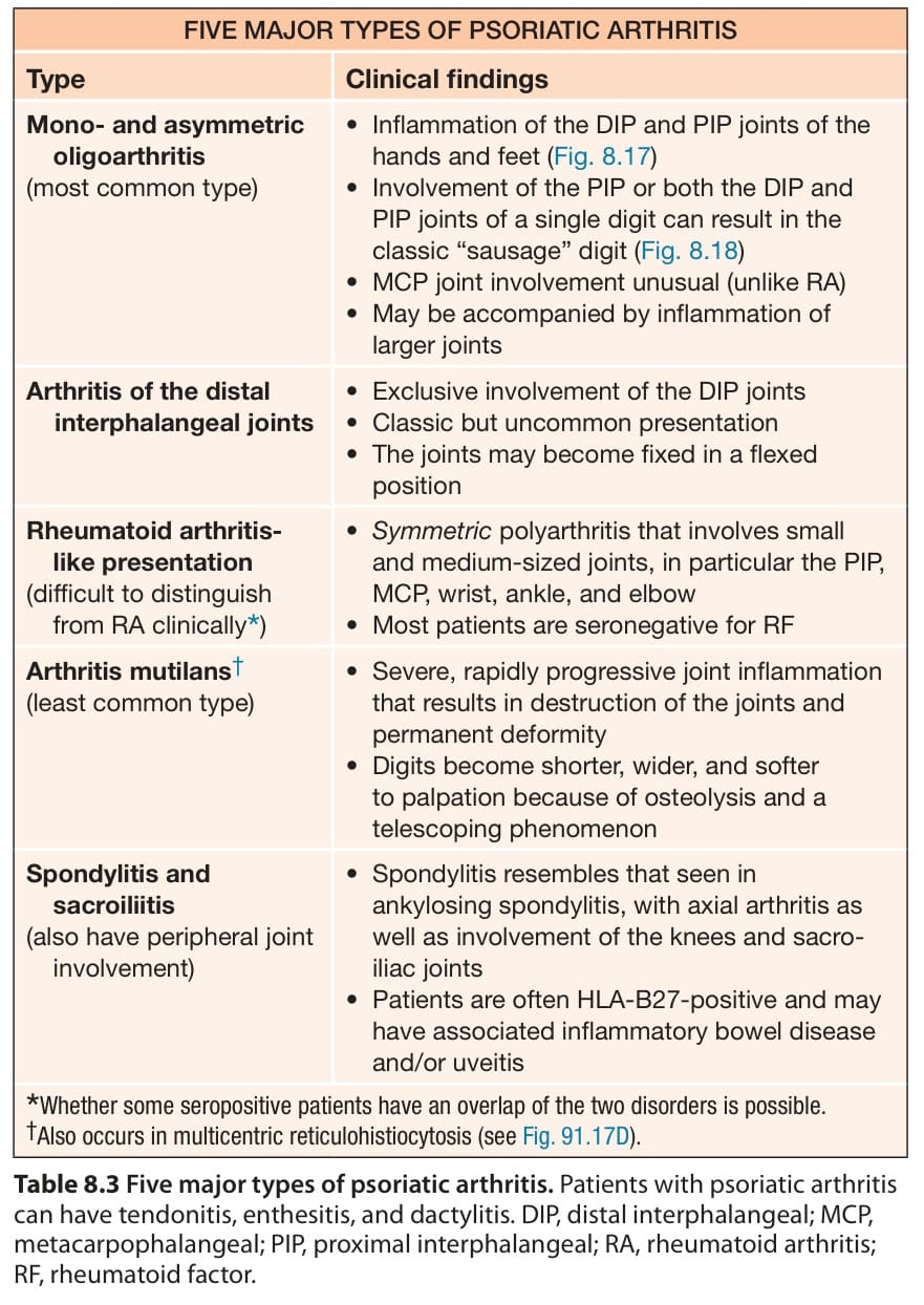
Table 8.3 Five major types of psoriatic arthritis. Patients with psoriatic arthritis can have tendonitis, enthesitis, and dactylitis. DIP, distal interphalangeal; MCP, metacarpophalangeal; PIP, proximal interphalangeal; RA, rheumatoid arthritis; RF, rheumatoid factor.