DIFFERENTIAL DIAGNOSIS
Although both seborrheic dermatitis (see above) and LSC are in the differential diagnosis of psoriasis, it is important to remember that either can coexist with psoriasis. Due to the Koebner phenomenon, psoriasis can also develop within areas of contact dermatitis. When there is a single or limited number of erythematous plaques, especially if they are treatment-resistant, the possibility of SCC in situ (e.g. Bowen disease, erythroplasia of Queyrat) needs to be excluded via histologic examination. Occasionally, a biopsy is necessary to distinguish chronic plaque psoriasis from mycosis fungoides. Clinical features suggestive of the latter include wrinkling due to epidermal atrophy and progression to infiltrated plaques. Dermatomyositis can involve the scalp, elbows and knees, as well as the hands, and initially may be diagnosed as psoriasis.
When plaques of psoriasis involve the shins, they may be misdiagnosed as hypertrophic lichen planus, but characteristic violaceous lesions elsewhere and mucosal involvement usually point to the correct diagnosis. There is clinical overlap between palmoplantar plaque psoriasis and keratotic eczema of the palms and soles, and both may have fissures and be exacerbated by repeated trauma. Sharp margination of the lesions favors psoriasis and examination of the remainder of the skin surface can provide clues to the diagnosis, e.g. involvement of the scalp or intergluteal fold with psoriasis. Elsewhere, chronic lesions of dermatitis may develop an appearance that is similar (clinically and histologically) to partially treated psoriasis and vice versa. However, over time, characteristic lesions often become apparent. When plaques of psoriasis develop pronounced hyperkeratosis (rupioid psoriasis), the possibility of concomitant hypothyroidism should be considered.
In addition to psoriasis, there are other causes of erythroderma, including Sézary syndrome, pityriasis rubra pilaris, and drug reactions (see Ch. 10). For guttate psoriasis, the differential diagnosis may include small plaque parapsoriasis, pityriasis lichenoides chronica, secondary syphilis, and pityriasis rosea. The lesions of guttate psoriasis rarely involve the palms or soles and are often more erythematous than those of parapsoriasis. When lesions are limited in number or have an annular configuration, the possibility of tinea corporis is raised, and when the upper trunk is the predominant site of involvement, pemphigus foliaceus may be considered.
Psoriasis of the flexures is one cause of intertrigo (see Figs. 13.4 & 13.12). Other etiologies include seborrheic dermatitis, contact dermatitis, cutaneous candidiasis, tinea incognito, erythrasma, and extramammary Paget disease. Although KOH examination of associated scale allows a narrowing of the differential diagnosis, psoriasis and candidiasis can coexist (see above). In infants (more so than adults), the possibility of Langerhans cell histiocytosis needs to be considered. In these patients, there may also be scalp involvement with scaling and crusts. Occasionally, tinea capitis is in the differential diagnosis of scalp psoriasis.
When there are widespread pustules on a background of erythema, the possibility of a pustular drug eruption, also referred to as AGEP, needs to be considered (in addition to generalized pustular psoriasis). The histologic findings of these conditions can be similar, including spongiform pustules of Kogoj and microabscesses in the stratum corneum. If eosinophils are also present, a pustular drug eruption is favored. Autoinflammatory disorders, in particular DIRA and DITRA (deficiency of IL-1 or -36 receptor antagonist), can also present with a phenotype similar to pustular psoriasis (see Tables 45.6 & 45.7). In patients with pustulosis of the palms and soles and acrodermatitis continua, the initial evaluation includes the exclusion of a dermatophyte infection or secondarily infected dermatitis. The differential diagnosis of the annular form of pustular psoriasis includes Sneddon–Wilkinson disease and other causes of subcorneal pustules (Table 8.4). Reactive arthritis needs to be considered in any patient with the diagnosis of arthritis in the setting of psoriasiform skin lesions.
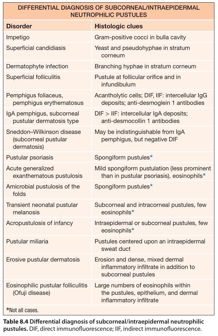
Table 8.4 Differential diagnosis of subcorneal/intraepidermal neutrophilic pustules. DIF, direct immunofluorescence; IIF, indirect immunofluorescence.