PATHOLOGY
Papulosquamous Lesions
Initial lesion
In the initial lesion, i.e. a pinhead-sized papule, the histopathologic features are not yet diagnostic. A superficial perivascular infiltrate of lymphocytes and histiocytes is seen in the dermis along with papillary edema and a dilation of capillaries. In acute eruptive guttate lesions, mast cell degranulation is a constant feature.
There is mild epidermal acanthosis without parakeratosis and the keratinocytes have a swollen appearance. Histiocytes (detected by CD68 staining) and lymphocytes appear within the epidermis, accompanied by mild focal spongiosis. In this early phase, neutrophils are not yet detectable.
Active lesion
A fully developed guttate lesion or the marginal zone of an enlarging psoriatic plaque is designated as an “active lesion”. The histopathologic findings in an active lesion are diagnostic for psoriasis.
In the papillary dermis, the capillaries are increased in number and length and they have a tortuous appearance. Edema is seen, especially at the tops of the papillae. There is a mixed perivascular infiltrate of lymphocytes, histiocytes, and neutrophils.
The epidermis is acanthotic with focal accumulations of neutrophils and lymphocytes. At these sites, the epidermis is variably spongiotic. Above these foci, the granular layer is absent and the stratum corneum still contains flattened nuclei (parakeratosis). The accumulation of
neutrophils within a spongiotic pustule is referred to as a “spongiform pustule of Kogoj” (Fig. 8.21) and the accumulation of neutrophils and/or neutrophil remnants in the stratum corneum, surrounded by parakeratosis, as a “microabscess of Munro”. These two findings are pathognomonic for psoriasis and AGEP.
Stable lesion
In the papillary dermis, the capillaries are elongated and tortuous, extending upward into elongated club-shaped dermal papillae; only a small suprapapillary plate of epidermal cells covers the tip of these dermal papillae. This micromorphology explains the “Auspitz” phenomenon (see earlier). A modest perivascular infiltrate is seen that consists primarily of lymphocytes and histiocytes. The psoriatic lesion is heterogeneous, consisting of active areas (hot spots) and chronic nonspecific areas (cold spots).
The hyperproliferation of the epidermis now has reached its characteristic pattern (Fig. 8.22). The rete ridges are elongated and have a squared-off appearance. The bases of some of the rete ridges also coalesce. The horny layer has parakeratotic foci with an absence of the stratum granulosum. In some lesions, micropustules of Kogoj and microabscesses of Munro may be seen.
Pustular Psoriasis
In pustular psoriasis, accumulation of neutrophils is the predominant feature. The epidermis is usually acanthotic. Numerous neutrophils accumulate between eosinophilic strands of keratinocytes. In the stratum corneum, large accumulations of neutrophils are observed, surrounded by parakeratosis (see Fig. 0.21B). As a result, exaggerated spongiform pustules of Kogoj and microabscesses of Munro, the histologic hallmarks of “active” psoriasis, are seen in pustular psoriasis.
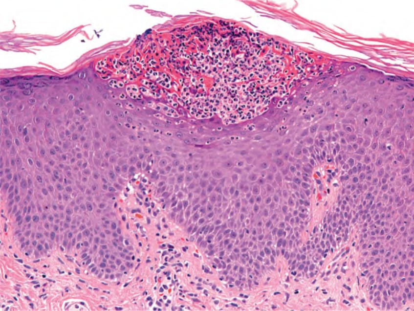
Fig. 8.21 Spongiform pustule of Kogoj. A collection of neutrophils is seen within the upper stratum spinosum. Courtesy Lorenzo Cerroni, MD.
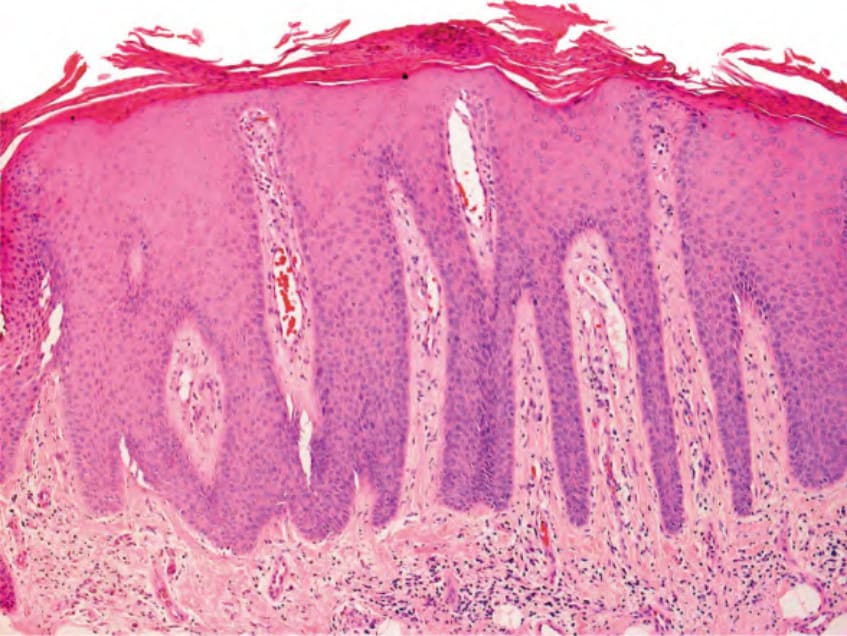
Fig. 8.22 Stable plaque psoriasis. Hyperplasia of the epidermis with “squaredoff” rete ridges, elongation of the dermal papillae, dilated superficial blood vessels, hypogranulosis, and parakeratosis plus remnants of neutrophils (microabscess of Munro). Courtesy Lorenzo Cerroni, MD.