PITYRIASIS LICHENOIDES
Synonyms: Pityriasis lichenoides et varioliformis acuta (PLEVA) Mucha–Habermann disease (PLEVA) Pityriasis lichenoides chronica (PLC) Guttate parapsoriasis (PLC)
Key features
Pityriasis lichenoides et varioliformis acuta (PLEVA) and pityriasis lichenoides chronica (PLC) are two ends of a disease spectrum
Both entities are characterized by recurrent crops of spontaneously regressing erythematous papules
Lesions of PLEVA are crusted and occasionally vesiculopustular, whereas in PLC they are scaly
Individual patients may show intermediate or mixed lesions
Histologically, an interface dermatitis with necrotic keratinocytes is seen
The infiltrate is predominantly T cells that are often monoclonal
Introduction
Pityriasis lichenoides is an uncommon disorder that encompasses pityriasis lichenoides et varioliformis acuta (PLEVA), the febrile ulceronecrotic Mucha–Habermann disease (FUMHD) variant of PLEVA, and pityriasis lichenoides chronica (PLC). These acute and chronic forms of pityriasis lichenoides exist on a disease spectrum with variable presentations that can pose diagnostic and therapeutic challenges. They are papular, often clonal T cell disorders that may rarely be associated with MF.
History
The two major variants of pityriasis lichenoides – PLEVA and PLC – were described as the 19th century ended and the 20th century began. Pityriasis lichenoides was included in Brocq’s 1902 treatise on a group of chronic, idiopathic dermatoses that he termed “parapsoriasis”. Nowadays, however, it is considered a separate entity.
Epidemiology
Pityriasis lichenoides is more prevalent in the pediatric population, but it affects patients in all age groups, races, and geographic regions. There is a male predominance.
Pathogenesis
The etiology of pityriasis lichenoides is unknown. It has been postulated to be a response to foreign antigens such as infectious agents and drugs. There is some evidence to support PLEVA being associated with viral infections, especially varicella–zoster virus, and in a few reports, HIV, parvovirus B19, and Epstein–Barr virus. Additional reports have described its association with medications (e.g. estrogen– progesterone, infliximab, adalimumab, statins), radiocontrast dye, and vaccines.
Both PLEVA and PLC contain lesional T cell infiltrates, sometimes with dominant T cell clonality. The concept of pityriasis lichenoides as a T cell lymphoproliferative disorder may help to explain its occasional association with other lymphoproliferative disorders such as MF. In addition, this would account for the variants of pityriasis lichenoides that have numerous CD30+ cells or γδ T cells as well as the subgroups that have been referred to as atypical pityriasis lichenoides and lymphomatoid pityriasis lichenoides. Because differentiation from T cell lymphomas is sometimes difficult, patients with pityriasis lichenoides and an aberrant T cell phenotype should undergo longitudinal evaluation, with the final diagnosis based upon repeated clinicopathologic correlation, including phenotypic and molecular studies of T cell clonality.
Clinical Features
Pityriasis lichenoides presents as recurrent crops of spontaneously regressing erythematous to purpuric papules (Fig. 9.2). The acute form (PLEVA) and the chronic form (PLC) exist on a disease continuum. Many patients have intermediate or mixed manifestations, either serially or concurrently. In patients with PLEVA, individual lesions develop crusts, ulcers, and occasionally vesicles or pustules, which
may heal with varioliform scars if dermal damage is extensive. Lesions are usually asymptomatic and typically resolve within weeks. Disease manifestations are confined to the skin, except rarely, when acute lesions are associated with malaise, fever, lymphadenopathy, arthritis, and/or bacteremia. The term febrile ulceronecrotic Mucha–Habermann disease (FUMHD) has been used to refer to such severe variants in which large, confluent necrotic skin lesions (see Fig. 9.2F) as well as mucosal, gastrointestinal, and pulmonary involvement can be observed. In the latter group, an associated mortality rate of up to 15% has been reported.
In PLC, the papules are erythematous to red–brown and scaly (see Fig. 9.2D). They pursue a more indolent course, regressing over weeks to months. When these lesions subside, there may be residual hypopigmented macules; the latter are more obvious in individuals with darker skin phototypes and may be the presenting complaint (Fig. 9.3). Pityriasis lichenoides can resolve spontaneously after weeks to months or it may pursue a chronic relapsing course, sometimes interspersed with long periods of remission. Some studies have suggested that the distribution of skin lesions is more important than their acute or chronic nature in predicting outcome. The average course of disease was shorter in patients with a diffuse distribution, while those with a peripheral distribution had the longest clinical course. Compared to adults, children tend to have a more persistent course and widespread distribution of lesions.
Pathology
Pityriasis lichenoides exhibits a superficial perivascular interface dermatitis in all cases (Fig. 9.4). Those lesions at the acute end of the spectrum contain a denser infiltrate that may be top-heavy and wedge-shaped. There is commonly an infiltration of lymphocytes into the adnexal epithelia, both follicular and eccrine. Lymphocytes predominate in the infiltrate, although neutrophils may be admixed. The epidermis shows parakeratosis and evidence of damage ranging from edema to extensive epidermal necrosis in well-developed lesions. Extravasation of erythrocytes is a frequent finding. While a perivascular lymphocytic infiltrate is a constant feature, a true lymphocytic vasculitis with fibrinoid necrosis of blood vessels is uncommon, occasionally occurring in PLEVA. All of these changes are blunted in the more chronic skin lesions, where the principal microscopic features include parakeratosis and a milder interface lymphocytic infiltrate that is accompanied by focal keratinocyte necrosis and mild erythrocyte extravasation.
Lymphoid atypia is not a standard feature of pityriasis lichenoides. What has been referred to as atypical pityriasis lichenoides was defined by conventional clinical and histologic features but with an aberrant T cell phenotype. Some pathologists allow occasional atypical lymphocytes or CD30+ lymphocytes, whereas others regard this as a sign of lymphomatoid papulosis. Patients with small papules that resemble pityriasis lichenoides clinically but have Pautrier microabscesses histologically were described as having “pityriasis lichenoides-like mycosis fungoides”, but no patients in that series evolved into typical MF. Controversies regarding T cell lymphoproliferative disorders, however, still remain.
Differential Diagnosis
The diagnosis of pityriasis lichenoides is made by the correlation of clinical features with lesional histopathology. The principal differential diagnostic considerations for pityriasis lichenoides are listed in Table 9.3. For the more acute form (PLEVA), the primary entities in the clinical and histopathologic differential diagnosis include lymphomatoid papulosis, arthropod reactions, cutaneous small vessel vasculitis, varicella, and drug eruptions. For the chronic form (PLC), the principal differential diagnosis includes guttate psoriasis, lichen planus (exanthematous), pityriasis rosea, secondary syphilis, lymphomatoid papulosis, drug eruptions, and papular dermatitis. All of these alternative diagnoses can usually be excluded on the basis of key historical, clinical, pathologic, and laboratory findings. More than one biopsy specimen, T cell immunophenotyping, and molecular studies of T cell clonality may be required to achieve clinicopathologic correlation. PCR, direct fluorescent antibody (DFA), and serologic testing as well as immunohistochemical staining are helpful in excluding varicella and syphilis.
Treatment
All treatments for pityriasis lichenoides are based primarily on uncontrolled case series, case reports, or anecdotes (Table 9.4). If a drug association is suspected, trial discontinuance of the suspected agent is warranted. First-line therapy includes topical corticosteroids (limited efficacy), tetracyclines, macrolide antibiotics (e.g. erythromycin, azithromycin) and various types of phototherapy (most effective; e.g. narrowband UVB). Oral antibiotics are used for their anti- inflammatory effects, with macrolides favored in younger children. A several-month course is often required, followed by a gradual taper.
More fulminant cases may require low-dose weekly methotrexate. Those rare cases associated with fever and arthritis may benefit from systemic corticosteroids, IVIg or cyclosporine, once infection has been excluded. There are also reports of the efficacy of TNF inhibitors.
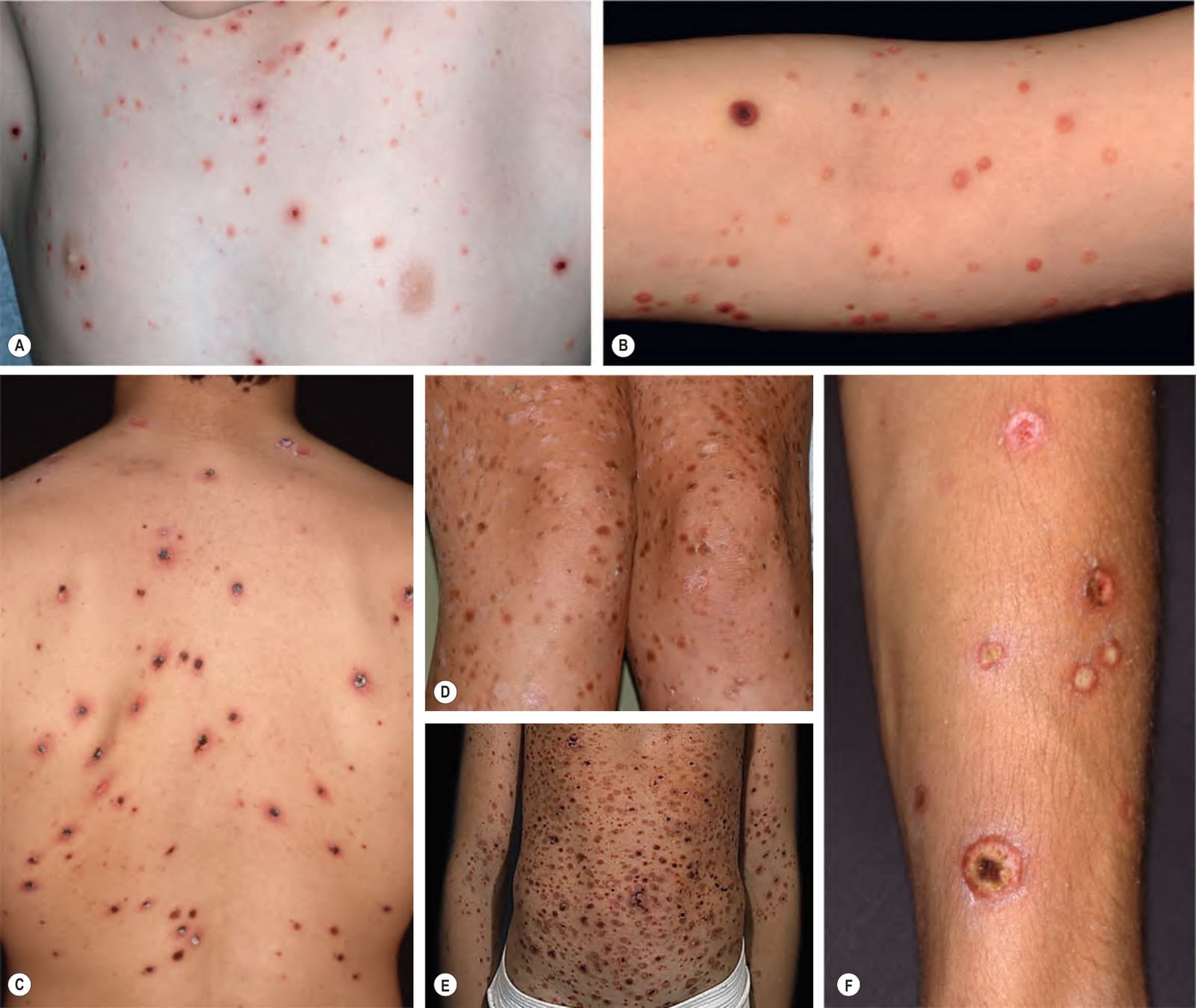
Fig. 9.2 Clinical spectrum of pityriasis lichenoides.A–C In the acute form (PLEVA), widespread erythematous papules and papulovesicles are admixed with crusted lesions; sometimes, there can be an ulcerative component (C). D Individuals with the chronic form (PLC) develop multiple dull red–brown papules, some of which have scale and may be hypopigmented. E In some patients the majority of lesions are characteristic of one end of the spectrum, e. g. PLEVA, but with scattered lesions suggestive of PLC. F Rarely, a more severe potentially life-threatening form of PLEVA, termed febrile ulceronecrotic Mucha–Habermann disease (FUMHD), may occur de novo or in the setting of pre-existing PLEVA. A, D, Courtesy Julie V. Schaffer, MD; B, Courtesy Thomas Schwarz, MD; C, F, Courtesy Lorenzo Cerroni, MD.
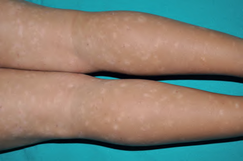
Fig. 9.3 Chronic form of pityriasis lichenoides (PLC) presenting as macules of hypopigmentation. This is more common in individuals with darker skin phototypes. Courtesy Antonio Torrelo, MD.
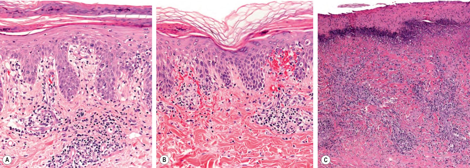
Fig. 9.4 Pityriasis lichenoides – histopathologic features.A In the chronic form (PLC), parakeratosis may be more prominent than in the acute form, but the epidermal destruction is less intense. B In the acute form (PLEVA), scattered necrotic keratinocytes are seen in addition to parakeratosis. The perivascular lymphoid infiltrates in the dermis are accompanied by extravasated erythrocytes, which are even present within the epidermis. As with the clinical presentations, there is a continuous histologic spectrum. C In the ulceronecrotic form, there is complete ulceration covered by crust. An intense lymphocytic infiltrate is seen within the dermis. Courtesy Lorenzo Cerroni, MD.
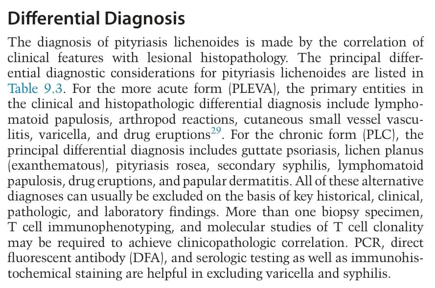
Table 9.3 Differential diagnosis of pityriasis lichenoides.
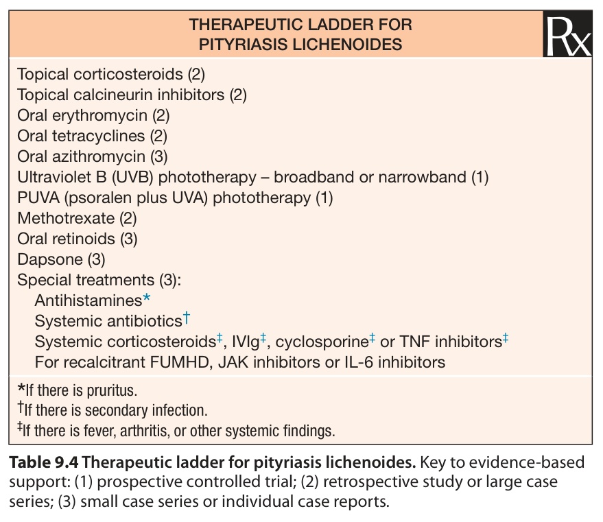
Table 9.4 Therapeutic ladder for pityriasis lichenoides. Key to evidence-based support: (1) prospective controlled trial; (2) retrospective study or large case series; (3) small case series or individual case reports.