PITYRIASIS RUBRA PILARIS
Synonyms: Lichen ruber pilaris Devergie disease Lichen ruber acuminatus
Key features
Follicular papules with an erythematous base are a characteristic finding, including on the proximal dorsal fingers
There is a coalescence of orange–red plaques, but with obvious islands of sparing
An orange–red waxy keratoderma of the palms and soles is often seen
Of the six types described, the classic adult form is the most common
The classic types (adult and juvenile) typically resolve within 3–5 years
Type V, the atypical juvenile form, is frequently due to CARD14 mutations
Histologically, there is alternating ortho- and parakeratosis, both vertically and horizontally
History
Pityriasis rubra pilaris (PRP) was first described in 1835 by Claudius Tarral as a variant of psoriasis and it was given several names during the 19th century. Devergie called it “pityriasis pilaris”, and later Besnier recommended the name “pityriasis rubra pilaris” in the first comprehensive illustrated article on the disease.
Epidemiology
Although PRP shows no sex bias, affecting both men and women equally, ethnic variations may exist. The incidence of this uncommon disease has two major peaks, one during the first and second decades and the second during the sixth decade.
Most cases are acquired, but there are familial forms, primarily autosomal dominant and less frequently autosomal recessive. The dominantly inherited form of PRP is associated with heterozygous gain-of-function mutations in CARD14, which encodes the caspase recruitment domain family member 14. Except for the latter group of patients and those with the non-classic forms, PRP appears to be a self-limited disease process, resolving within 3 to 5 years in almost all cases.
Pathogenesis
Excluding inherited PRP due to CARD14 mutations, infections, UV exposure, and various minor traumas to the skin have been reported to precede the onset of PRP. Autoimmunity (e.g. associated thyroid disease) or an abnormal immunologic response to particular antigens may play a role, as in patients with HIV-associated PRP (type VI). Paraneoplastic PRP has also been described in individuals with solid-organ malignancies such as renal cell, bronchogenic, and hepatocellular. Rarely, drugs or vaccines have been implicated in triggering PRP, including infliximab, tyrosine kinase inhibitors (e.g. ponatinib, imatinib), and antivirals (e.g. sofosbuvir).
Clinical Features
The clinical features of classic PRP are helpful in making the diagnosis. Follicular hyperkeratosis on an erythematous base is a key finding (Fig. 9.5). This results in rough papules, especially on the dorsal aspect of the proximal fingers, reminiscent of a nutmeg grater. These papules are also seen on the trunk and extremities, and they can coalesce to form large salmon-colored to orange–red plaques with distinctive “islands of sparing”. The plaques can then progress to an erythrodermic appearance with varying degrees of exfoliation (see Figs. 10.7 & 10.8). The palms and soles are commonly involved with a distinctive orange–red waxy keratoderma (Fig. 9.6). Erythema with a fine diffuse scale is often seen on the scalp. Of note, scalp disease that mimics seborrheic dermatitis is a common initial clinical manifestation of adult PRP, and rapid progression to erythroderma can occur over several weeks.
Nail involvement is characterized by a thickened plate with a yellow– brown discoloration and subungual debris. The mucous membranes are rarely involved, but they may show features similar to oral lichen planus.
The most common form of PRP (type I) usually begins in the head and neck region and then progressively advances caudally. Pruritus and burning have been reported in 20% of patients with PRP. Although less common than in atopic dermatitis or Darier disease, Kaposi varicelliform eruption can occur in patients with PRP. As with other forms of erythroderma (see Ch. 10), eruptive seborrheic keratoses may appear. Both photoaggravated and phototriggered forms of PRP can also occur (see Fig. 9.5C). Resolving PRP may have a clinical appearance that resembles erythema gyratum repens (see Fig. 19.9).
The classification of the different forms of PRP is based upon age, duration, and type of cutaneous involvement (Fig. 9.7). Type I (classic adult) PRP is the most common form and is typically self-limited, clearing within 3 years in 80% of patients. Type II differs from type I by a palmoplantar keratoderma with a coarse and lamellated scale, a more ichthyosiform scaling on the lower extremities, occasional alopecia, and a protracted clinical course.
PRP types III–V are seen in children and adolescents. Type III, the classic juvenile form, closely resembles the classic adult form and
also typically clears within 3 years. Type IV (circumscribed) is the most common of the juvenile forms, and its focal nature sets it apart from the other clinical presentations (Fig. 9.8). Type V, the atypical juvenile form, is frequently due to CARD14 mutations; it has features similar to type II but with more ichthyosiform scaling and a chronic course (see Fig. 8.9). Patients with CARD14 mutations tend to present with clinical features observed in both PRP (often resembling type V PRP but sometimes with classic “islands of sparing”) and psoriasis. These patients have been referred to as having “CARD14- associated papulosquamous eruption (CAPE)”. In addition to a family history of PRP or psoriasis, characteristic features of CAPE include an early age of onset; prominent involvement of the cheeks, chin, and ears; involvement of the palms and soles; and a chronic course with a variable response to conventional treatments. Gain-of-function CARD14 mutations have rarely been detected in other types of PRP.
PRP type VI is associated with HIV infection, and while it often fails to respond to conventional therapy for PRP, it may improve with antiretroviral therapy. These patients can have erythematous follicular papules and keratotic spines as well as nodulocystic lesions of acne conglobata and hidradenitis suppurativa. In addition, coalescence of inflammatory plaques into an erythroderma may occur, as in PRP type I.
Pathology
Psoriasiform dermatitis with irregular hyperkeratosis and alternating vertical and horizontal ortho- and parakeratosis (“checkerboard pattern”) is distinctive (Fig. 9.9A). The hair follicles are dilated and filled with a keratinous plug, while the “shoulder” of stratum corneum surrounding the follicular opening frequently shows parakeratosis (Fig. 9.9B). The interfollicular epidermis often exhibits hypergranulosis as well as thick, shortened rete pegs. A sparse lymphohistiocytic perivascular infiltrate is seen in the underlying dermis. Acantholysis and focal acantholytic dyskeratosis within the epidermis have also been described and can be helpful in distinguishing PRP from psoriasis. In CAPE, histopathologic features are more similar to PRP than to psoriasis, including alternating checkerboard parakeratosis and orthokeratosis and follicular plugging.
Differential Diagnosis
The major entity in the differential diagnosis is psoriasis. The distinctive orange–red palmoplantar keratoderma plus the keratotic follicular papules with a nutmeg grater appearance, the classic islands of sparing of the trunk, and a finer scale (see Fig. 10.7), help differentiate PRP from psoriasis. The presence of oil-drop changes, small pits, and marginal onycholysis of the nails favor psoriasis. Microscopically, acantholysis and focal acantholytic dyskeratosis, if present, are especially useful in diagnosing PRP rather than psoriasis. A thickened granular cell layer, short thicker rete pegs, limited vascular dilation in the dermal papillae, and the absence of both neutrophilic migration towards the epidermis and Munro microabscesses further help distinguish PRP from psoriasis. Histopathologic features of PRP may be seen in patients with what has been described as facial discoid dermatosis.
Early PRP of the scalp can mimic seborrheic dermatitis. The relative recalcitrant nature of scalp PRP may serve as a clue to the diagnosis. A PRP-like eruption can be seen in patients with dermatomyositis (Wong type) and cutaneous T cell lymphoma, as can erythema and scaling of the scalp. Children with acute-onset PRP may be misdiagnosed as having Kawasaki disease, and, occasionally, PRP is confused with erythrokeratodermia variabilis et progressiva or ichthyosis with confetti.
Keratotic follicular papules are also seen in keratosis pilaris and lichen spinulosus, but the lesions are more limited in scope and the
other manifestations of PRP are absent. The various disorders that can present as an erythroderma are discussed in Chapter 10.
Treatment
Therapy for PRP is primarily empiric, based upon case series and case reports, and often involves a combination of topical and systemic therapies. Topical therapies alone are typically insufficient for most patients with PRP, unless there is limited disease. Topical high-potency corticosteroids, tar, calcipotriene (calcipotriol), keratolytics (e.g. urea, salicyclic acid), and retinoids (e.g. tazarotene, tretinoin) can be used as adjuncts to systemic therapy.
Systemic retinoids represent a first-line treatment for PRP in both adults and children; an excellent response is seen in >40% of patients. Isotretinoin (0.5 to 1 mg/kg/day) and acitretin (0.5 mg/kg/day) are the two most commonly prescribed retinoids, with scattered reports of some efficacy with alitretinoin. There are reports of the occasional use of higher doses of isotretinoin, but in the opinion of the authors, this is not recommended. Significant clearing is usually seen within 3–6 months of instituting therapy.
Methotrexate (5–25 mg/week) is considered second-line therapy for PRP, in particular for patients who have failed or cannot tolerate systemic retinoids. A response rate of >90% and complete clearance in ~40% of patients has been observed. As with retinoids, clinical responses are usually seen within 3–6 months. Hepatotoxicity and myelosuppression are well-known potential side effects (see Ch. 130).
In severe cases of PRP, a combination of methotrexate (5–25 mg/week) plus a systemic retinoid (acitretin) has been utilized. Both agents can be given at the onset of therapy, or the second medication can be added if the response to the first is inadequate. In this situation, an increased risk for hepatotoxicity must be kept in mind.
Based upon case reports and small case series, TNF inhibitors (e.g. etanercept, infliximab, adalimumab), secukinumab, and ustekinumab have led to improvement of PRP. A marked to complete response was noted in 50%–80% of patients. In a small series of patients, ixekizumab was reported to be moderately successful. Ustekinumab, often at higher doses, has been reported as beneficial in patients with CAPE. Due to limited data as well as the natural history of PRP, precise efficacy is difficult to assess.
Other anecdotal potential therapies include systemic immunosuppressives (e.g. azathioprine, prednisone, cyclosporine), apremilast, and IVIg, with variable results observed. Although UV light therapy runs the risk of exacerbating the disease, there are some reports of success with narrowband UVB, UVA1, or PUVA in combination with an oral retinoid.
Less toxic regimens are recommended for the classic juvenile form of PRP, given the high likelihood of spontaneous remission and the potential long-term effects of aggressive systemic therapies.
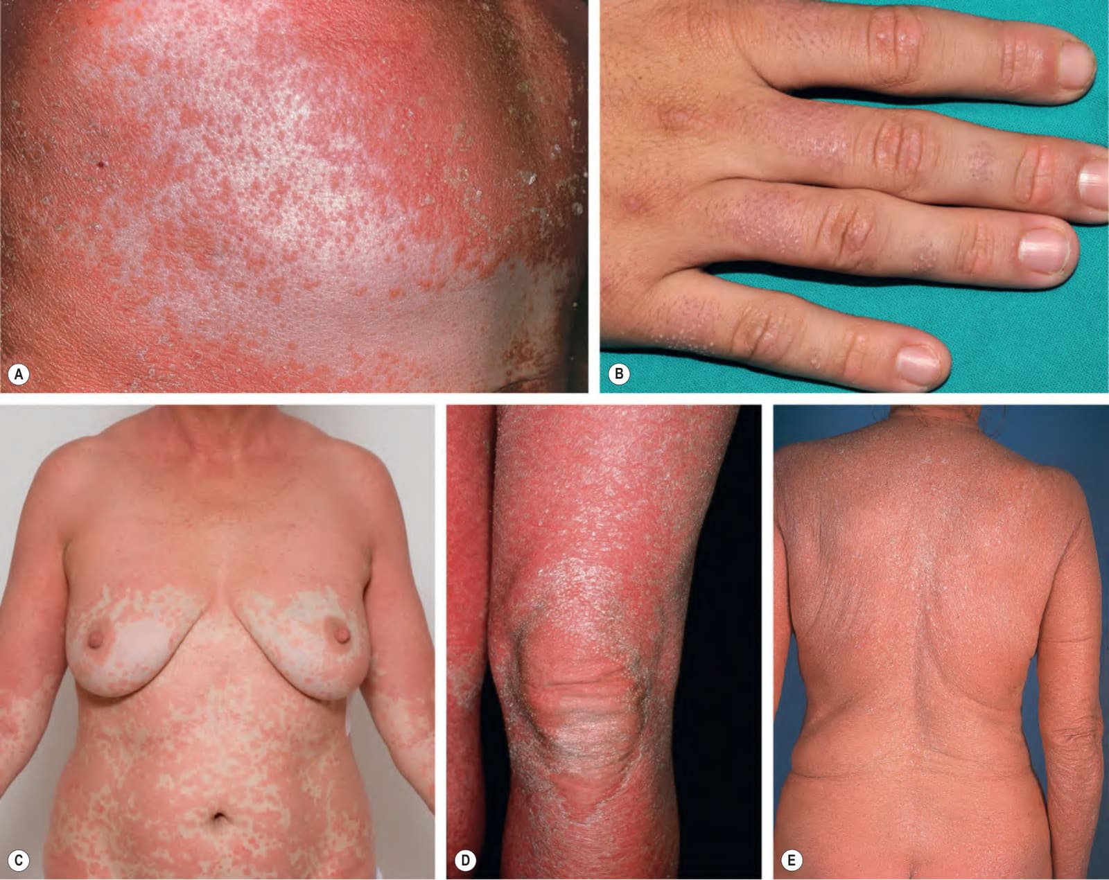
Fig. 9.5 Pityriasis rubra pilaris.A Orange–red follicular papules with coalescence into large scaly plaques, in addition to islands of sparing; follicular hyperkeratosis is often easier to appreciate within the relatively spared islands. B On the dorsal aspects of the digits, clustered keratotic follicular papules can resemble a “nutmeg grater”. C The islands of sparing may be sharply demarcated; note the salmon color of the plaques. Both photoaggravated and phototriggered forms of PRP exist. Patients can eventually become diffusely erythrodermic, but follicular accentuation of the scale (D) and small islands of sparing (E) can provide clues. B, Courtesy Antonio Torrelo, MD; C, Used with permission of the Mayo Foundation for Medical Education and Research.
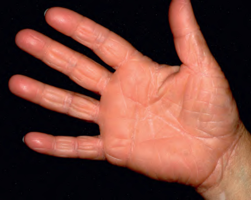
Fig. 9.6 Pityriasis rubra pilaris. The orange–red color of the skin, including that of the palmar keratoderma, aids in the clinical diagnosis. Courtesy Luis Requena, MD.
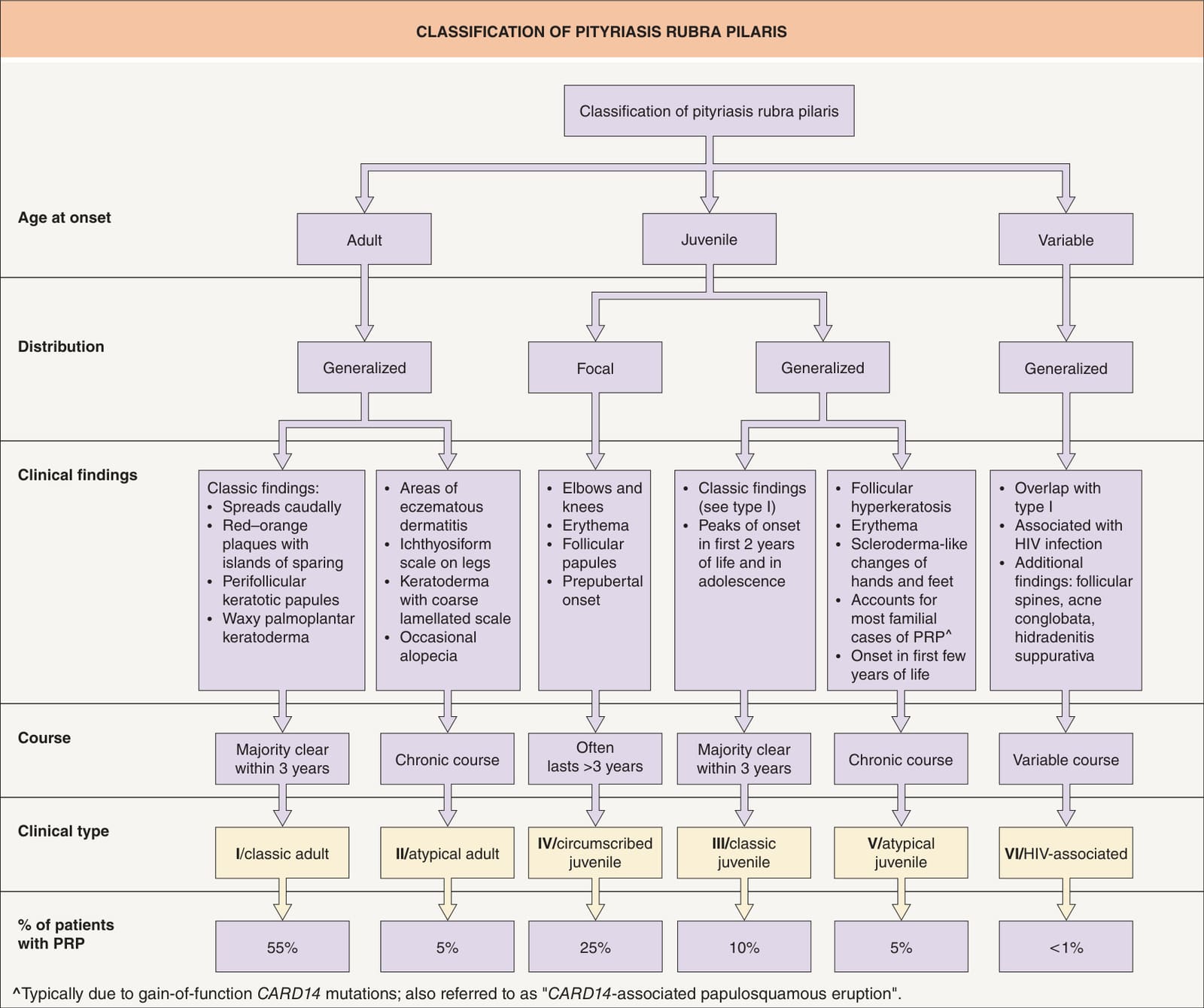
Fig. 9.7 Classification of pityriasis rubra pilaris (PRP). Type VI PRP is also referred to as HIV-associated follicular syndrome.
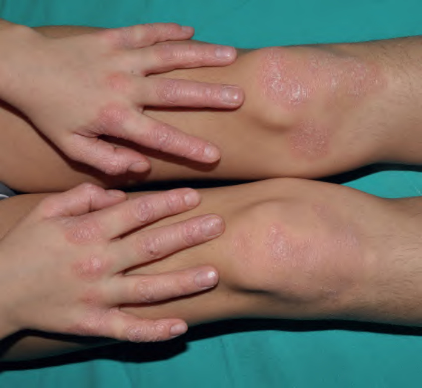
Fig. 9.8 Circumscribed juvenile pityriasis rubra pilaris (type IV). Symmetric pink plaques with scale are present on the dorsal aspect of the fingers, metacarpophalangeal joints, and knees. There are follicular keratotic papules on the knees. Courtesy Antonio Torrelo, MD.
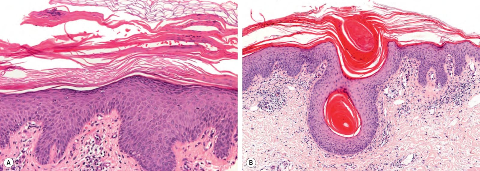
Fig. 9.9 Pityriasis rubra pilaris – histopathologic features. Psoriasiform dermatitis with alternating vertical and horizontal ortho- and parakeratosis (“checkerboard pattern”) (A), as well as follicular hyperkeratosis with parakeratosis in the shoulders of the follicular plug (B). Courtesy Lorenzo Cerroni, MD.