PITYRIASIS ROSEA
Synonym: Pityriasis rosea Gibert
Key features
A self-limited papulosquamous eruption that is occasionally pruritic
Seen primarily in adolescents and young adults, favoring the trunk and proximal extremities
Herald patch followed by lesions that are usually oval in shape and oriented with their long axes along Langer cleavage lines
Less common variants include inverse, vesicular, purpuric, and pustular
Introduction
Pityriasis rosea is a common, “acute”, self-limited papulosquamous eruption that favors otherwise healthy adolescents and young adults. Its striking appearance often prompts a request for medical evaluation. There is an absence of significant systemic manifestations, and its spontaneous resolution provides great consolation to the patient. Although the classic presentation is readily recognized, atypical forms may present a greater challenge. A viral etiology has been postulated, but this remains unproven.
History
Robert Willan described “roseola annulata” in 1798 as a self-limited eruption in otherwise healthy children. In 1860, the French physician Camille Melchior Gibert first named the condition pityriasis (scaly) rosea (pink). This aptly conveyed the clinical features and remains the name that is still used today.
Epidemiology
Most cases of pityriasis rosea occur in young healthy persons, the majority of whom are between the ages of 10 and 35 years. Its peak incidence is during adolescence, and it is rarely diagnosed before the age of 2 years. There is no racial predilection and it is found worldwide. There may be a slight female predominance. The typical eruption lasts 6 to 8 weeks, though exceptionally it can persist for 5 months or longer. Some authors report a modest seasonal variation with peaks in the spring and fall or in some countries during the rainy season.
Pathogenesis
The precise cause of pityriasis rosea remains elusive, and although a viral etiology is frequently proposed, this remains controversial. While attention has focused on human herpesvirus-7 (HHV-7) and, less so, on HHV-6, some studies have shown no difference in the prevalence of DNA from HHV-6 or HHV-7 in peripheral blood mononuclear cells of patients with pityriasis rosea. An association with HHV-8, H1N1 influenza virus, and SARS-CoV-2 has also been reported. Proponents of a viral etiology point to the prodromal symptoms experienced by some patients, the clustering of cases, and the rarity of recurrent episodes, suggesting an immunologic defense against an infectious agent. Pityriasis rosea is considered a prototypic paraviral exanthem, i.e. a characteristic skin disease associated with a possible viral infection or reactivation, but without virus identified in the cutaneous lesions.
Clinical Features
Although classic pityriasis rosea is usually easily recognized, the more unusual variants can be more challenging to diagnose. In the classic presentation, a solitary lesion appears on the trunk and enlarges over several days (Fig. 9.10). Less often, this initial lesion is seen on the neck or proximal extremities. It predates the remainder of the eruption by hours to days and is referred to as the “herald patch” because it heralds the onset of the disease. Its incidence varies, but in most series it has been seen in over 50% of cases. Multiple herald patches have also been reported.
The herald patch is a pink- to salmon- to pink–brown-colored patch or plaque with a slightly raised advancing margin (Fig. 9.11). The size commonly varies from 2 to 4 cm, but it can be as small as 1 cm or as large as 10 cm. The center shows the characteristic small fine scales
Typically, within the next few days, there is a blossoming of lesions on the trunk and proximal extremities. These numerous smaller thin papules and plaques are reminiscent of the larger herald patch that predated them; in more darkly pigmented skin, the lesions tend to be more papular, folliculocentric, and hyperpigmented (Fig. 9.12). Again, there is a subtle advancing border, central fine scaling, and the characteristic collarette of scale (Fig. 9.13A,B). The lesions are usually round to oval in shape, with their long axis following Langer cleavage lines (Fig. 9.13C). On the posterior trunk, such an orientation leads to what is often referred to as a “fir tree” or “Christmas tree” pattern. Minute pustules can also be seen during this initial phase of pityriasis rosea.
The face, palms, and soles are usually (but not always) spared. When all these features are present, the diagnosis is easiest to establish.
The eruption of pityriasis rosea usually persists for 6–8 weeks and then spontaneously resolves; however, occasional patients have lesions that may last 5 months or longer. In the latter situation, the possibility of pityriasis lichenoides chronica arises. Most patients with pityriasis rosea experience few symptoms, but in some individuals, pruritus is noted that is mild to severe.
Atypical forms of pityriasis rosea require a more astute observer. Inverse pityriasis rosea involves the axillae and inguinal areas (see Fig. 9.13B) and sometimes the face. It is more common in younger children and in those with darker skin phototypes. Oral lesions are also uncommon, but they may be underreported and palmoplantar involvement is unusual. Urticarial, erythema multiforme-like, vesicular, pustular, and purpuric variants have also been described.
Pathology
The microscopic features of pityriasis rosea include spongiotic dermatitis, along with elongation of the rete ridges and enlargement of the dermal papillae. Small mounds of parakeratosis and a predominantly papillary dermal infiltrate comprised of lymphocytes and histiocytes are also common features (Fig. 9.13D). There may be mild erythrocyte extravasation with scattered intraepidermal erythrocytes. In severe cases, these features are more pronounced, including the rare formation of intraepidermal pustules.
Most patients with pityriasis rosea do not have biopsies performed, because the clinical picture is quite characteristic.
Differential Diagnosis
Pityriasis rosea can sometimes be difficult to distinguish from secondary syphilis. The presence of cutaneous signs such as split papules and condyloma lata points to syphilis, as does the history or presence of a primary chancre. Patients with secondary syphilis usually have more systemic complaints, and they often have peripheral lymphadenopathy. Histologically, the absence of plasma cells favors pityriasis rosea and immunohistochemical staining for spirochetal antigens can also be done (see Ch. 0). Serologic testing allows the diagnosis of secondary syphilis to be confirmed.
There are drug eruptions that can mimic pityriasis rosea; they have been reported in patients receiving angiotensin-converting enzyme (ACE) inhibitors, β-blockers, metronidazole, and isotretinoin, as well as arsenic, bismuth, gold, barbiturates, clonidine, hydrochlorothiazide, imatinib, omeprazole, terbinafine, TNF inhibitors, NSAIDs, and multiple vaccines. A drug-induced pityriasis rosea-like eruption is often slower to resolve than the idiopathic form.
The herald patch or generalized eruption can resemble tinea corporis, tinea versicolor, or nummular dermatitis. The presence of the collarette of scale, the orientation of the lesions, and the history can help distinguish pityriasis rosea from nummular dermatitis. Guttate psoriasis usually has a thicker scale, smaller size, and lacks the fir-tree distribution. Other papulosquamous disorders such as pityriasis lichenoides should also be considered, especially when lesions last longer than 4 months.
Treatment
Because pityriasis rosea is often asymptomatic and self-limited, patient education and reassurance represent a satisfactory treatment plan. In patients with pruritus, counterirritant antipruritic lotions or low- to medium-strength topical corticosteroids may be needed for symptomatic relief. In more severe cases, UVB phototherapy (broadband or narrowband) or natural sunlight exposure and oral antihistamines can be used. Rarely, a brief course of systemic corticosteroids may be required. In a 2019 Cochrane review, oral acyclovir was found to lead to “good or excellent” improvement of lesions with inconclusive effect on pruritus and oral erythromycin was shown to decrease pruritus. However, a pathogenetic explanation for these findings is not clear.
Lastly, in pregnant women, there is a possible association between the development of pityriasis rosea and spontaneous abortion when there is active HHV-6 infection, in particular during the first 15 weeks of gestation.
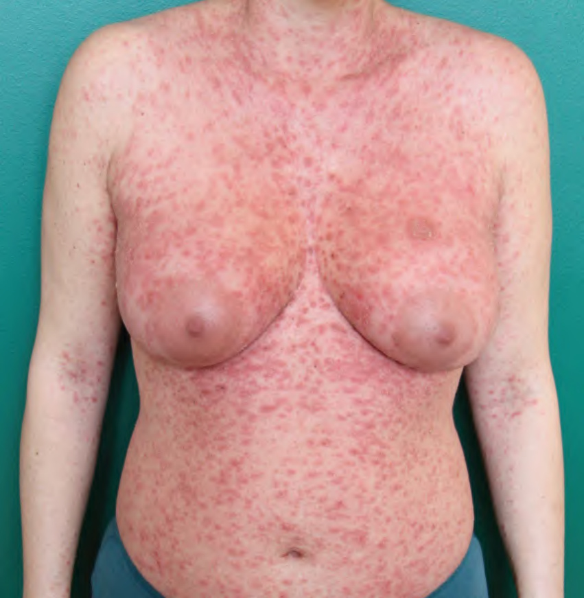
Fig. 9.10 Pityriasis rosea with a herald patch on the left breast. Numerous symmetrically distributed pink papules on the trunk that are round or oval in shape; associated scale is most obvious on the neck and breasts. Fewer lesions are noted on the flexor surfaces of the arms.
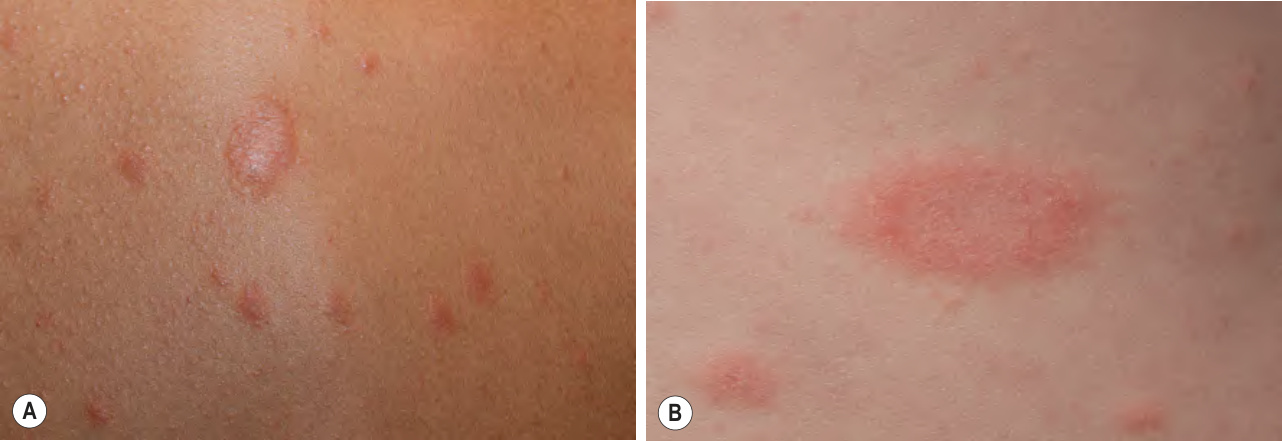
Fig. 9.11 Pityriasis rosea.A, B Herald patches in two patients with different skin phototypes. Either central or peripheral white scale can be seen. A, Courtesy Kalman Watsky, MD; B, Used with permission of the Mayo Foundation for Medical Education and Research of pityriasis rosea, and the margin has a larger, more obvious trailing collarette of scale with the free edge pointing inwards. There may be a mild prodrome with headache, fever, arthralgias, or general malaise. On occasion, the herald patch appears simultaneously with the more widespread eruption.
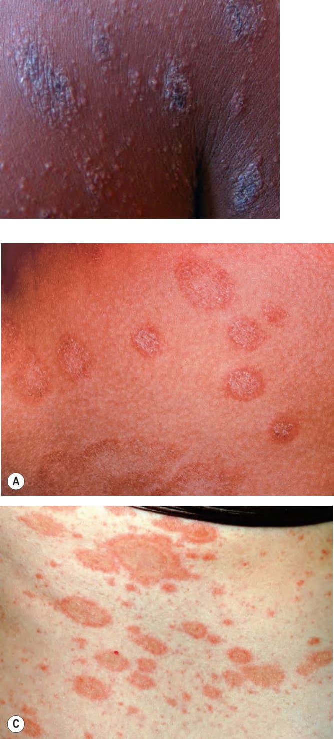
Fig. 9.12 Follicular accentuation of pityriasis rosea in darkly pigmented skin. Note the central hyperpigmentation and the small follicular papules. Courtesy Aisha Sethi, MD.
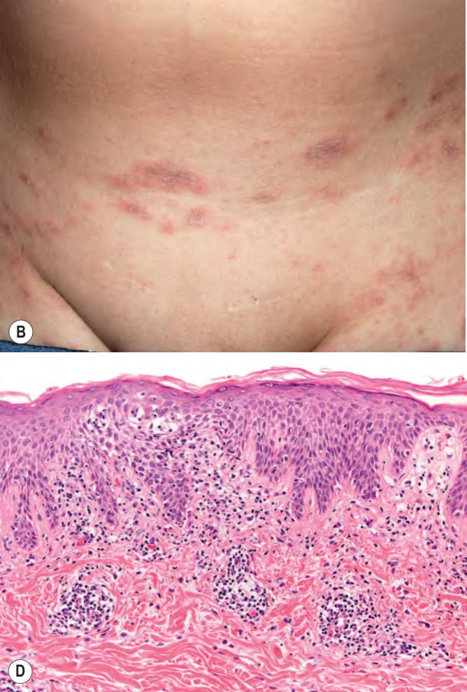
Fig. 9.13 Pityriasis rosea.A Both oval and round plaques can be seen as well as central fine scale. B A collarette of scale can also be seen and when lesions are limited to the pelvic region and/or axillae, the pattern of involvement is termed inverse. C A more inflammatory border may be seen and the long axes of the oval plaques follow skin cleavage lines. D Histopathologically, parakeratosis, mild spongiosis associated with exocytosis of lymphocytes into the epidermis, perivascular dermal lymphocytic infiltrates, and extravasated red blood cells are seen. B, Courtesy Luis Requena, MD; C, Courtesy Julie V. Schaffer, MD; D, Courtesy Lorenzo Cerroni, MD.