PITYRIASIS ROTUNDA
Synonyms: Tinea circinata Pityriasis circinata
Key features
Asymptomatic dermatosis seen primarily in the Far East and
Mediterranean basin and in individuals of African descent
There are large circular and polycyclic scaly patches with sharp margins that are typically hyperpigmented > hypopigmented and localized to the trunk and proximal extremities
Histologic features are similar to those of ichthyosis vulgaris
Introduction
Pityriasis rotunda is an unusual dermatosis that can occur alone or in association with a number of unrelated conditions. There are familial cases that may represent a minor acquired ichthyosis.
History
Pityriasis rotunda was first described by Toyama in 1906 as “tinea circinata” and was later named pityriasis rotunda by Matsura.
Epidemiology
Pityriasis rotunda is most commonly observed in adults in their 30s and in children and has a slight female predominance. It has been described in the Far East (Japan and China), the Mediterranean basin (Morocco, Italy, and Israel), and in Africans, African-Americans, and Afro-Caribbeans.
Pathogenesis
No clear unifying cause has been identified. In adults, possible associations include malnutrition, mycobacterial diseases (tuberculosis and leprosy), malignancies (e.g. hepatocellular carcinoma, gastric carcinoma, multiple myeloma), and hepatic cirrhosis. However, the majority of
patients, especially children, are in good health. An absence of profilaggrin N-terminal domain and decreased expression of filaggrin-2 have been observed in some patients.
Clinical Features
Pityriasis rotunda is typically asymptomatic and is characterized by large circular and polycyclic lesions that are often 10 cm (but may be up to 30 cm) in diameter (Fig. 9.14). They have associated fine scale and are moderately hyperpigmented with a sharp margin and no inflammation. The trunk and extremities are favored. A hypopigmented halo has been described in some patients, and sometimes the entire lesion can be hypopigmented.
Grimalt proposed categorizing pityriasis rotunda into two groups. Type I is seen primarily in Black and Asian individuals as hyperpigmented lesions, possibly in association with malnutrition or internal malignancy but with no family history. Type II occurs in younger White individuals with numerous (>30) lesions who sometimes have a family history, but no associated diseases.
Pathology
Pityriasis rotunda has microscopic features of ichthyosis vulgaris, i.e. an absent or diminished granular cell layer and moderate hyperkeratosis without parakeratosis. Increased pigmentation of the basal cell layer, pigment incontinence, a perivascular lymphohistiocytic infiltrate, and follicular plugging may also be observed. Special stains for fungi are negative.
Differential Diagnosis
The differential diagnosis includes tinea corporis, tinea versicolor, leprosy, and MF. The combination of fungal culture and routine histology excludes major entities in the differential diagnosis. The hypopigmented variant of pityriasis rotunda can resemble one variant of progressive macular hypomelanosis, which has no associated scale and lacks the histologic features of ichthyosis vulgaris.
Treatment
Pityriasis rotunda is relatively difficult to treat. Trials with topical lactic acid, urea, tars, emollients, and corticosteroids have provided little benefit. Topical tretinoin cream can result in modest improvement and systemic retinoids warrant consideration for patients with more extensive disease. Spontaneous remission is possible.
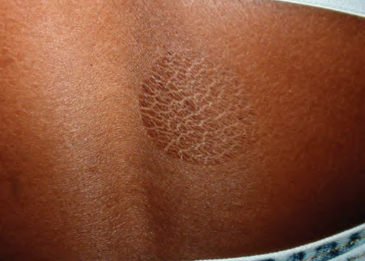
Fig. 9.14 Pityriasis rotunda. Circular, well demarcated, thin plaque with scale. Courtesy Richard M. Haber, MD.