GRANULAR PARAKERATOSIS
Synonyms: Axillary granular parakeratosis Intertriginous granular parakeratosis
Key features
Brownish-red keratotic papules that can coalesce into plaques
Adult form occurs more often in women and favors the axillae over other intertriginous areas
Infantile form is associated with diaper wearing or chemical irritation (e. g. use of benzalkonium chloride) and presents as bilateral inguinal plaques or erythematous geometric plaques underneath diaper pressure points
Histopathology is distinct, with retention of basophilic keratohyaline granules within areas of parakeratosis in the stratum corneum
A defect in processing profilaggrin to filaggrin is a proposed mechanism
Introduction
Granular parakeratosis is an acquired, typically pruritic disorder of the axillae and other intertriginous sites, e.g. the groin and inframammary and abdominal folds. It has distinct histopathologic findings, suggesting a disorder in keratinization.
History
Northcutt and colleagues first described granular parakeratosis in
1991. Multiple cases have been described since then in various clinical settings, suggesting a multifactorial reactive condition.
Epidemiology
Granular parakeratosis is observed most commonly in middle-aged women. Although it is less common in young adults, there are reports in infants and children linked to diaper-wearing and exposure to benzalkonium chloride in laundry rinse aids. Granular parakeratosis has been observed in all skin phototypes.
Pathogenesis
Granular parakeratosis is an acquired keratotic dermatosis originally linked to maceration and the use of various personal hygiene products, such as deodorants and antiperspirants. More recently, occlusion from diapers or chemical irritation from benzalkonium chloride (see above) has been implicated.
A disorder of keratinization characterized by a basic defect in the processing of profilaggrin to filaggrin has also been proposed, based upon ultrastructural and immunohistochemical studies, i.e. retention of keratohyalin granules within the stratum corneum due to a defect in filaggrin metabolism. This suggests a distinctive reactive pattern rather than a specific entity.
Clinical Features
The primary lesions are keratotic, brownish-red papules that can have a conical shape. They may coalesce into larger, well-demarcated plaques with various degrees of maceration secondary to local occlusion (Fig. 9.15). Lesions can persist for months or longer and recur. Pruritus is the most common complaint, but irritation is also a problem if there are erosions or fissures. Some patients experience a flare with an increase in ambient temperature and sweating. The axilla is the most common site of involvement, followed in frequency by other major body folds (e.g. inguinal, inframammary). Occasionally granular parakeratosis develops in the genital area.
In infants, bilateral plaques within the inguinal folds or erythematous geometric plaques underlying pressure points from the diaper can be seen. Larger lesions in other intertriginous sites have been observed in older children.
Pathology
The characteristic feature is an unusual form of parakeratosis. The stratum corneum is thickened and compacted with increased eosinophilic staining (Fig. 9.16). Retained nuclei are present throughout this keratin layer, creating parakeratosis. The most unusual feature is the
visible retention of basophilic keratohyalin granules within these areas of parakeratosis. Because clinically similar lesions may not have these characteristic histologic features, the encompassing term “hyperkeratotic flexural erythema” has been proposed.
Differential Diagnosis
The differential diagnosis includes the most common causes of intertrigo (e.g. seborrheic dermatitis, candidiasis, inverse psoriasis, erythrasma) as well as Hailey–Hailey disease, Darier disease, and pemphigus vegetans. Irritant or allergic contact dermatitis also needs to be considered in some patients. A biopsy will confirm the diagnosis of granular parakeratosis.
Treatment
Based upon case reports and small series, therapeutic success has been reported with topical corticosteroids, vitamin D analogues, retinoids, ammonium lactate, and antifungals. In addition, cryotherapy, laser therapy, and oral isotretinoin or antibiotics have been used. Spontaneous resolution has also been observed (including in infants), as have relapses.
Additional figures available in our eBook (see inside front cover for access code).
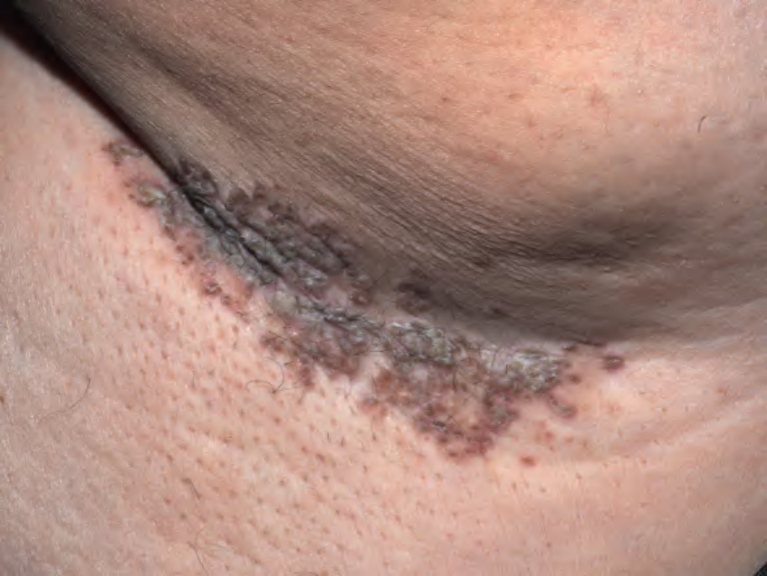
Fig. 9.15 Granular parakeratosis involving the axilla. Coalescing brown papules with hyperkeratosis and slight maceration. Courtesy Kalman Watsky, MD.
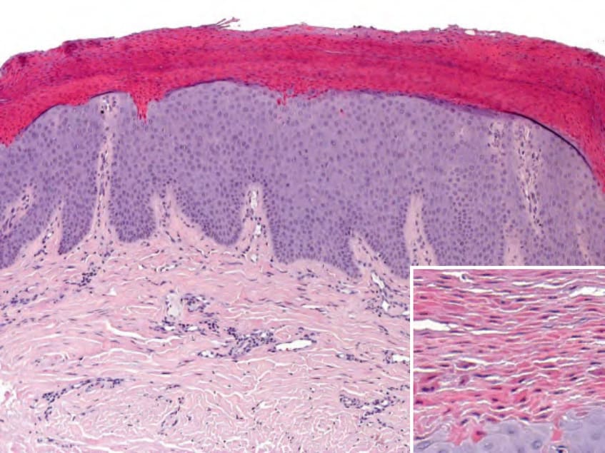
Fig. 9.16 Axillary granular parakeratosis – histopathologic findings. Marked, compact parakeratosis with small bluish granules within the stratum corneum representing keratohyaline granules (inset). Courtesy Lorenzo Cerroni, MD.